Automated Determination of Lymph Nodes in Scanned Images

a lymph node and scanned image technology, applied in the field of automatic determination of lymph nodes in noninvasive scanned images of the body, can solve the problems of inappropriate treatment of cancer patients, difficult and tedious detection of lymph nodes, and the inability to identify a structure as a lymph node which is not cancerous, and achieve the effect of avoiding cancer and avoiding cancerous patients

Inactive Publication Date: 2008-12-25
THE TRUSTEES OF COLUMBIA UNIV IN THE CITY OF NEW YORK
View PDF11 Cites 65 Cited by
  • Summary
  • Abstract
  • Description
  • Claims
  • Application Information

AI Technical Summary

Benefits of technology

[0020]In other embodiments, an apparatus, or logic encoded in one or more tangible media, or instructions en

Problems solved by technology

The detection of lymph nodes, especially small lymph nodes, is difficult and tedious for a radiologist, because lymph nodes are “intertwined” with normal anatomic structures such as blood vessels and the bowel.
This can result in inappropriate treatment for their cancer, for instance, by subjecting the patient to unnecessary surgery that would not have been performed if it had been recognized that the tumor had involved a lymph node.
Identification of a structure as a lymph node which is not, could have similar dire consequences.
Lacking correct treatment, the patient's cancer may spread unnecessarily and threaten the patient's health and life.
However, the measurement of tumors on medical imaging is a manual and subjective task performed by a trained radiologist according to current clinical practice.
Older model CT scanners produced transverse plane images (called slices) with tw

Method used


Image

  • Automated Determination of Lymph Nodes in Scanned Images
  • Automated Determination of Lymph Nodes in Scanned Images
  • Automated Determination of Lymph Nodes in Scanned Images

Examples


Embodiment Construction

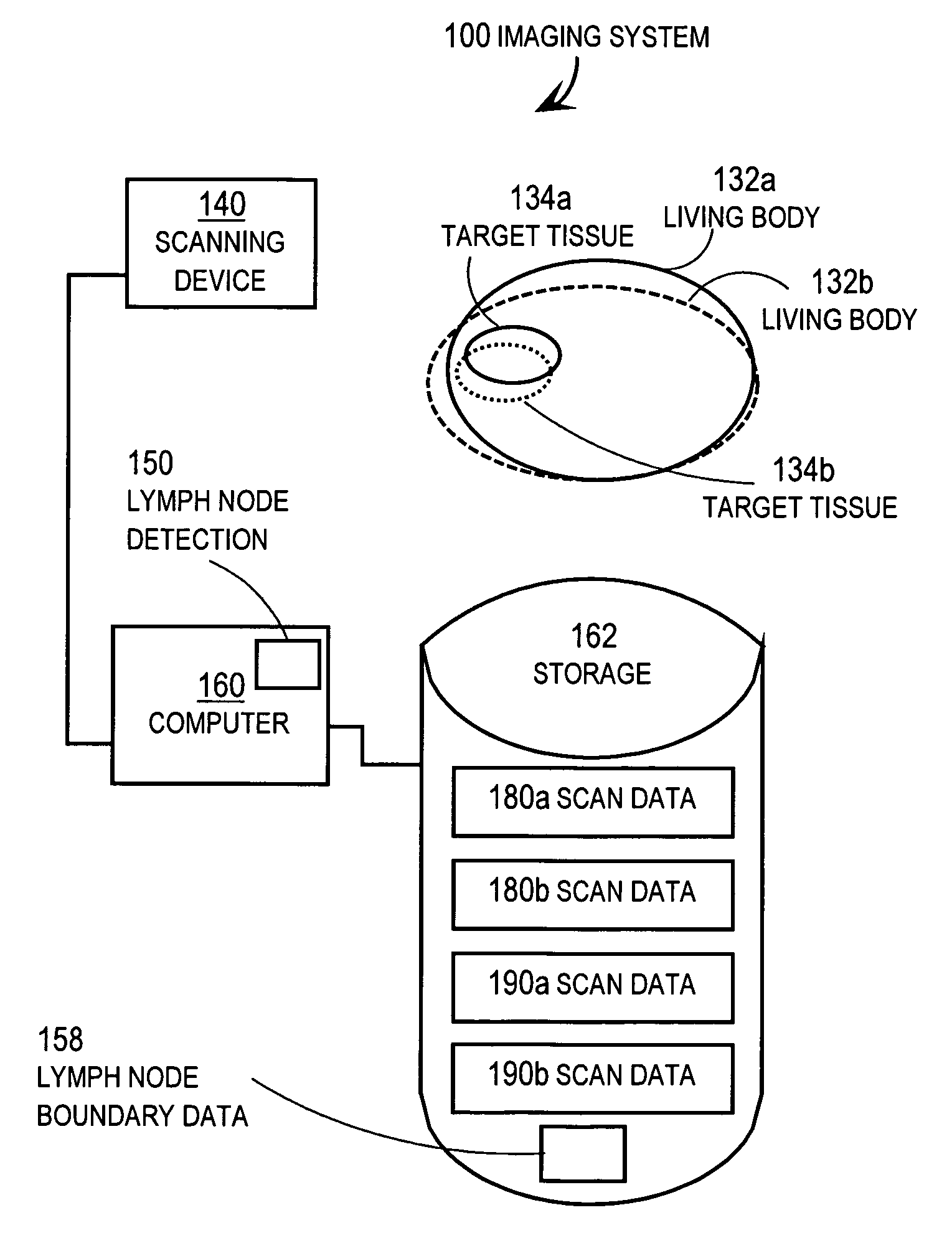
[0051]A method and apparatus are described for automatically detecting or segmenting lymph nodes, or both, in non-invasive scanned images of a body. In the following description, for the purposes of explanation, numerous specific details are set forth in order to provide a thorough understanding of the present disclosure. It will be apparent, however, to one skilled in the art that an embodiment may be practiced without these specific details. In other instances, well-known structures and devices are shown in block diagram form in order to avoid unnecessarily obscuring the present disclosure.

[0052]Some embodiments are described in the context of lymph nodes in CT scanned images of a human body. However, the invention is not limited to this context. In other embodiments lymph nodes or spatially related anatomical objects, such as the aorta, liver and inferior vena cava, are detected or segmented in scans of human or animal bodies using any scanning device, such as CT scanners, MRI sc...


PUM


Abstract

Techniques include automatically detecting a lymph node in a scanned image of a body without human intervention, using one or more of three approaches. First, a subset of scanned images is determined, which belongs to one anatomical domain. A search region for lymph tissue is in a particular spatial relationship outside an anatomical object in the domain. Second, scanned images are segmented without human intervention to determine a boundary of a particular lymph node. The scanned images and outline data are received. Some of these embodiments automatically segment by determining an external marker, based on the outline data, and an internal marker, based on a geometric center of the outline data or thresholds determined automatically inside detected edges, or both, for a marker-controlled watershed algorithm. Third, based on lymph node data at a particular time, a second scanned image at a different time is segmented automatically, without human intervention.

Description

CROSS-REFERENCE TO RELATED APPLICATIONS[0001]This application claims benefit of Provisional Appln. No. 60 / 945,333, filed Jun. 20, 2007, the entire contents of which are hereby incorporated by reference as if fully set forth herein, under 35 U.S.C. §119(e).BACKGROUND[0002]1. Technical Field[0003]The present disclosure relates generally to automated determination of lymph nodes in non-invasive scanned images of a body, such as in X-ray computed tomography (CT) scans and magnetic resonance imagery (MRI) scans of a human body.[0004]2. Background[0005]One way that tumors spread is through a patient's lymphatic system. Lymph node metastasis is a significant prognostic factor in many types of primary solid and hematologic malignancies. A patient's appropriate staging for either surgical or medical therapy depends upon the recognition and proper evaluation of lymph nodes throughout the body. A patient's disease prognosis also depends on the extent of lymph node involvement with a tumor at t...

Claims


Application Information

Patent Timeline
IPC IPC(8): A61B5/00G06V10/26
CPCA61B6/03G06K9/34G06K2209/053G06T7/0012G06T2207/30096G06V10/26G06V2201/032
Inventor SCHWARTZ, LAWRENCE H.ZHAO, BINSHENGYAN, JIAYONG
Owner THE TRUSTEES OF COLUMBIA UNIV IN THE CITY OF NEW YORK
Why Eureka
Try Eureka