Process for Decellularizing Soft-Tissue Engineered Medical Implants, and Decellularized Soft-Tissue Medical Implants Produced

a technology of engineered medical implants and soft tissues, which is applied in the direction of biocide, prosthesis, drug compositions, etc., can solve the problems of proteolytic degradation of the matrix structure of the processed tissues, prohibitively time-consuming prior art processing methods, and easy requiring numerous days

Inactive Publication Date: 2009-02-12
LIFENET HEALTH
View PDF34 Cites 41 Cited by
  • Summary
  • Abstract
  • Description
  • Claims
  • Application Information

AI Technical Summary

Problems solved by technology

The prior art processing methods are prohibitively time consuming, easily requiring numerous days, for example anywhere from eight to twenty-one days total processing time.
Such long processing times result in proteolytic degradation of the matrix structures of the processed tissues.
While vascular prostheses are available for clinical use, they have met with limited success due to cellular and immunological complications, and the inability to remain patent and function.
These problems are especially pronounced for small diameter prostheses, i.e. less than about 6 mm.

Method used


Image

  • Process for Decellularizing Soft-Tissue Engineered Medical Implants, and Decellularized Soft-Tissue Medical Implants Produced
  • Process for Decellularizing Soft-Tissue Engineered Medical Implants, and Decellularized Soft-Tissue Medical Implants Produced
  • Process for Decellularizing Soft-Tissue Engineered Medical Implants, and Decellularized Soft-Tissue Medical Implants Produced

Examples


example 1

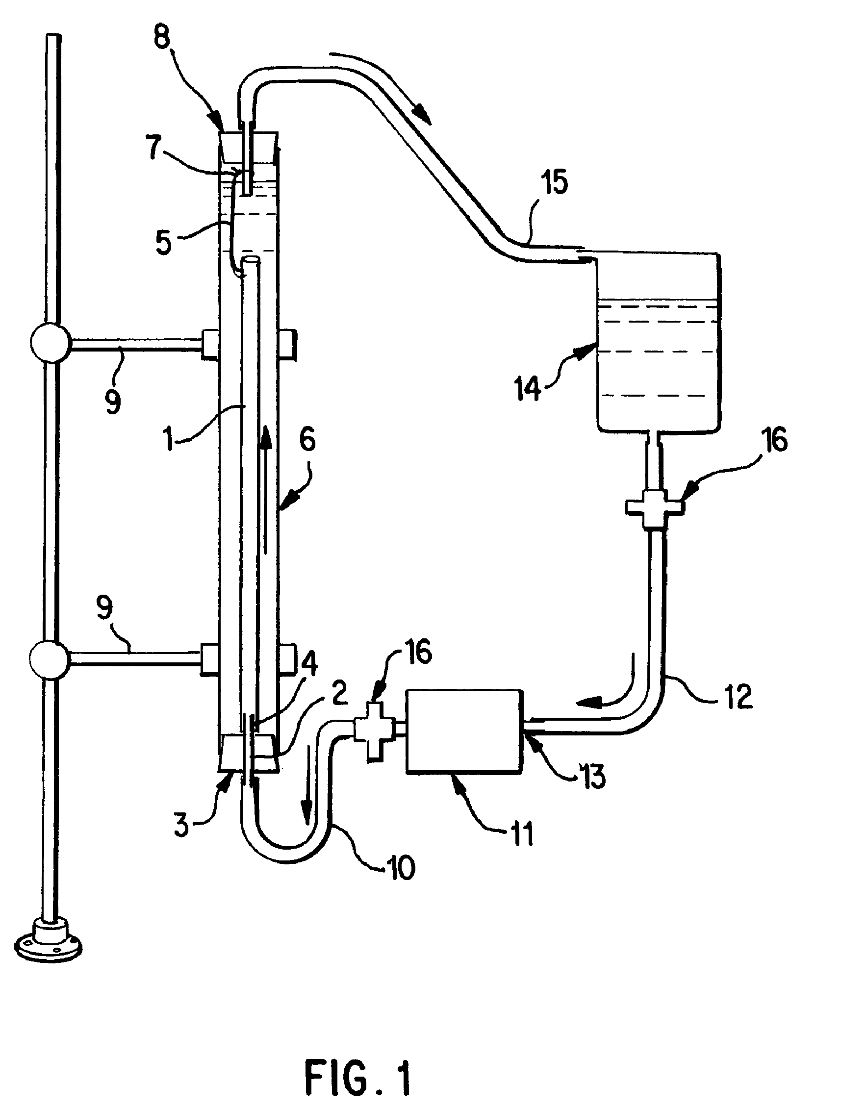
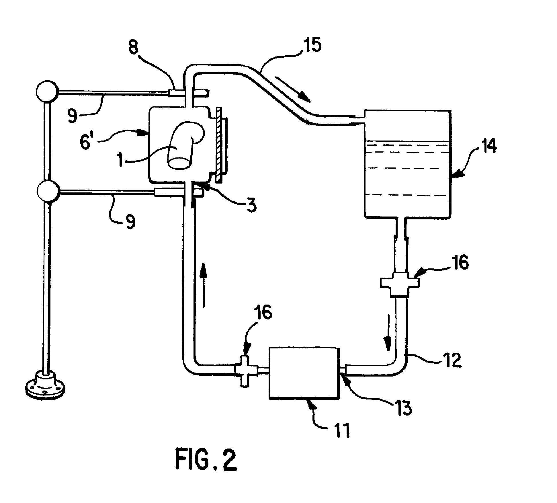
[0058]Saphenous vein tissues (two) from each leg of an acceptable human donor were carefully dissected under sterile conditions to remove all visible fat deposits and the side vessels were tied off using nonresorbable suture materials such that the ties did not occur in close proximity to the long run of the vessel. Sutures can restrict the decellularization process and the tissues under the sutures were removed following decellularization. For long vein grafts (40-60 cm) (FIG. 1), the distal ends of the veins were cannulated onto the ribbed attachment of the inlet port(s) and single sutures used to secure each vein. Additional suture lines were attached to the proximal ends of the veins. The veins were then removed from the dissecting solution (ultrapure water containing 50 mM Tris-HCl (pH 7.2), 5 μm EDTA, and one or more antibiotics) and transferred to the processing vessel which had been temporarily inverted. The second suture line along with the vein was passed through the proce...

example 2

[0059]Saphenous vein tissues (two) from each leg of an acceptable human donor were carefully dissected under sterile conditions to remove all visible fat deposits and side vessels were tied off using nonresorbable suture materials such that the ties did not occur in close proximity to the long run of the vessel. Sutures can restrict the decellularization process and the tissues under the sutures were removed following decellularization. For these long vein grafts (33 and 28 cm) (FIG. 1), the distal ends of the veins were cannulated onto the ribbed attachment of the inlet port(s) and single sutures used to secure each vein. Additional suture lines were attached to the proximal ends of the veins. At this point, the veins were removed from the dissecting solution (ultrapure water containing 50 mM Tris-HCl (pH 7.2), 5 mM EDTA, and one or more antibiotics and transferred to the processing vessel which had been temporarily inverted. The second suture line along with the vein was passed th...

example 3

[0060]Internal mammary artery tissues (two) from an acceptable human donor were carefully dissected under sterile conditions to remove all visible fat deposits and side vessels were tied off using nonresorbable suture materials such that the ties did not occur in close proximity to the long run of the vessel. Sutures can restrict the decellularization process and the tissues under the sutures were removed following decellularization. For short artery grafts (11 and 8 cm) (FIG. 1), one end of each artery were cannulated onto the ribbed attachment of the inlet port(s) and single sutures used to secure each arteries. The arteries were then removed from the dissecting solution (ultrapure water containing 50 mM Tris-HCl (pH 7.2), 5 mM EDTA, and one or more antibiotics) and transferred to the processing vessel which had been temporarily inverted. Prior to closing the processing vessel, a portion of the first processing (extracting) solution was gently added to the processing vessel and th...


PUM

PropertyMeasurementUnit
processing timeaaaaaaaaaa
diameteraaaaaaaaaa
thicknessaaaaaaaaaa

Abstract

The invention provides methodologies and apparatus for producing acellular soft-tissue implants, both in small quantities and in commercializable quantities. Such soft-tissue implants include vascular graft substitutes. An acellular graft is produced by subjecting the tissue sample to an induced pressure mediated flow of an extracting solution, followed by inducing a pressure mediated flow of a treating solution, then washing the treated tissue to produce the acellular graft. The acellular grafts produced are uniform and non-immunogenic. The inventive method allows for the production of multiple decellularized soft tissue implants, where processing time is significantly less than prior art processes and the number of implants produced per day is increased over prior art processes. In clinical use, the decellularized grafts produced exhibit significantly improved in long-term durability and function.

Description

[0001]This application is a continuation-in-part application of U.S. patent application Ser. No. 09 / 327,240, filed Jun. 7, 1999, hereby incorporated herein in its entirety.FIELD OF THE INVENTION[0002]The invention is directed toward methodologies and apparatus for use in the preparation of acellular, i.e. essentially lacking in living cells andi or non-living cells, soft-tissue implants, in small quantities and commercializable quantities. Such soft-tissue implants include vascular graft substitutes. These implants can be derived from tissue engineered soft tissue devices, tissue products derived from animal or human donors that contain or are devoid of cells, and that contain or are devoid of valve structures useful in directing the flow of fluids through tubular vascular devices, and / or combinations of natural tissue products and tissue engineered soft-tissue products. The invention includes methodologies and apparatus for producing uniform, gently processed, decellularized multip...

Claims


Application Information

Patent Timeline
Patent Type & Authority Applications(United States)
IPC IPC(8): A61K35/12A61P43/00A61K35/32A61K35/36A61K35/44A61L27/36C12N5/00C12N5/02C12N5/06
CPCA61K35/12A61K35/32A61K35/36A61K35/44A61L27/3633A61L27/3687A61L2430/40A61L27/3625A61L27/3604A61P43/00A61L2430/34
Inventor WOLFINBARGER, JR., LLOYDLANGE, PERRYLINTHURST JONES, ALYCEMOORE, ERICNOLF, BARRY
Owner LIFENET HEALTH
Why Eureka
Try Eureka