Bioluminescent Endoscopy Methods And Compounds

a bioluminescent and endoscopy technology, applied in the field of methods, can solve the problems of inability to see, damage to the ureter, and difficult observation of certain anatomical structures in conventional endoscopy under normal visible lighting conditions, and achieve the effects of preventing damage to these structures, facilitating detection of leakage or injury, and excellent instantaneous visualization

Active Publication Date: 2009-03-26
BIOLUME
View PDF51 Cites 11 Cited by
  • Summary
  • Abstract
  • Description
  • Claims
  • Application Information

AI Technical Summary

Benefits of technology

[0015]We have found that instillation of a bioluminescent solution into the bile duct, intestinal anastomosis, or ureter during surgery allows excellent instantaneous visualization to the surgeon, potentially preventing damage to these structures. These techniques may also facilitate recognition of leaks or injuries, greatly expediting

Problems solved by technology

However, certain anatomical structures are difficult to observe in conventional endoscopes under normal visible lighting conditions.
Similarly, the ureter can be damaged in surgery, especially in pelvic operations.
Although these injuries can be repaired, they may not be apparent until after the conclusion of the procedure.
Furthermore, the operator and assistants must put on lead gowns making this procedure inconvenient and time consuming.
However, selective intraoperative cholangiography as compared to routine intraoperative cholangiography (IOC) techniques in itself increases the risk of injury to the common bile duct (Flum D R, Dellinger E P, Cheadle A, Chan L, Koepsell T (2003

Method used


Image

  • Bioluminescent Endoscopy Methods And Compounds
  • Bioluminescent Endoscopy Methods And Compounds
  • Bioluminescent Endoscopy Methods And Compounds

Examples


example 1

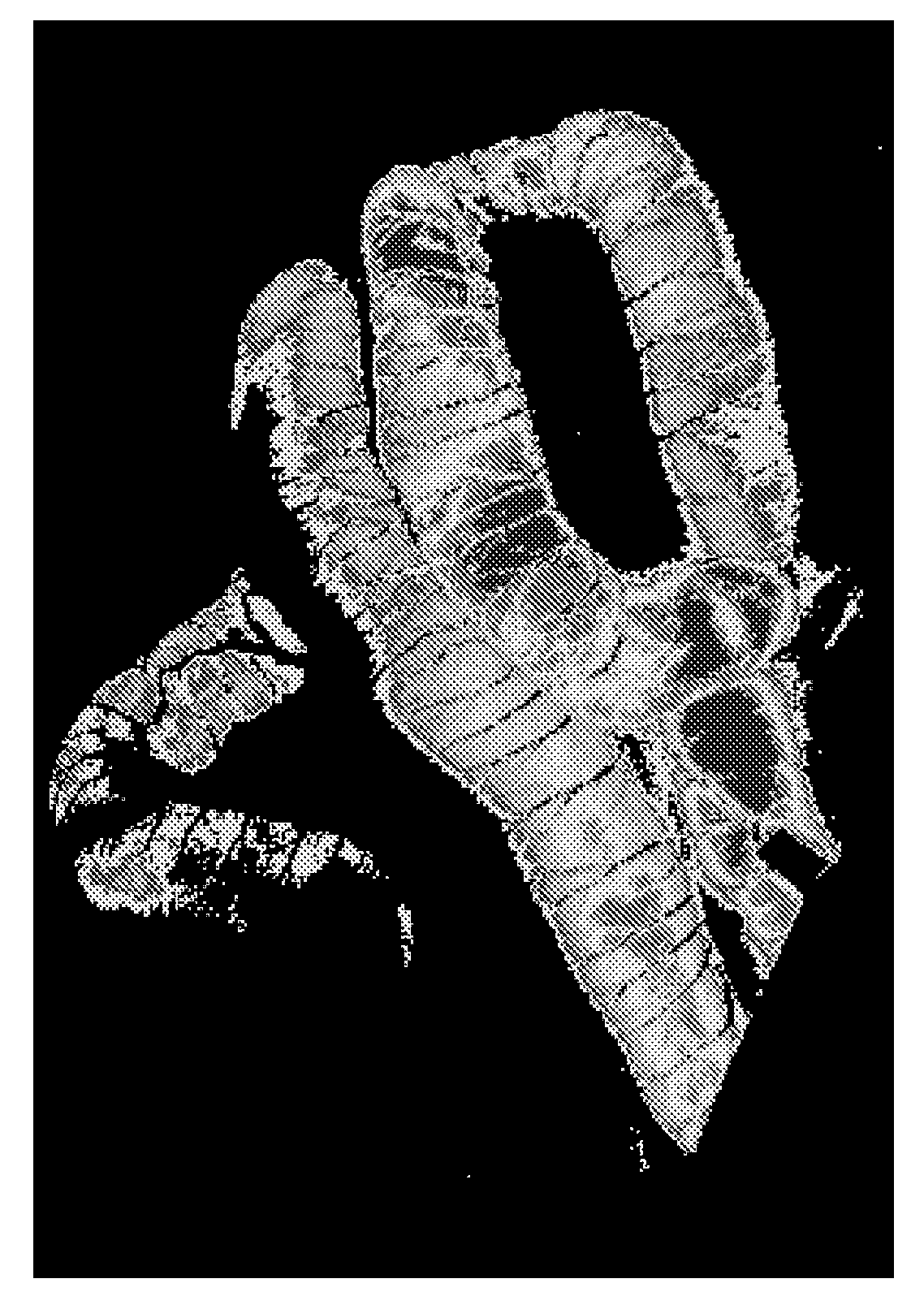
[0205]A rat was equipped with bilateral jugular cannulas. To one was applied a solution of coelenterazine (50 mM) in Hank's balanced salt solution and to the opposite jugular cannula was administered Renilla luciferase, 5 mg / ml in Hank's balanced slat solution. An image was obtained and was pseudocoloured with Scion Image according to the light level obtained by a Princeton Instruments camera and is shown at FIG. 19.

example 2

[0206]A rat was equipped with bilateral jugular cannulas. To one was applied a solution of coelenterazine (50 mM) in Hank's balanced salt solution and to the opposite jugular cannula was administered Renilla luciferase, 5 mg / ml in Hank's balanced slat solution. An image was obtained and was pseudocoloured with Scion Image according to the light level obtained by a Princeton Instruments camera and is shown at FIG. 20. FIG. 20 illustrates patchy contrast regions in the liver:

example 3

[0207]A rat was equipped with bilateral jugular cannulas. To one was applied a solution of coelenterazine (50 mM) in Hank's balanced salt solution and to the opposite jugular cannula was administered Renilla luciferase, 5 mg / ml in Hank's balanced slat solution. An image was obtained and was pseudocoloured with Scion Image according to the light level obtained by a Princeton Instruments camera and is shown at FIG. 21. FIG. 21 perfusion of bioluminescence in the whole animal:


PUM


Abstract

Bioluminescent endoscopy methods and compounds, wherein an anatomical object is examined by means of filling, perfusing, intubating, injecting, or otherwise administering a solution containing a bioluminescent substance or a mixture of luciferin and luciferase which produces bioluminescence, wherein a color or monochrome image of the object is constituted using the images and information based on bioluminescent emitted by the bioluminescent substance. Procedures are demonstrated which allow bioluminescent solutions to be perfused into certain tissue regions, such as but not limited to the common biliary duct, genitourinary tract, gastro-intestinal tract, cardiovascular system and lymphatic system wherein said structures may be conveniently visualized during surgery to avoid damage to these structures. Such images may also be combined with visual light images. Methods of detection of cancer cells using bioluminescence are provided. Preferred embodiments disclosed include membrane permeant coelenterazine analogs.

Description

CROSS-REFERENCE TO RELATED APPLICATIONS[0001]The present application claims the benefit under 35 U.S.C. §119(e) of the U.S. Provisional Patent Application Ser. No. 60 / 968,772, filed on Aug. 29, 2007, the content of which is incorporated herein by reference.FIELD OF THE INVENTION[0002]The present invention relates to methods and compositions for in vivo endoscopic diagnosis and surgical treatment in a living animal, bird, or man. Specifically, the present invention relates to a bioluminescent endoscopy methods, and compounds for use in bioluminescent endoscopy, by which images may be obtained by the use of appropriate cameras and signal processing algorithms, if necessary, from tissues which contain a bioluminescent compound and / or mixture of compounds which are injected, administered, or by any means whatsoever placed within said tissue.BACKGROUND OF THE INVENTION[0003]In recent years endoscopes capable of viewing a patient or test subject have been widely employed. An endoscope can...

Claims


Application Information

Patent Timeline
IPC IPC(8): A61K49/00C07D487/02C12Q1/66
CPCC07D487/04G01N21/763G01N21/6456
Inventor NEFF, PHILIP T.MURPHY, RANDALL B.BRYAN, BRUCE
Owner BIOLUME
Why Eureka
Try Eureka