
Method for Treating Disseminated Cancer

Inactive Publication Date: 2009-03-26
SENTOCLONE INT AB
View PDF15 Cites 9 Cited by
  • Summary
  • Abstract
  • Description
  • Claims
  • Application Information

AI Technical Summary

Benefits of technology

[0057]As mentioned above the first phase i) is initiated by adding at least one substance having agonistic activity towards the IL-2 receptor. The function of such substances is to stimulate T-lymphocytes via the IL-2 receptor to promote cell division of T-lymphocytes, thereby preventing cell death.
[0105]Accordingly, the present invention relates to a method for the preparation of memory T-lymphocytes. Normally, when a culture of tumour-reactive T-lymphocytes are expanded according to the present invention from about 35% to about 90% of tumour-reactive T-lymphocytes of the memory type, such as, e.g. from about 40% to about 90%, from about 50% to about 80% or from about 60% to about 70%, will be obtained. The present inventors speculates that the fact that the lymphocytes in phase i) are allowed to regenerated before tumour antigen is added, together with the relatively slow expansion phase leads to formation of a high ratio of memory lymphocytes to effector lymphocytes.

Problems solved by technology

However, up till now, it has not been known that sequential lymphatic drainage involving a certain individual lymph node applies to metastasis as well, i.e. that for a metastasis one or more lymph nodes draining the metastasis area can be detected.

Method used


Image

  • Method for Treating Disseminated Cancer
  • Method for Treating Disseminated Cancer
  • Method for Treating Disseminated Cancer

Examples


example 1

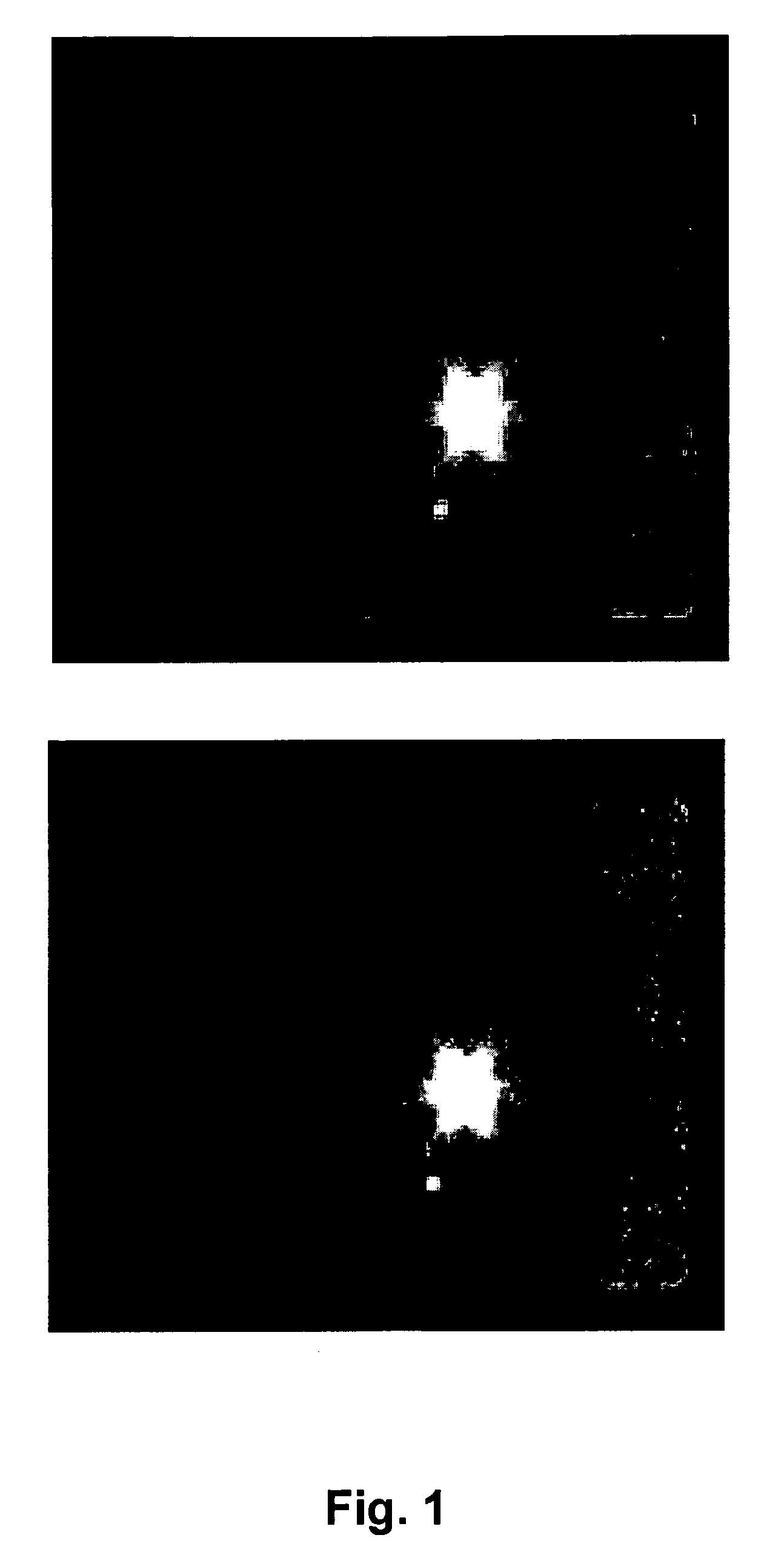
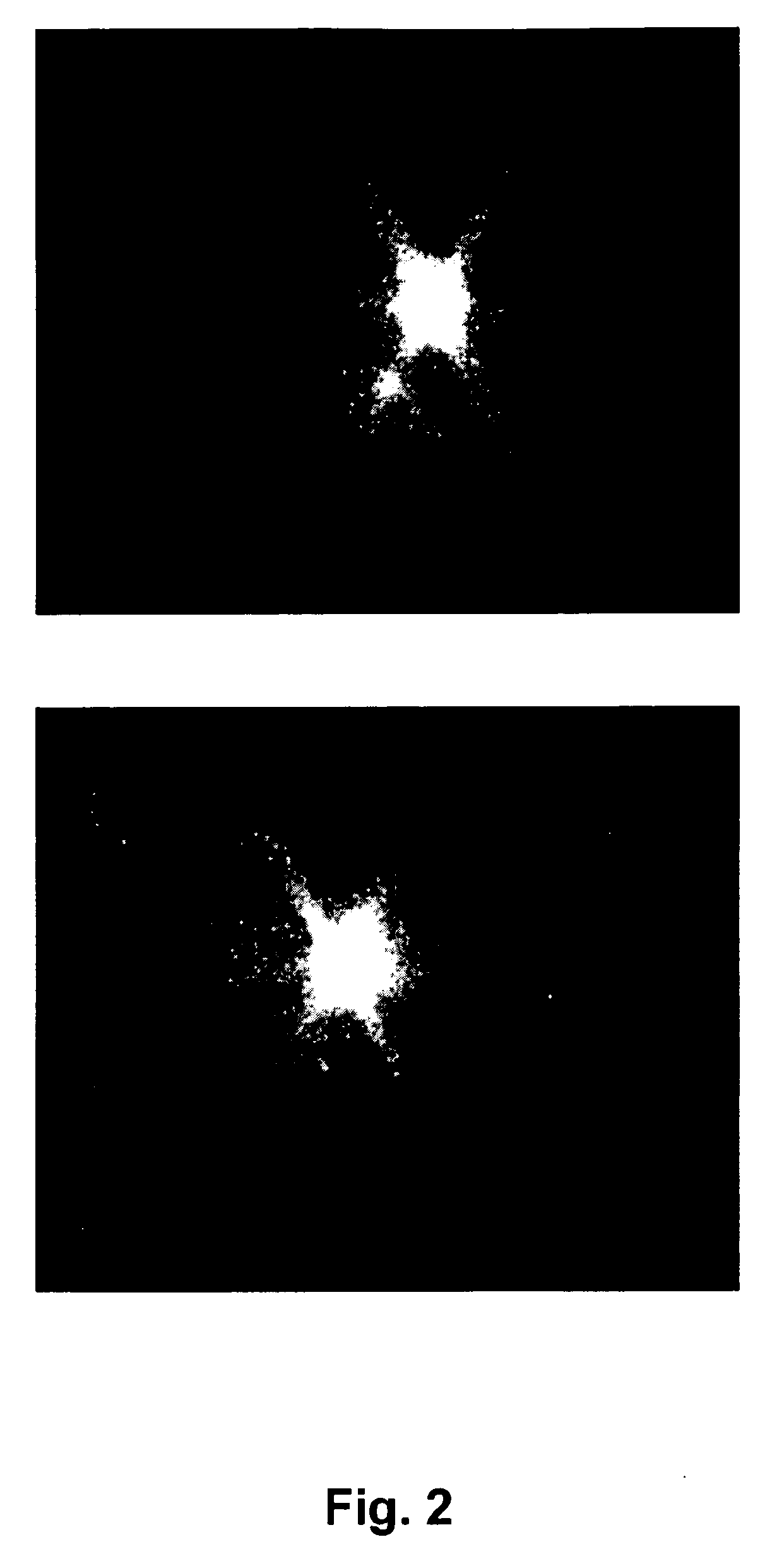
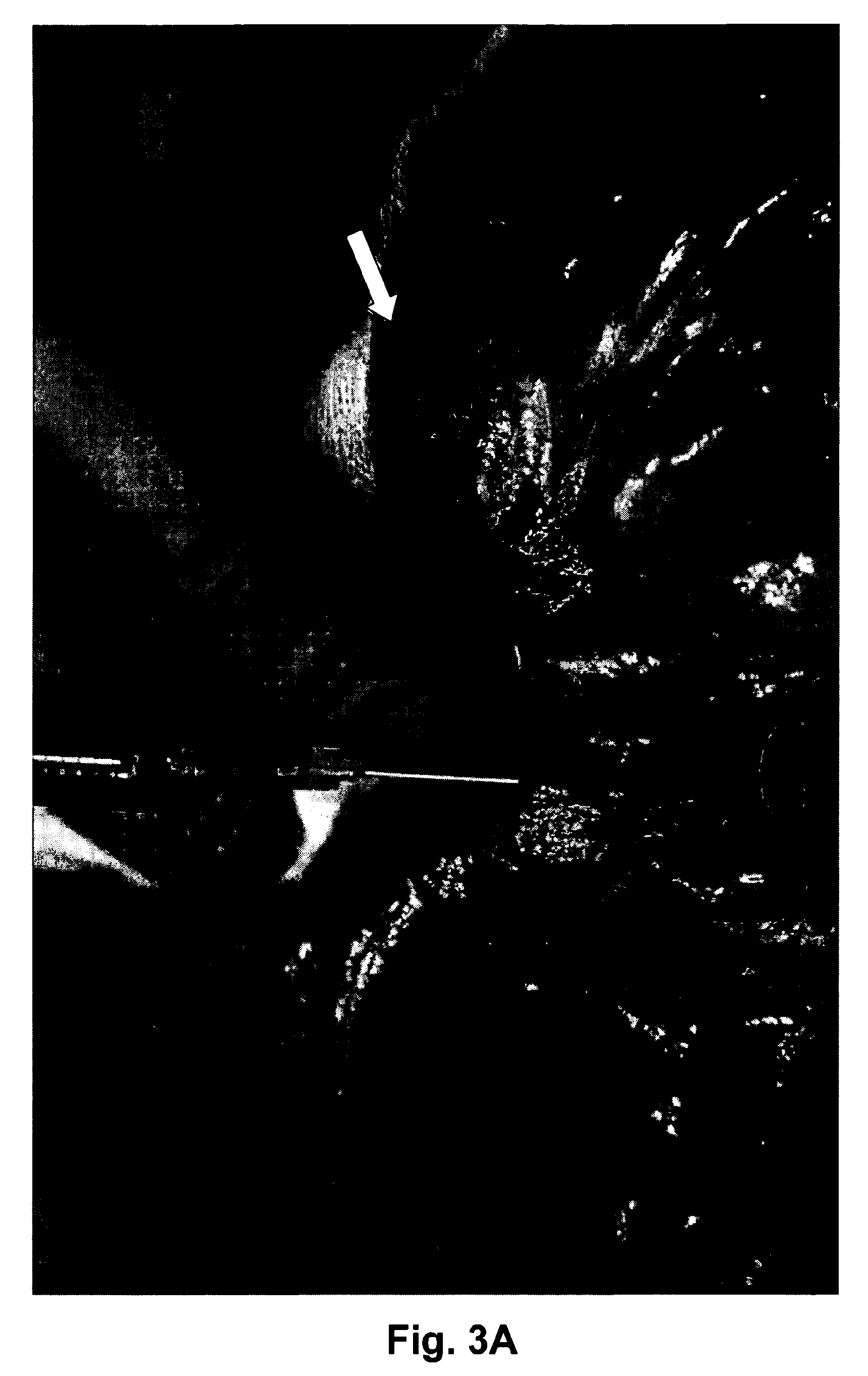
Identification of Metinel Lymph Nodes for Metastasis

[0136]Eight patients (four women and four men) were included in the study (see Table 1); the average age was 61.4 years. One of them underwent two operations with localisations of metinel nodes at both procedures. Metinel nodes were located by three principally different ways. A lymph node locator was injected around and close to abdominal recurrences, parenchymatous liver metastases, or groin lymph node metastases.

TABLE 1Overview of patients in studyNumber ofPos / Neg forPatientYearsPrimary tumourOrigin ofmetinelmetastaticno.oldSexsitemetinel nodesnodesdisease148MCaecumLocal recurrence3Pos254MRectumLiver metastases2Neg377FColonLiver metastases3Negsigmoideum473MColon ascendensLiver metastases4Neg566MColon ascendensLiver metastases4Neg651FOvarian cancerLiver metastases3Pos763FOvarian cancerGroin lymph2Posnode metastases859FPancreatic cancerLocal recurrence4Pos

[0137]The first patient was a 48-year old man who had earlier been operated ...

example 2

Expansion of Tumour-Reactive T-Lymphocytes

[0147]Identification of metinel nodes was done using the method described herein.

[0148]The metinel- and non-metinel lymph nodes were cut in half and one part of the node was sent for histopathological examination according to routine procedure. A part of the metastasis including a sample of the invasive margin was removed to use as an antigen source during the further procedure.

Cell Culture

Phase I, Initial Activation

[0149]The metinel node material was kept on ice and immediately taken care of using AIM V® Media (Invitrogen) at all times. Single cell suspensions of metinel node lymphocytes was obtained through gentle homogenisation in a loose fit glass homogenisator, and following homogenisation cells were washed twice in medium. The metinel node lymphocytes were put in cell culture flasks at 4 million cells / ml and interleukin-2 (IL-2) (Proleukin®, Chiron) was added to a concentration of 240 IU / ml medium.

[0150]Autologous metastasis extract wa...

example 3

Treatment of Metastases by Administering Expanded Metastasis-Reactive T-Lymphocytes

[0158]The following three cases tend to illustrate that T-lymphocytes obtained and expanded from metinal lymph nodes may be used for treating disseminated cancer.

[0159]A 47 year old man had earlier been operated on for a colon cancer in the caecum with a right-sided hemicolectomy. One year later he developed a 5 cm large intraabdominal recurrence in the mesenteric fat outside the area of the anastomosis. During surgery, patent blue dye was injected close in the fat surrounding the metastasis and three metinel nodes along the medial colic artery, including one apical node in the root of the mesentery, were identified. A resection of the anastomotic region and the recurrence was made en bloc. The metinel nodes and an invasive part of the metastasis were dissected postoperatively and processed as described herein. Metastasis-reactive lymphocytes were expanded to high numbers. The patient had a transfusio...


PUM

PropertyMeasurementUnit
Fractionaaaaaaaaaa
Fractionaaaaaaaaaa
Fractionaaaaaaaaaa

Abstract

The present invention discloses an immunotherapeutic method for treating a patient suffering from a disseminated cancer by administering expanded tumour-reactive CD4+ helper and / or CD8+ T-lymphocytes obtainable from one or more metastasis-draining lymph nodes (metinel nodes) draining a metastasis. The method comprises identification of one or more metinel lymph nodes in a patient, resection of the one or more nodes and, optionally all or part of the metastases, isolation of metastasis-reactive T-lymphocytes from said lymph nodes, in vitro expansion of said metastasis-reactive T-lymphocytes, and administration of the thus obtained T-lymphocytes to the patient, wherein the T-lymphocytes are CD4+ helper and / or CD8+ T-lymphocytes.

Description

FIELD OF THE INVENTION[0001]The invention relates to an immuno-therapeutically method for treating patients suffering from disseminated cancer, by administering expanded tumour-reactive CD4+ helper and / or CD8+ T-lymphocytes obtainable from one or more metinel lymph nodes draining a metastasis, wherein the T-lymphocytes are not CD4+ CD25+Hi lymphocytes, i.e. the present invention does not cover regulatory T-lymphocytes.BACKGROUND OF THE INVENTION[0002]The lymph system contains a network of vessels that carry lymph, a colorless, watery fluid originating from interstitial fluid. The vessels transport excess fluids away from interstitial spaces in body tissue and return it to the bloodstream, while preventing backflow of the lymph fluid. The three main types of lymphatic vessels are lymph capillaries, lymphatics, and lymph ducts. Lymph capillaries join to form larger vessels called lymphatics or lymph veins. These resemble blood-conducting veins but have thinner walls and relatively lar...

Claims


Application Information

Patent Timeline
IPC IPC(8): A61K35/12A61P35/04A61K39/00C12N5/078C12N5/0783
CPCA61K39/0011A61K2039/5158C12N2501/24C12N5/0651C12N2501/23C12N5/0636A61P35/00A61P35/04A61K39/4644A61K2239/46A61K39/4611
Inventor WINQVIST, OLATHORN, MAGNUS
Owner SENTOCLONE INT AB
Learn More