Method for the processing of images in interventional radioscopy

a radioscopy and interventional technology, applied in the field of medical imaging, can solve the problems of long recovery and hospitalization time, high cost, and high cost, and achieve the effects of low contrast-to-noise ratio, enhanced false positives, and easy implementation

Active Publication Date: 2009-05-28
GENERAL ELECTRIC CO
View PDF24 Cites 30 Cited by
  • Summary
  • Abstract
  • Description
  • Claims
  • Application Information

AI Technical Summary

Benefits of technology

[0023]Use of the confidence map during the fusion enables the re-injection of the pixels having low contrast-to-noise ratios into the de-noised image. It also enables the enhancing of only those pixels of the guiding equipment that have been identified with a high confidence value so as not to

Problems solved by technology

As compared with surgery, this interventional approach can be done without any major cuts or incision and results in far shorter recovery and hospitalization time.
However, this type of radioscopy apparatus has drawbacks.
This total intensity of X-rays received may cause excessive irradiation.
In the short term and the long term, such irradiation causes health problems for the patient as well as for the medical staff.
The immediate consequence of the use of a relatively low intensity is a lowering of the quality of the image as compared with radiography.
This drop in quality results in significant deterioration of the signal-to-noise ratio of the content of the images.
This is observed from the scintillation and graininess of the image, which commonly results in a noisy image.
Furthermore, the reduction of the intensity of the X-ray beam lowers the visibility of the guiding instrumentation equipment.
The time spent in performing the operation is increased by the fact that it is difficult for the physician to see his guiding equipment clearly.
The detection of the pixels belonging to the guiding equipment is therefore d

Method used


Image

  • Method for the processing of images in interventional radioscopy
  • Method for the processing of images in interventional radioscopy
  • Method for the processing of images in interventional radioscopy

Examples


Embodiment Construction

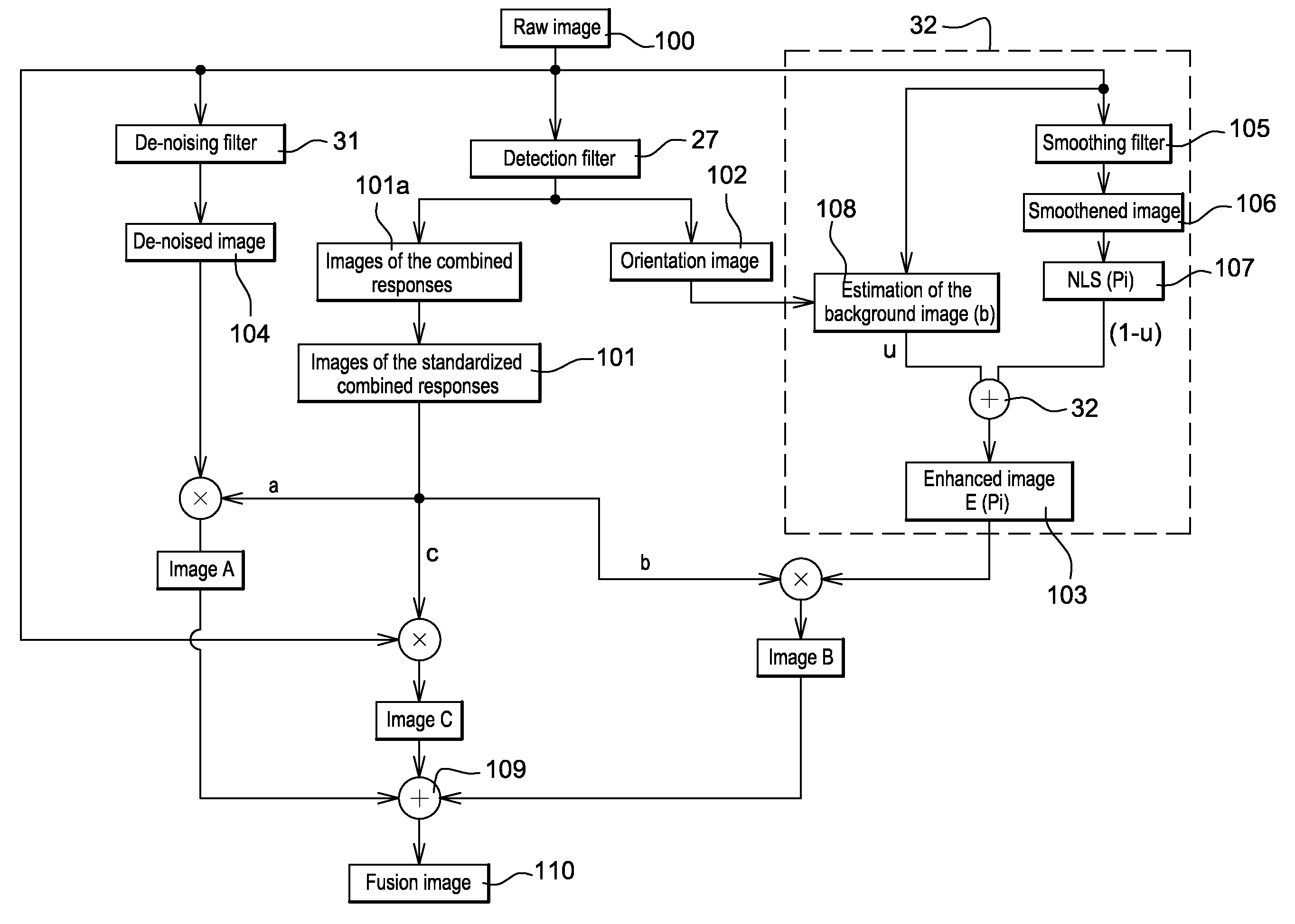
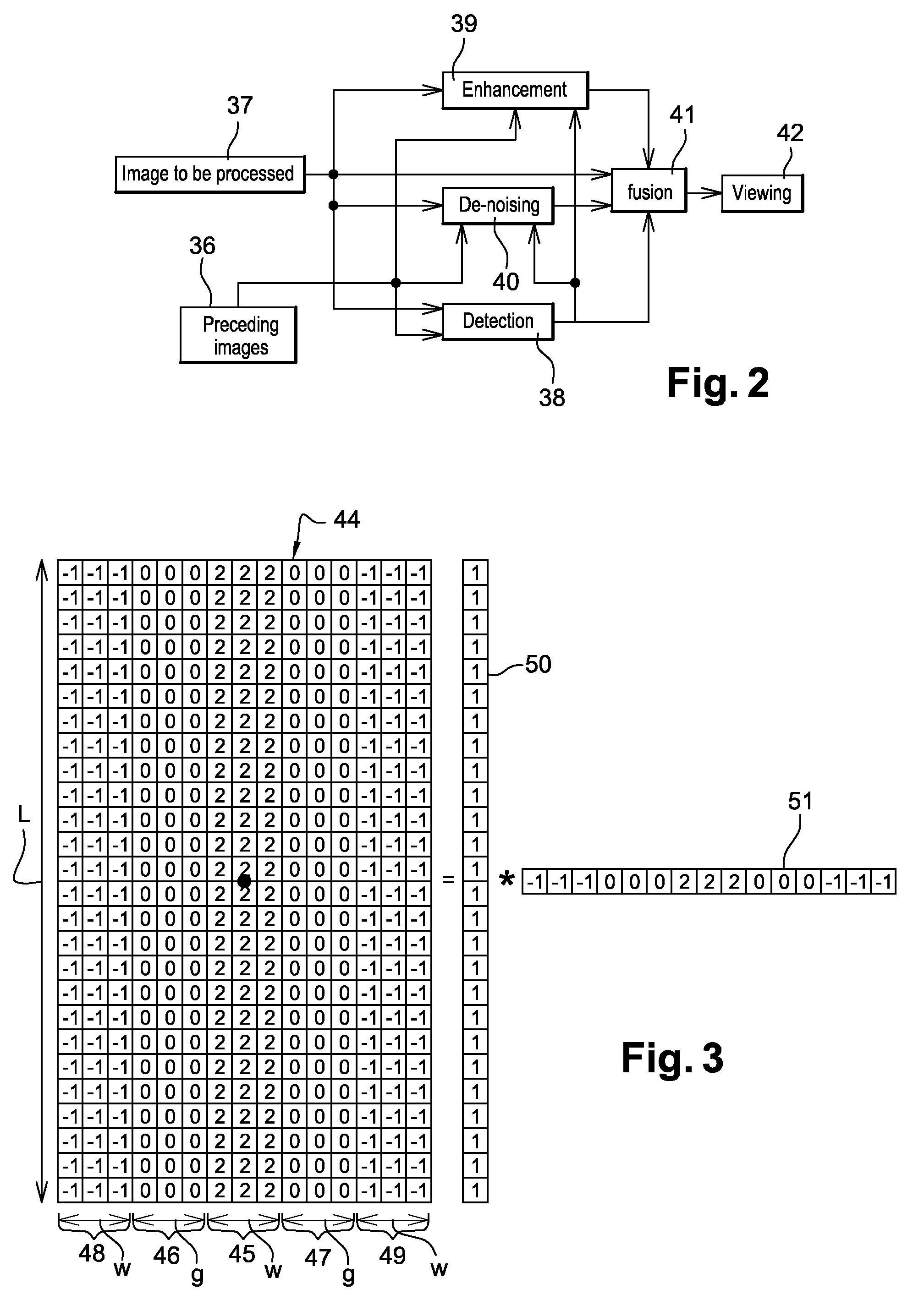
[0033]In order to make it easier to understand the description of the method of the invention, reference is made to the elements 100 to 110 of FIG. 8 in the description of FIGS. 1 to 7.

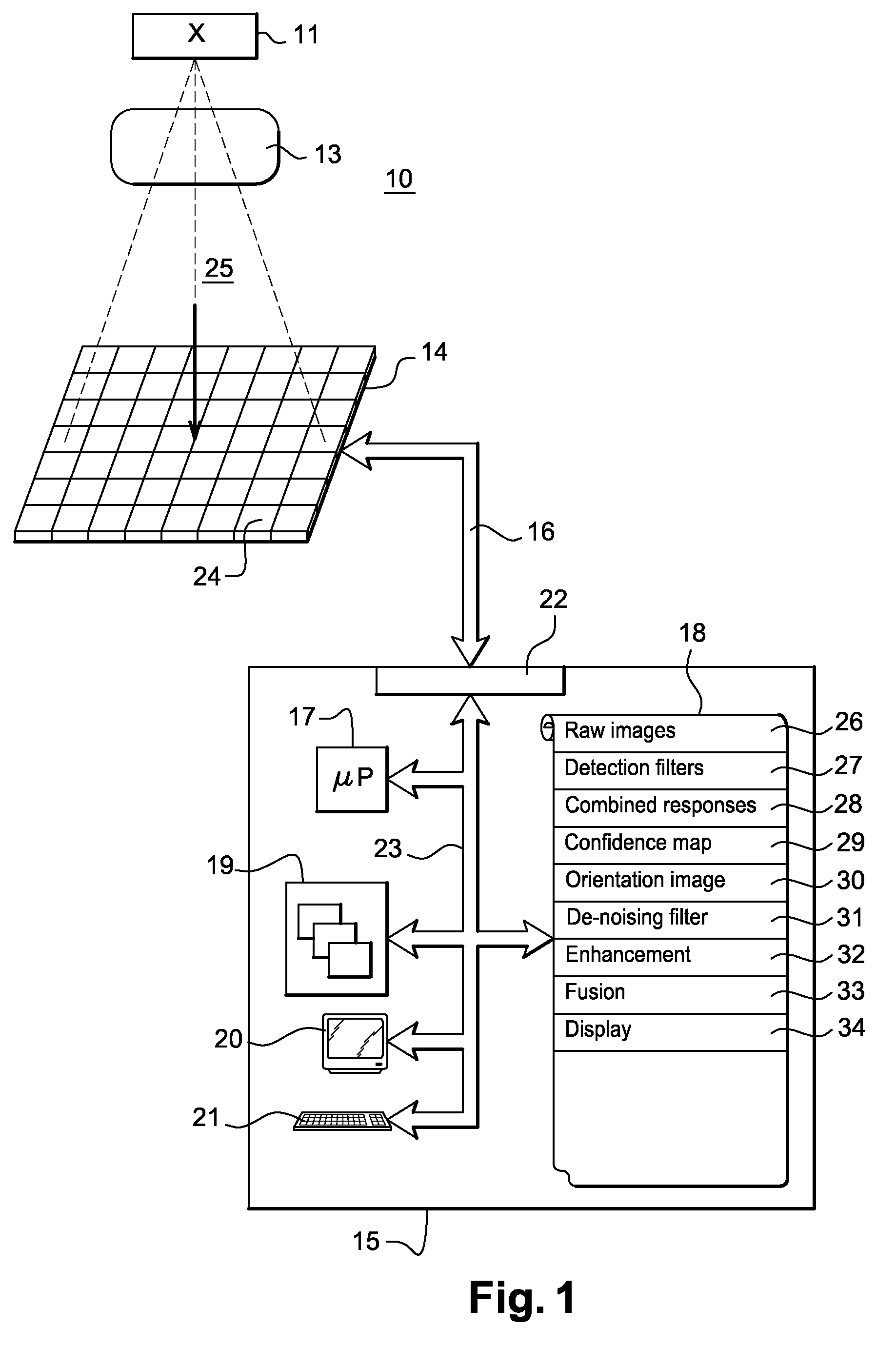
[0034]FIG. 1 illustrates a radioscopic image-production apparatus implementing a radioscopic image-processing method according to the invention. Radioscopy is a medical imaging system used to obtain a dynamic radiological image, i.e. a sequence of images or a video of the patient. This tool is used for diagnostics and interventional purposes, i.e. during the treatment of the patient, as an aid in an interventional operation on the patient. Radioscopy is for example used to assist in angiography (diagnostics) or angioplasty (interventional action).

[0035]The images produced by the production apparatus 10 result from detecting an incident irradiation coming from a radiation source 11 to which the patient 13 is exposed. The apparatus 10 also has an image detector 14 and a control circuit 15.

[0036]The imag...


PUM


Abstract

A method for the processing of images in interventional radioscopy to produce a sequence of de-noised images in which elongated objects of guiding instrumentation equipment are preserved and enhanced. The method of the invention uses a tool for indicating the confidence to be placed in a given pixel as regards its belonging to a piece of guidance equipment. This tool is used to create a combination algorithm capable of re-injecting the pixels having a low contrast-to-noise ratio into the de-noised image. This combination algorithm enables the enhancement solely of the pixels of the guidance equipment with a high confidence value so as not to produce artifacts with the enhancement of false positives.

Description

CROSS-REFERENCE TO RELATED APPLICATIONS[0001]This application claims priority under 35 U.S.C. §119(a)-(d) to prior-filed, co-pending French patent application serial number 0759245, filed on Nov. 23, 2007, which is hereby incorporated by reference in its entirety.FIELD OF THE INVENTION [0002]The present invention can be applied to particularly advantage but not exclusively in medical imaging and more especially in radiology. An embodiment of the present invention provides a method for processing images in interventional radioscopy to produce a sequence of de-noised images in which elongated objects of guiding instrumentation equipment are preserved and enhanced. An embodiment of the invention also provides a medical image reviewing station comprising a software program for updating an image-processing method of this kind.DESCRIPTION OF RELATED ART [0003]Radioscopy today is widely used for diagnostic and therapeutic operations performed under imaging control. Radioscopy is a method o...

Claims


Application Information

Patent Timeline
IPC IPC(8): G06K9/40
CPCG06T5/002G06T2207/10116G06T2207/20221G06T2207/20182G06T2207/20064
Inventor BISMUTH, VINCENTAUVRAY, VINCENTVAILLANT, REGISLIENARD, JEAN
Owner GENERAL ELECTRIC CO
Why Eureka
Try Eureka