
Stabilized, sterilized collagen scaffolds with active adjuncts attached

a technology of stabilized and sterilized collagen and active adjuncts, which is applied in the direction of prosthesis, peptide/protein ingredients, drug compositions, etc., can solve the problems of not being able to achieve the effect of infusion method on less porous bioimplant devices such as heart valves, skin grafts, tendon, bone and ligament repair tissues, and unable to achieve diffusion release of growth factors

Active Publication Date: 2009-08-06
SYNOVIS LIFE TECH
View PDF19 Cites 0 Cited by
  • Summary
  • Abstract
  • Description
  • Claims
  • Application Information

AI Technical Summary

Benefits of technology

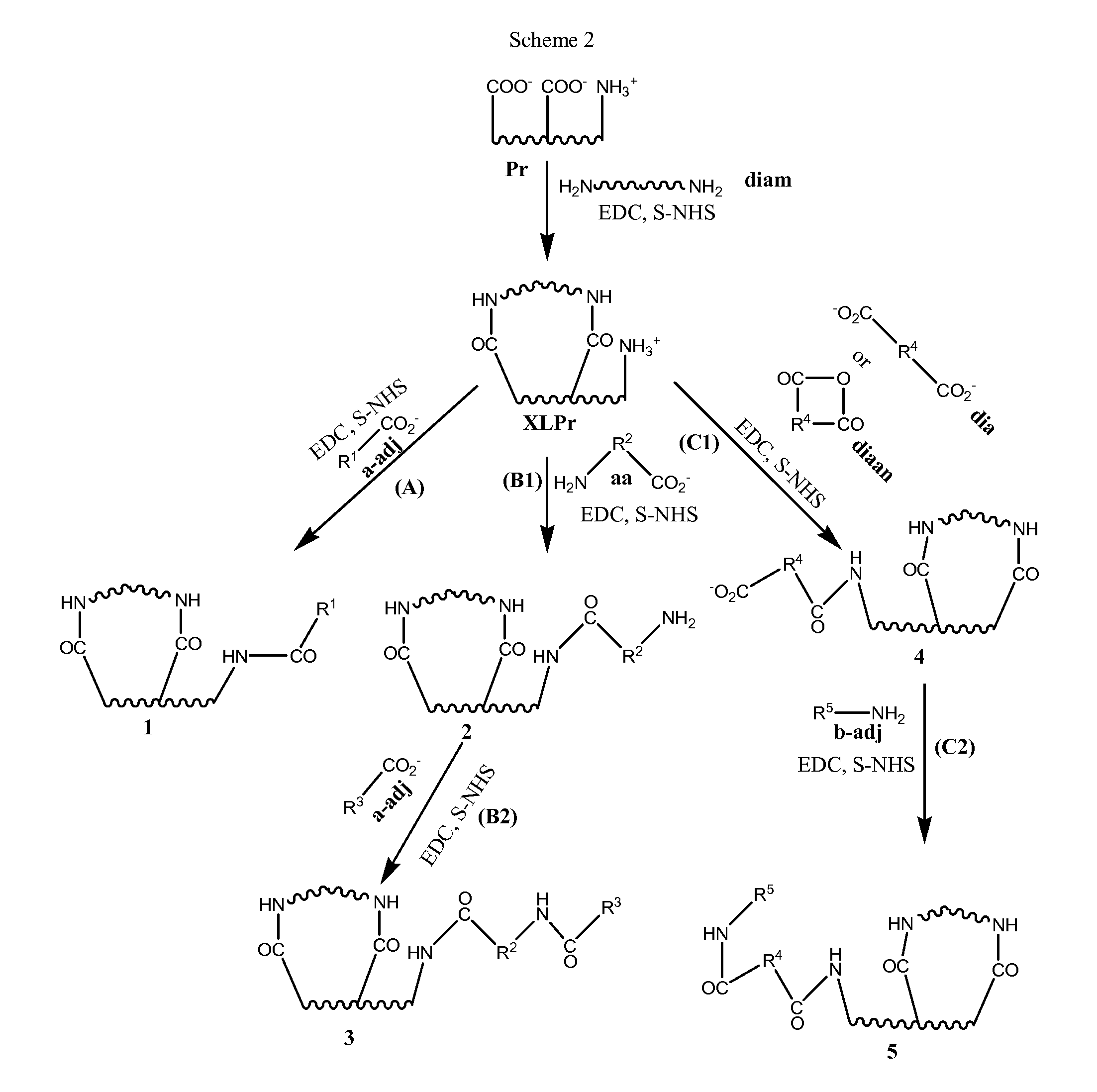
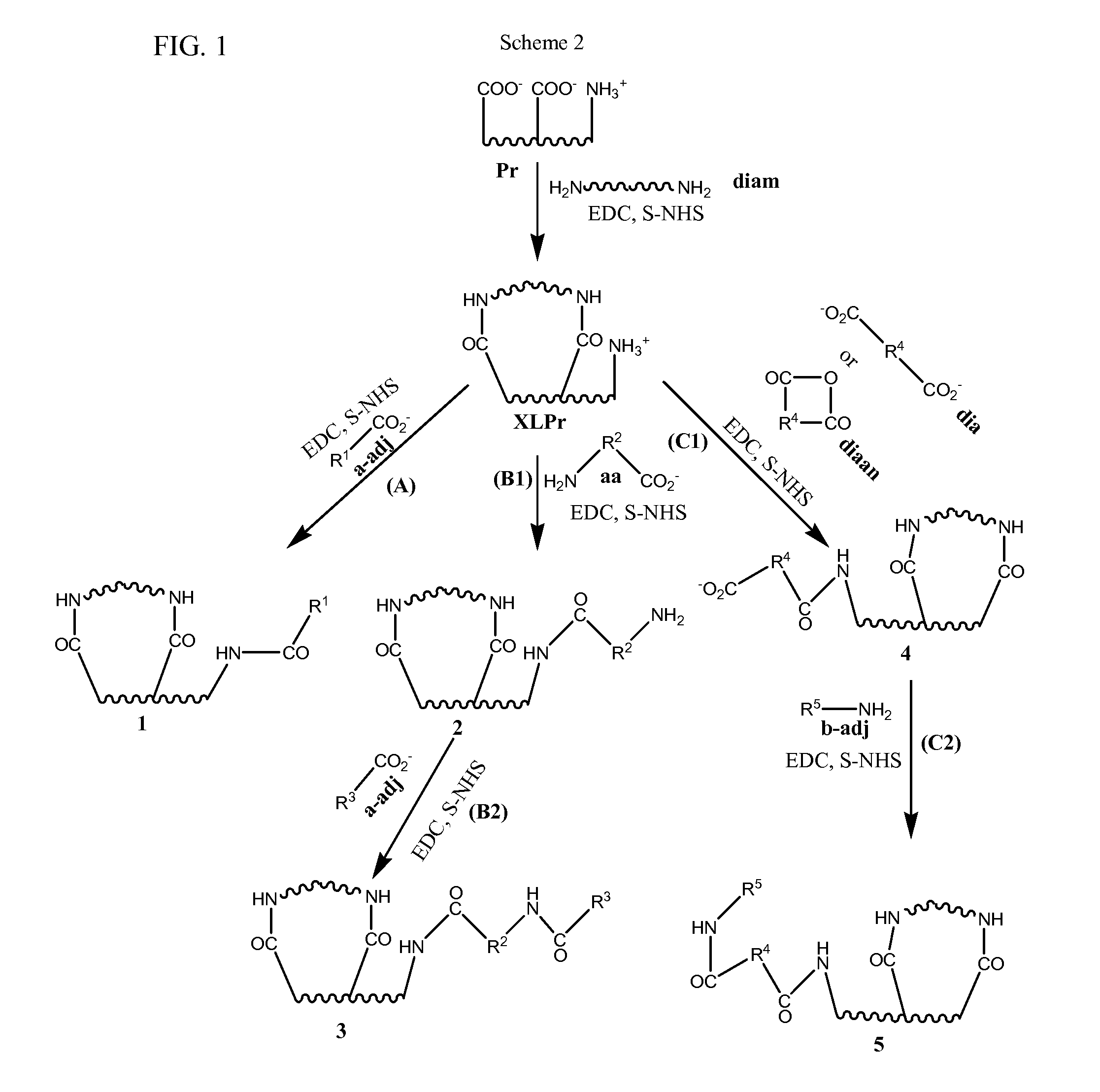
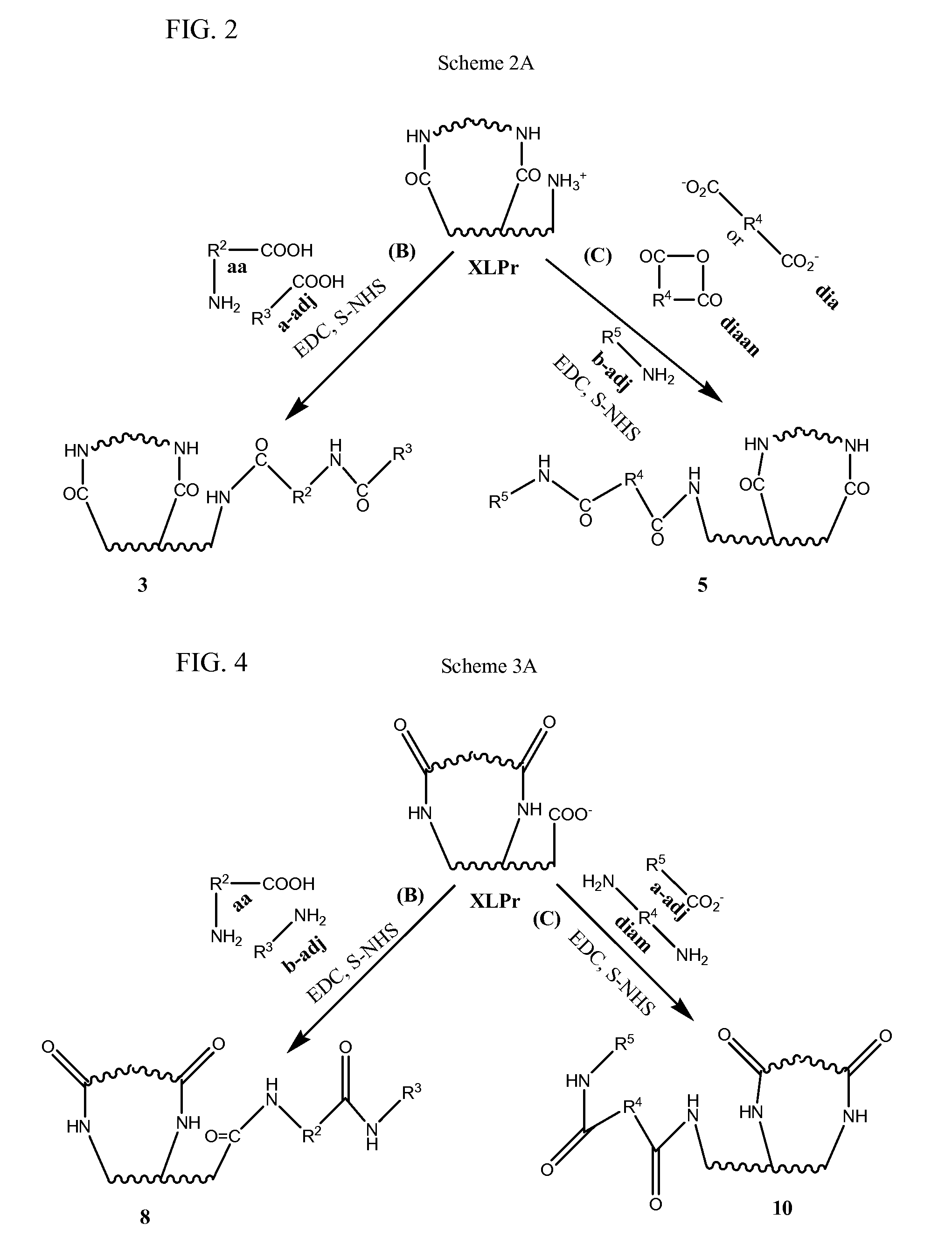
"The invention provides a method for making a sterilized biological implant by chemically sterilizing a biological tissue with a water soluble carbodiimide and covalently conjugating an adjunct to the tissue. The sterilization is carried out in the presence of a penetration enhancer, such as an alcohol, to improve the effectiveness of the sterilizing agent. The resulting implant is suitable for use in various medical applications."

Problems solved by technology

Despite the improvements in surgical outcomes that have already been provided by growth factor infused bioimplants, many challenges remain to be overcome.
Thus, the infusion method is not considered effective for less porous bioimplant devices such as heart valves, skin grafts, tendon, bone and ligament repair tissues, etc.
Another limitation is that release of the growth factor is by diffusion.
While diffusion can in some instances be a useful method of release, in other circumstances diffusion may result in too high an initial rate of release and thus too low a later rate of release.
Thus, one disadvantage of diffusive release is that the effective release period may be shorter than desired, unless excess growth factor is infused into the bioimplant at the start.
However, this may not always be feasible or even possible.
Moreover, even if it were possible to infuse excess growth factor into the bioimplant, a disadvantage arising out of this approach may be that the local concentration of growth factor may cause diffusion of the growth factor into surrounding tissue, including capillaries, veins and arteries, where it may bring about deleterious local or systemic effects.

Method used


Image

  • Stabilized, sterilized collagen scaffolds with active adjuncts attached
  • Stabilized, sterilized collagen scaffolds with active adjuncts attached
  • Stabilized, sterilized collagen scaffolds with active adjuncts attached

Examples


example 1

Attachment of Glycosaminogycan to Pericardial Tissues

[0231]Pericardial tissue was stabilized (cross-linked) using the techniques defined in U.S. Pat. No. 5,447,536, and U.S. patent application Ser. No. 11 / 276,398, filed Feb. 27, 2006, each of which is expressly incorporated herein by reference in its entirety. Following the cross-linking step, pericardial tissues were exposed to a solution of the glycosaminoglycan (GAG) chondroitin sulfate (0.5%-2.0%) in the presence of EDC for defined periods of time (2 h-overnight). Tissues were subsequently rinsed, and sterilized using the methods described in U.S. Pat. Nos. 6,521,179; 6,506,339; and 5,911,951, each of which is expressly incorporated herein by reference in its entirety. The levels of GAG (glycosaminoglycan) attachment in the tissues were assessed by histological means (differential staining of sections with PAS-Alcian Blue, where GAGs stain blue, while collagen stains pink). The pictures in FIGS. 5A-5C, indicate attachment of GAG...

example 2

Evaluation of Attachment Steps

[0232]The various processing steps (cross-linking, sterilization, etc.,) were then evaluated to determine the tissue processing steps during which the adjuncts could be attached to the tissues. Specifically, this example indicates the various methods of generating a sterile tissue with stably attached adjuncts. A Type 1 collagen sponge that had been subjected to various intermediary processing steps was contacted with a solution of chondroitin sulfate (as an adjunct example) and EDC. FIGS. 6A and 6B show low- and high-magnification histological sections of GAG-attached Type 1 collagen sponge, wherein GAG-attachment was carried out during tissue sterilization. FIG. 7B shows GAG-attached Type 1 collagen sponge, wherein GAG was attached to the tissue during cross-linking. As is apparent from FIGS. 6A, 6B and 7B, attachment of GAG to collagen sponge during sterilization resulted in a diffused attachment, whereas attachment of GAG to tissue during cross-link...

example 3

Attachment of Adjuncts to Different Tissue Types

[0233]In order to demonstrate the applicability of the methods of the present invention to various collagen-based tissue types, three different tissue types were treated in accordance with the present invention. Three different tissue types (pericardium, demineralized cancellous bone, and a collagen sponge made from solubilized Type I collagen) were first cross-linked and subsequently exposed to chondroitin sulfate in the presence of EDC. Tissues were subsequently rinsed, and sterilized using the methods described in U.S. Pat. Nos. 6,521,179; 6,506,339; and 5,911,951. The levels of GAG attachment in the tissues were assessed by histological means (differential staining of sections with PAS-Alcian Blue). The pictures in FIGS. 7A and 7B indicate uniform attachment of GAGs in cancellous bone and collagen sponge tissues.


PUM

PropertyMeasurementUnit
reaction temperatureaaaaaaaaaa
temperatureaaaaaaaaaa

Abstract

Bioimplants and methods of making the bioimplants are provided. The bioimplants comprise biological tissues having conjugated thereto adjunct molecules. The biological tissues are sterilized with a chemical sterilizing agent, such as a water soluble carbodiimide. The processes of making the bioimplants include a process in which an adjunct molecule is conjugated to a biological tissue during the sterilization process.

Description

PRIORITY CLAIM AND CROSS-REFERENCE TO RELATED APPLICATIONS[0001]This application is a divisional of U.S. patent application Ser. No. 11 / 686,859, filed Mar. 15, 2007, which claims the benefit of U.S. provisional patent application 60 / 743,542, filed Mar. 17, 2006, which is incorporated herein by reference in its entirety.BACKGROUND OF THE INVENTION[0002]Natural tissue bioimplants are gaining acceptance as advantageous alternatives to synthetic implants in many surgical procedures. Among other advantages, bioimplants more closely resemble in size, shape and performance the biological structures that they are designed to replace than do synthetic implants. Thus bioimplants are, in many circumstances, considered the devices of choice for replacement or structural augmentation of internal tissues and organs.[0003]The sources of bioimplants include non-human and human donors. In general, the choice of donor depends on a number of factors, including the relative sizes of the donor and recip...

Claims


Application Information

Patent Timeline
Patent Type & Authority Applications(United States)
IPC IPC(8): A61K9/00A61K35/12A61K38/16A61P43/00A61K35/32A61K35/34A61K35/36
CPCA61F2/28A61F2/30756A61F2002/2817A61F2002/30677A61F2002/30766A61F2310/00359A61K35/32A61K35/34A61K35/36A61L17/08A61L27/3683A61F2310/00365A61L17/005A61L27/3604A61L27/3687A61L27/54A61L33/0011A61L33/18A61L2300/42A61P43/00
Inventor NATARAJ, CHANDRASEKARANRITTER, GREGGSANDER, THOMAS
Owner SYNOVIS LIFE TECH
Learn More