3D imaging of live cells with ultraviolet radiation

a technology of live cells and ultraviolet radiation, applied in the field of optical projection tomography for 3d microscopy, can solve the problems of large degree of variability in this measurement, insufficient quantitative analysis, and insufficient phototoxicity of irradiated cells

Inactive Publication Date: 2009-08-20
VISIONGATE
View PDF91 Cites 87 Cited by
  • Summary
  • Abstract
  • Description
  • Claims
  • Application Information

AI Technical Summary

Problems solved by technology

Native absorbance and fluorescence imaging using deep ultraviolet (DUV) at 250 nm to 290 nm wavelengths has been technically challenging and causes phototoxicity in irradiated cells.
However, in these cases, the 3D image resulting from a stack of 2D images has about four times less axial resolution as the lateral resolution within each slice, thereby making quantitative analysis imprecise.
Furthermore, the label or stain is only an intermediary step toward the measurement of target protein or nucleotide (DNA) which adds a large degree of variability in this measurement.
Unfortunately, the use of DUV illumination has demonstrated phototoxicity in the past, due to the high dose of radiation required to stimulate a strong signal.

Method used


Image

  • 3D imaging of live cells with ultraviolet radiation
  • 3D imaging of live cells with ultraviolet radiation
  • 3D imaging of live cells with ultraviolet radiation

Examples


Embodiment Construction

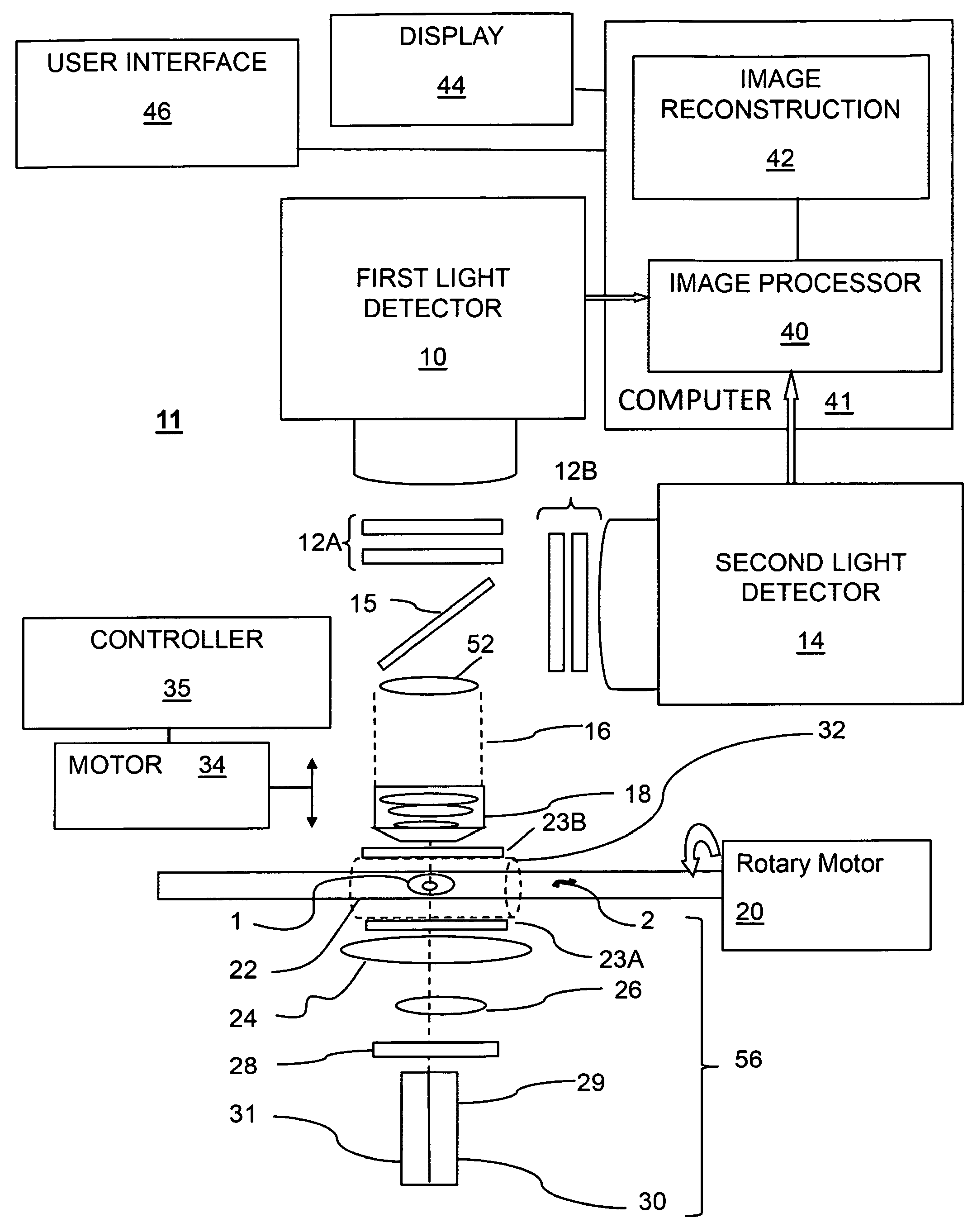
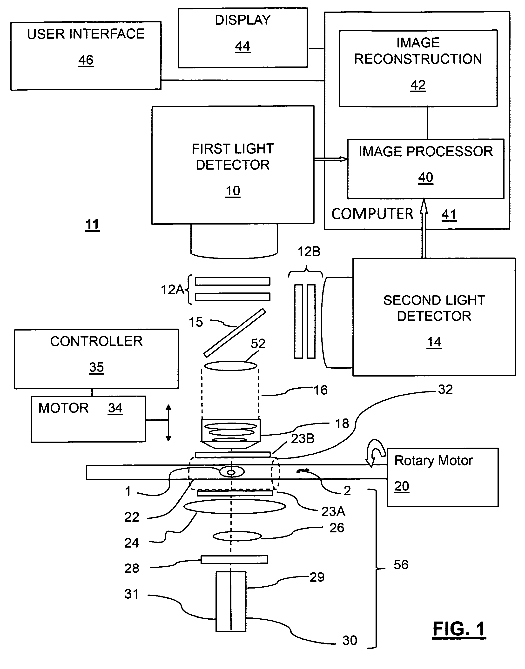
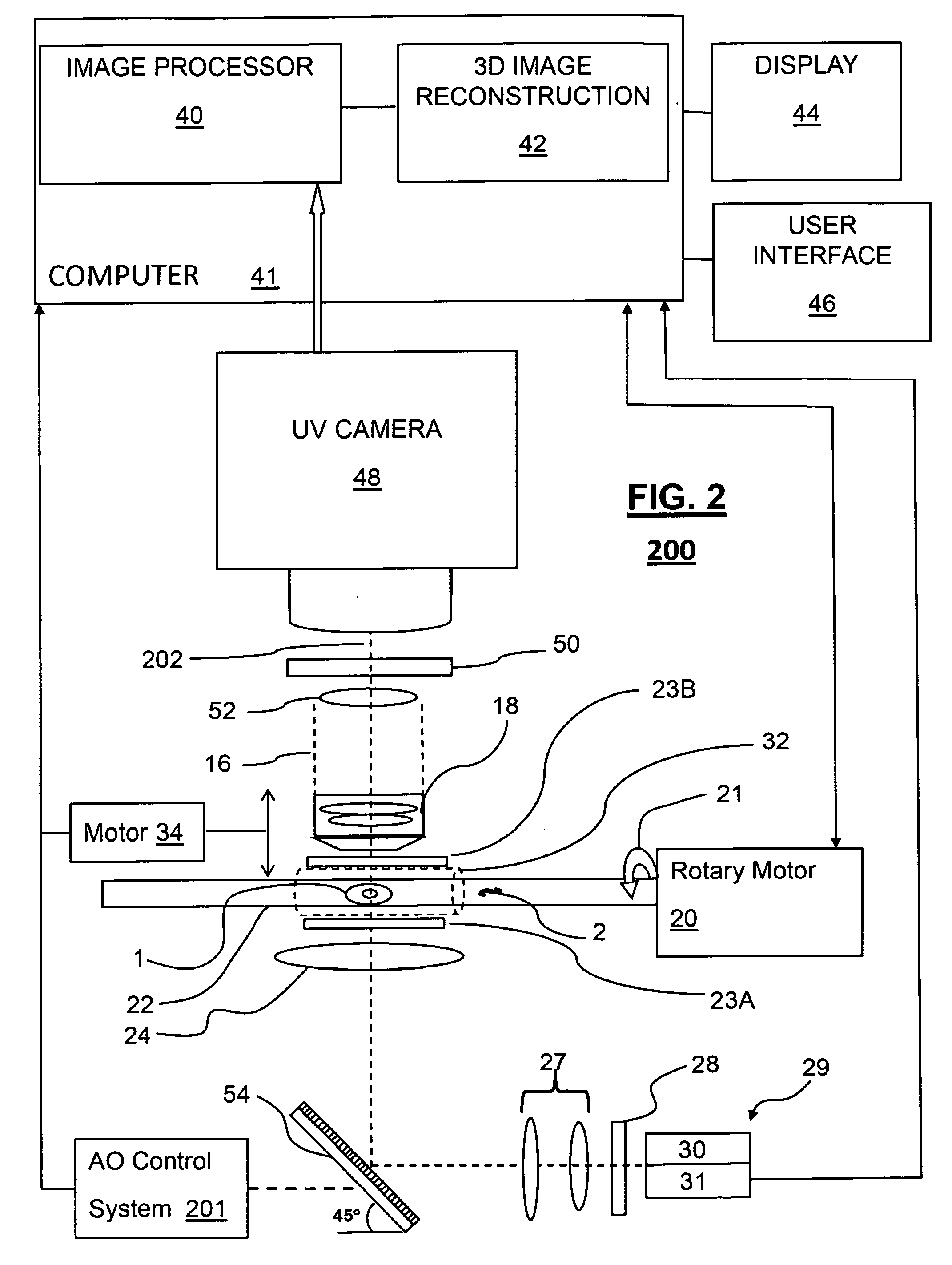
[0018]The following disclosure describes several embodiments and systems for imaging an object of interest. Several features of methods and systems in accordance with example embodiments of the invention are set forth and described in the Figures. It will be appreciated that methods and systems in accordance with other example embodiments of the invention can include additional procedures or features different than those shown in Figures. Example embodiments are described herein with respect to biological cells. However, it will be understood that these examples are for the purpose of illustrating the principals of the invention, and that the invention is not so limited.

[0019]Additionally, methods and systems in accordance with several example embodiments of the invention may not include all of the features shown in these Figures. Throughout the Figures, like reference numbers refer to similar or identical components or procedures.

[0020]Unless the context requires otherwise, through...


PUM


Abstract

A method for 3D imaging of cells in an optical tomography system includes moving a biological object relatively to a microscope objective to present varying angles of view. The biological object is illuminated with radiation having a spectral bandwidth limited to wavelengths between 150 nm and 390 nm. Radiation transmitted through the biological object and the microscope objective is sensed with a camera from a plurality of differing view angles. A plurality of pseudoprojections of the biological object from the sensed radiation is formed and the plurality of pseudoprojections is reconstructed to form a 3D image of the cell.

Description

TECHNICAL FIELD[0001]The present invention relates to optical tomographic imaging systems in general, and, more particularly, to optical projection tomography for 3D microscopy, in which a small object, such as a biological cell, is illuminated with ultraviolet radiation for pseudoprojection imaging and reconstruction into a 3D image.BACKGROUND[0002]Advances in imaging biological cells using optical tomography have been developed by Nelson as disclosed, for example, in U.S. Pat. No. 6,522,775, issued Feb. 18, 2003, and entitled “Apparatus and method for imaging small objects in a flow stream using optical tomography,” the full disclosure of which is incorporated by reference. Further developments in the field are taught in Fauver et al., U.S. patent application Ser. No. 10 / 716,744, filed Nov. 18, 2003 and published as US Publication No. US-2004-0076319-A1 on Apr. 22, 2004, entitled “Method and apparatus of shadowgram formation for optical tomography,” (Fauver '744) and Fauver et al....

Claims


Application Information

Patent Timeline
Patent Type & Authority Applications(United States)
IPC IPC(8): G06K9/00
CPCG01N21/4795G06T11/006G01N2015/1445G01N2021/1787G01N15/1436
Inventor SEIBEL, ERIC J.NELSON, ALAN C.FAUVER, MARK E.RAHN, J. RICHARD
Owner VISIONGATE
Why Eureka
Try Eureka