Inkjet Printing of Tissues and Cells

a tissue and inkjet printing technology, applied in the field of inkjet printing, can solve the problems of limited hydrogel printing technology, low mechanical strength, and difficulty in handling and in vivo application

Active Publication Date: 2009-08-20
WAKE FOREST UNIV HEALTH SCI INC
View PDF19 Cites 124 Cited by
  • Summary
  • Abstract
  • Description
  • Claims
  • Application Information

AI Technical Summary

Benefits of technology

[0010]Provided herein is an apparatus for printing cells, including: (a) an electrospinning device having a high voltage power supply; and / or (b) an inkjet printing device operatively associated with the electrospinning device. In some embodiments, the apparatus includes a three dimensional plotter operatively connected with the electrospinning device and / or inkjet printing device. In some embodiments, the apparatus includes a controller operatively connected to the electrospinning device and / or inkjet printing device. In some embodiments, the high voltage supply is conductively isolated from, e.g., the controller and / or plotter (e.g., by optics or wireless communication).

Problems solved by technology

However, most current printing technologies are limited to hydrogel as the primary scaffold for tissue constructs.
A major disadvantage of hydrogels is their low mechanical strength, which makes handling and in vivo application difficult, particularly for load-bearing implants.
However, these methods have some limitations.
For example, micro-fluidic approaches are usually limited to specific geometries because they require laminar fluid flow to produce microparticles (Khademhosseini et al., 2004, “Layer-by-layer deposition of hyaluronic acid and poly-L-lysine for patterned cell cocultures,” Biomaterials, 25 (17), pp.
These methods can only create small quantities of particles, since there are a limited number of micro-channels within these devices.
Furthermore, the costly equipment, specialized material, and extensive expertise required for operation of these devices may further limit the use of these methods in single cell particle fabrication.

Method used


Image

  • Inkjet Printing of Tissues and Cells
  • Inkjet Printing of Tissues and Cells
  • Inkjet Printing of Tissues and Cells

Examples


example 1

Printing Cartilage Constructs Using A Hybrid Printing Device

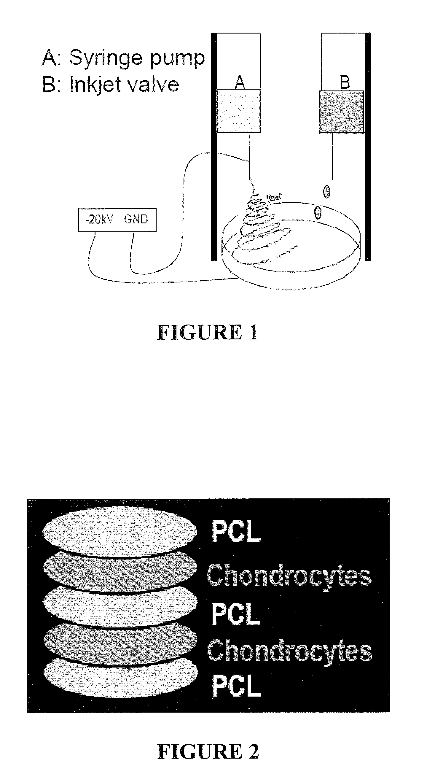
[0103]To demonstrate the feasibility of generating structured cartilage constructs using a combination of electrospinning and inkjet printing, a layer-by-layer electrospun PCL and inkjet printed chondrocyte scaffold was created (FIG. 1 and FIG. 2).

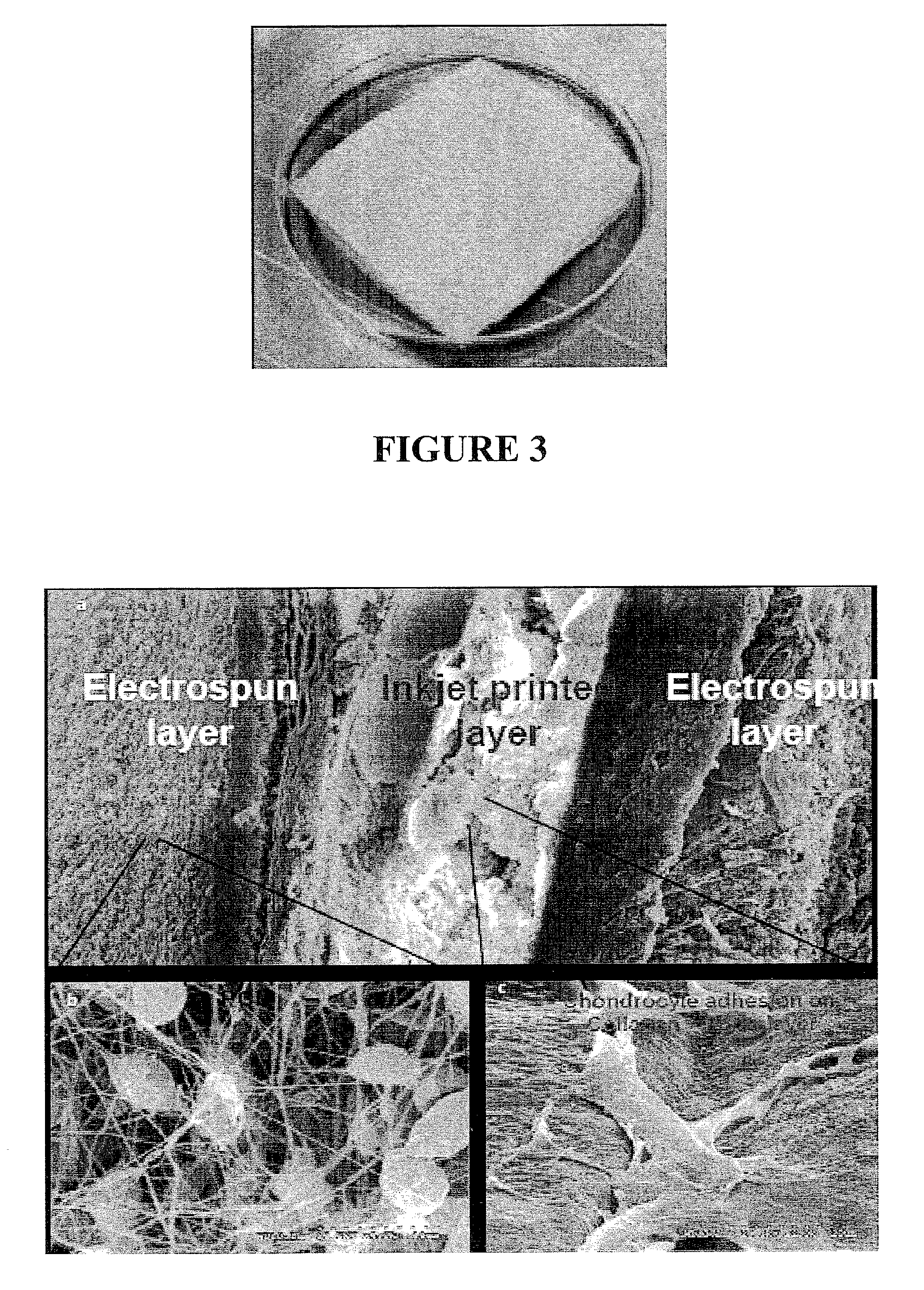
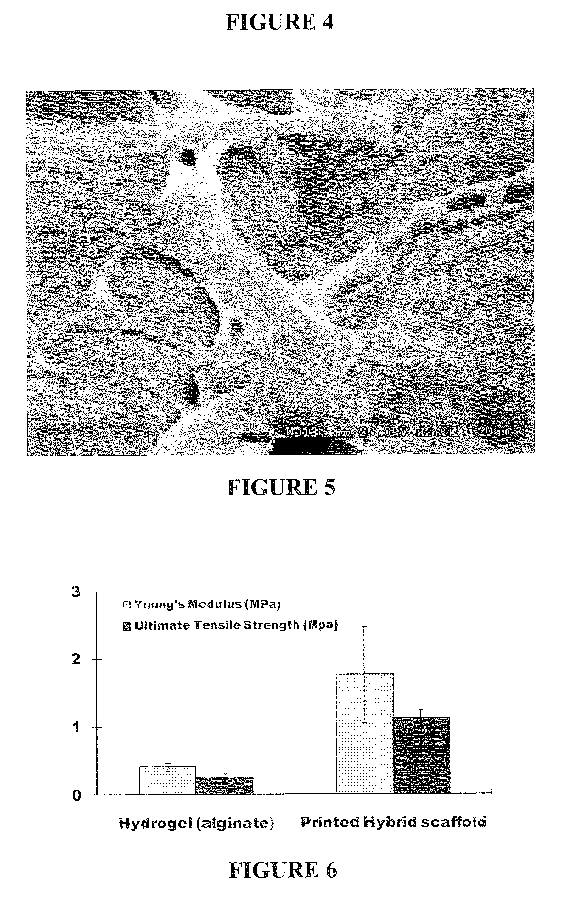
[0104]Electrospinning was first used to fabricate the PCL scaffold layer with polymeric nanofibers. Then the inkjet print head laid down rabbit elastic chondrocytes with the fibrin hydrogels on the electro-spun layer. By alternately applying electrospinning and inkjet printing, a 3 D cartilage construct containing multi-layers of cells and scaffolds was generated (FIG. 3). The multi-layered structures of the constructs were observed under SEM examination (FIG. 4). Printed chondrocytes attached to the collagen / elastin within the layer (FIG. 5).

[0105]Mechanical properties, cell viability, and cartilage production of the printed constructs were evaluated. The fabricated scaffolds dem...

example 2

High-Throughput Production of Single Cell Microparticles Using Inkjet Printing

[0124]An insulin-producing beta cell line (TC6) was obtained from American Type Culture Collection (ATCC, Manassas, Va.). The cells were maintained in Dulbecco's Modified Eagles Medium (ATCC) supplemented with heat-inactivated 10% fetal bovine serum (Invitrogen, Carlsbad, Calif.), 100 IU of penicillin (Invitrogen) and 100 μg / ml of streptomycin (Invitrogen) and incubated at 37° C. in a humidified 5% CO2 atmosphere.

[0125]Sodium alginate (MVG; Pronova Biomedical, Oslo, Norway) with an overall gluronic acid (G-block) content of 70% (as reported by the manufacturer) was mixed with PBS at different concentrations (0.5%, 1%, and 2% solution (w / v)) and autoclaved. Beta-TC6 cells were trypsinized, cell pellets collected and re-suspended in sodium alginate solutions at different final cell concentrations (2×10⁶, 6×10⁶, and 12×106 cells / ml).

[0126]HP DeskJet 550C printers were modified using previously described metho...


PUM

PropertyMeasurementUnit
diameteraaaaaaaaaa
vibrating frequencyaaaaaaaaaa
diameteraaaaaaaaaa

Abstract

Provided herein is an apparatus for printing cells which includes an electrospinning device and an inkjet printing device operatively associated therewith. Methods of making a biodegradable scaffold having cells seeded therein are also provided. Methods of forming microparticles containing one or more cells encapsulated by a substrate are also provided, as are methods of forming an array of said microparticles.

Description

RELATED APPLICATIONS[0001]This application claims the benefit under 35 U.S.C. § 119 (e) of U.S. Provisional Patent Application Ser. No. 61 / 028,761, filed Feb. 14, 2008, the disclosure of which is incorporated herein by reference in its entirety.FIELD OF THE INVENTION[0002]The present invention concerns inkjet printing of viable cells and arrays of cells so produced.BACKGROUND OF THE INVENTION[0003]In the interdisciplinary field of tissue engineering, powerful new therapies are being developed to address structural and functional disorders of human health by utilizing living cells as engineering materials. In some areas of tissue engineering, researchers are creating two- and three-dimensional tissues and organs from combinations of cells in order to repair or replace diseased or damaged tissues.[0004]Organ printing using inkjet printing is evolving to become more optimized by delivering multiple cell types and scaffolds to target specific regions. However, most current printing tech...

Claims


Application Information

Patent Timeline
Patent Type & Authority Applications(United States)
IPC IPC(8): A61K9/00C12N11/04A61K35/12C40B50/06B28B1/00C12N5/02A61F2/06
CPCA61F2/062A61L27/38A61L27/50A61L27/58B41J3/407A61K35/36C12N11/08D01D5/0007D01F6/625A61K35/32A61K35/34C12N11/04B33Y80/00C12N5/0655C12N2533/54C12N2533/56C12N11/096A61K9/5089
Inventor XU, TAOYOO, JAMES J.ATALA, ANTHONYDICE, DENNIS
Owner WAKE FOREST UNIV HEALTH SCI INC
Why Eureka
Try Eureka