Patient bed for pet/mr imaging systems

a technology of imaging system and patient bed, which is applied in the field of medical imaging arts, can solve the problems of long lag time, minimal disturbance of patients, and availability of both ct and spect imaging capabilities, and achieve the effect of convenient radio frequency cabling and without compromising ease of patient loading

Active Publication Date: 2009-08-20
KONINKLIJKE PHILIPS ELECTRONICS NV
View PDF19 Cites 30 Cited by
  • Summary
  • Abstract
  • Description
  • Claims
  • Application Information

AI Technical Summary

Benefits of technology

[0016]One advantage resides in providing a spatially compact hybrid imaging system.
[0017]Another advantage resides in providing spatial compactness without compromising ease of patient loading through the availability of height adjustment and access to the patient bed from an end of the bed.
[0018]Another advantage resides in advantageously placing the patient bed in space between a magnetic resonance scanner and a second modality imaging system that is provided to isolate the two imaging systems from one another.
[0019]Another advantage resides in providing convenient radio frequency cabling in a hybrid imaging system that includes a magnetic resonance scanner.
[0020]Another advantage resides in enabling magnetic resonance scanning followed by PET or other second modality imaging, or vice versa, without disturbing the subject except for short-range translational motion.

Problems solved by technology

Separate imaging systems have a longer lag time between studies, and make it difficult to minimally disturb the patient between studies.
Thus, both CT and SPECT imaging capability are available with limited modifications to either the CT gantry or the spatially separated gamma camera.
However, construction of a hybrid imaging system including a magnetic resonance (MR) scanner and a second modality imaging system employing high energy particles or photons (such as SPECT or PET) is challenging.
Problematically, the electronics for radiation detectors used in PET scanners or other imaging systems that detect high energy particles or photons typically generate high levels of radio frequency interference.
Consequently, when considering placement in the same room with close proximity, there is an inherent practical incompatibility between a magnetic resonance scanner and an imaging system that detects high energy particles or photons.
Unfortunately, this approach vitiates many of the benefits of a hybrid MR / PET system.
The patient may find such a long-distance transfer uncomfortable, and shifting or other movement of the patient during such a long transfer can introduce spatial registration errors in images acquired by the MR and PET.
Moreover, difficulties can arise in transferring local coils used in magnetic resonance imaging across the long rail distance.
However, this approach does not address the issue of radio frequency interference from the radiation detectors interfering with the magnetic resonance detection system.
Additionally, the integrated PET radiation detectors occupy valuable bore space in the MR scanner.
This approach reduces, but does not eliminate, MR bore space usage by PET components, and additionally introduces sensitivity issues in the PET system due to optical losses in the extensive fiber optical light coupling systems.
Moreover, while arranging the light detection electronics remotely is beneficial, some types of scintillation crystals exhibit spontaneous radioactivity that can still produce substantial radio frequency interference.
A disadvantage of existing hybrid approaches is that these approaches are not conducive to retrofitting an existing magnetic resonance scanner.
The approach of Cho et al. requires availability of a PET scanner room suitably located adjacent to the radio frequency isolation room of the magnetic resonance scanner, and further requires cutting a passthrough into the separating wall and adding a complex and bulky railway system for coupling the PET and MR scanners located in separate rooms.
Approaches that integrate the PET radiation detectors into the MR scanner bore similarly add complexity to the retrofitting process, and may be unworkable with some existing MR scanners.

Method used


Image

  • Patient bed for pet/mr imaging systems
  • Patient bed for pet/mr imaging systems
  • Patient bed for pet/mr imaging systems

Examples


Embodiment Construction

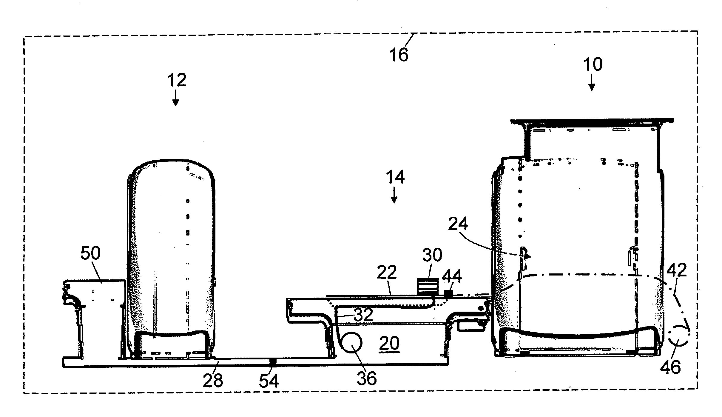
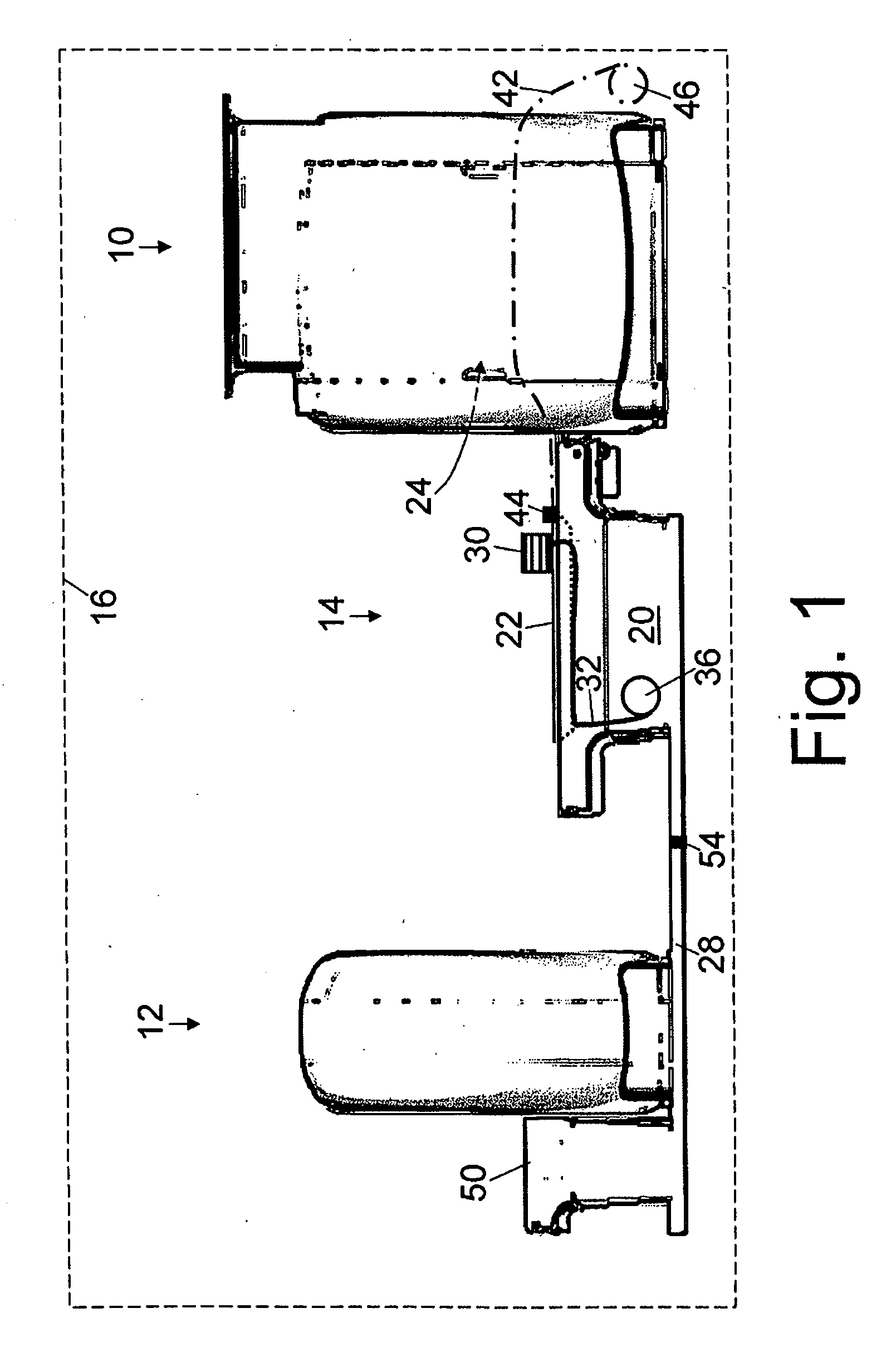
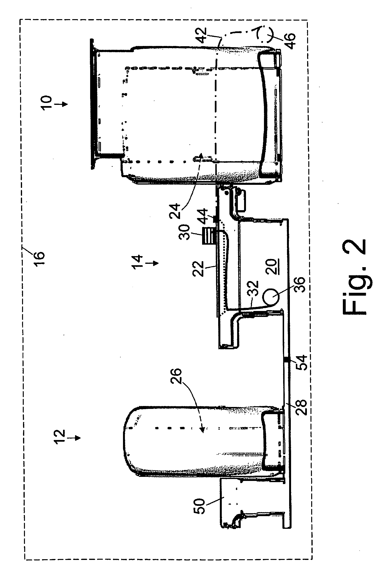
[0033]With reference to FIGS. 1-5, a hybrid imaging system includes a magnetic resonance scanner 10, a second modality imaging system 12, and a patient support, such as an illustrated patient bed 14, disposed between the magnetic resonance scanner 10, a second modality imaging system 12. A radio frequency shield substantially surrounds and defines a radio frequency isolated room or space 16. The magnetic resonance scanner 10, the second modality imaging system 12, and patient bed 14 are disposed within the radio frequency isolated room. The magnetic resonance scanner 10 in some embodiments is a commercial magnetic resonance scanner such as an Achieva or Intera magnetic resonance scanner available from Philips Medical Systems, Eindhoven, The Netherlands. More generally, the magnetic resonance scanner 10 can be substantially any type of scanner, such as the depicted horizontal cylindrical bore magnet scanner, an open bore scanner, or so forth.

[0034]The radio frequency isolated room 16...


PUM


Abstract

A hybrid imaging system and a patient bed for same are disclosed. The hybrid imaging system includes a magnetic resonance scanner and a second modality imaging system spaced apart from the magnetic resonance scanner by a gap. In some embodiments, the gap is less than seven meters. The patient bed is disposed at least partially in the gap between the magnetic resonance scanner and the second modality imaging system, and includes a linearly translatable patient support pallet aligned to be selectively moved into an examination region of the magnetic resonance scanner for magnetic resonance imaging and into an examination region of the second modality imaging system for second modality imaging. In some embodiments, a linear translation range of the linearly translatable pallet is less than five times a length of the patient support pallet along the direction of linear translation.

Description

CROSS REFERENCE TO RELATED APPLICATIONS[0001]This application is a continuation of PCT application number PCT / US2007 / 081458 filed Oct. 16, 2007 which claims the benefit of U.S. provisional application Ser. No. 60 / 863,637 filed Oct. 31, 2006, which is incorporated herein by reference.BACKGROUND OF THE INVENTION[0002]The present application relates to the medical imaging arts. It particularly relates to combined magnetic resonance (MR) and positron emission tomography (PET) imaging systems, and is described with particular reference thereto. The following relates more generally to imaging systems that combine the MR imaging modality with a modality employing energized particles, such as the aforementioned PET modality, single photon emission computed tomography (SPECT) modality, transmission computed tomography (CT) modality, a radiation therapy modality, or so forth.[0003]In a hybrid imaging system, two or more medical imaging modalities are integrated into the same facility or room,...

Claims


Application Information

Patent Timeline
Patent Type & Authority Applications(United States)
IPC IPC(8): A61B5/055A61B6/04
CPCA61B5/0555A61B6/032A61B6/037A61B6/4417A61B6/4429A61B5/0035A61B5/055
Inventor GAGNON, DANIELMORICH, MICHAEL A.BLAKELEY, DOUGLAS M.
Owner KONINKLIJKE PHILIPS ELECTRONICS NV
Why Eureka
Try Eureka