Recombinant vector expressing MDR1 shRNA and thymidine kinase and use thereof

a kinase and mdr1 technology, applied in ester active ingredients, tissue culture, enzymes, etc., can solve the problems of high mortality of cancer patients, cytotoxicity of most chemical anticancer drugs on normal cells, and death of individuals, so as to maximize the anti-tumor effect and achieve effective methods

Inactive Publication Date: 2009-08-27
KYUNGPOOK NAT UNIV IND ACADEMIC COOP FOUND
View PDF12 Cites 0 Cited by
  • Summary
  • Abstract
  • Description
  • Claims
  • Application Information

AI Technical Summary

Benefits of technology

[0010]As a result of a variety of extensive and intensive studies and experiments to solve the problems as described above and develop an effective method for treatment of tumor, the inventors of the present invention discovered that the combined therapy of ganciclovir and an anticancer drug can be achieved to maximize anti-tumor effects by cloning of a thymidine kinase gene and MDR1 siRNA into one vector system and consequent intracellular incorporation of such a vector into a host cell, and it is also possible to obtain a nuclear medical image of tumor lesions by means of thymidine kinase-green fluorescent protein (TK-GFP) fusion gene inserted into the vector. The present invention has been completed based on these findings.

Problems solved by technology

Cancer cells initially proliferate, then invade and destroy adjacent tissues, and finally spread into the circulatory system and metastasize to distant sites where they can continue their destructive processes, thereby resulting in death of individuals.
However, most of chemical anticancer drugs exhibit cytotoxicity on normal cells.
Further, since cancer is characterized by the propensity of tumor cells to spread from the primary lesion site to other normal tissues, the development and spread (i.e., metastasis) of the cancer disease leads to high mortality of cancer patients, despite remarkable advancement in the anticancer therapy including surgical operation, radiotherapy, and chemotherapy.
P-glycoprotein-associated drug resistance is thought to be one of the obstacles in cancer chemotherapy.
Unfortunately, therapeutic effects of these substances on cancer are insignificant.

Method used


Image

  • Recombinant vector expressing MDR1 shRNA and thymidine kinase and use thereof
  • Recombinant vector expressing MDR1 shRNA and thymidine kinase and use thereof
  • Recombinant vector expressing MDR1 shRNA and thymidine kinase and use thereof

Examples


example 1

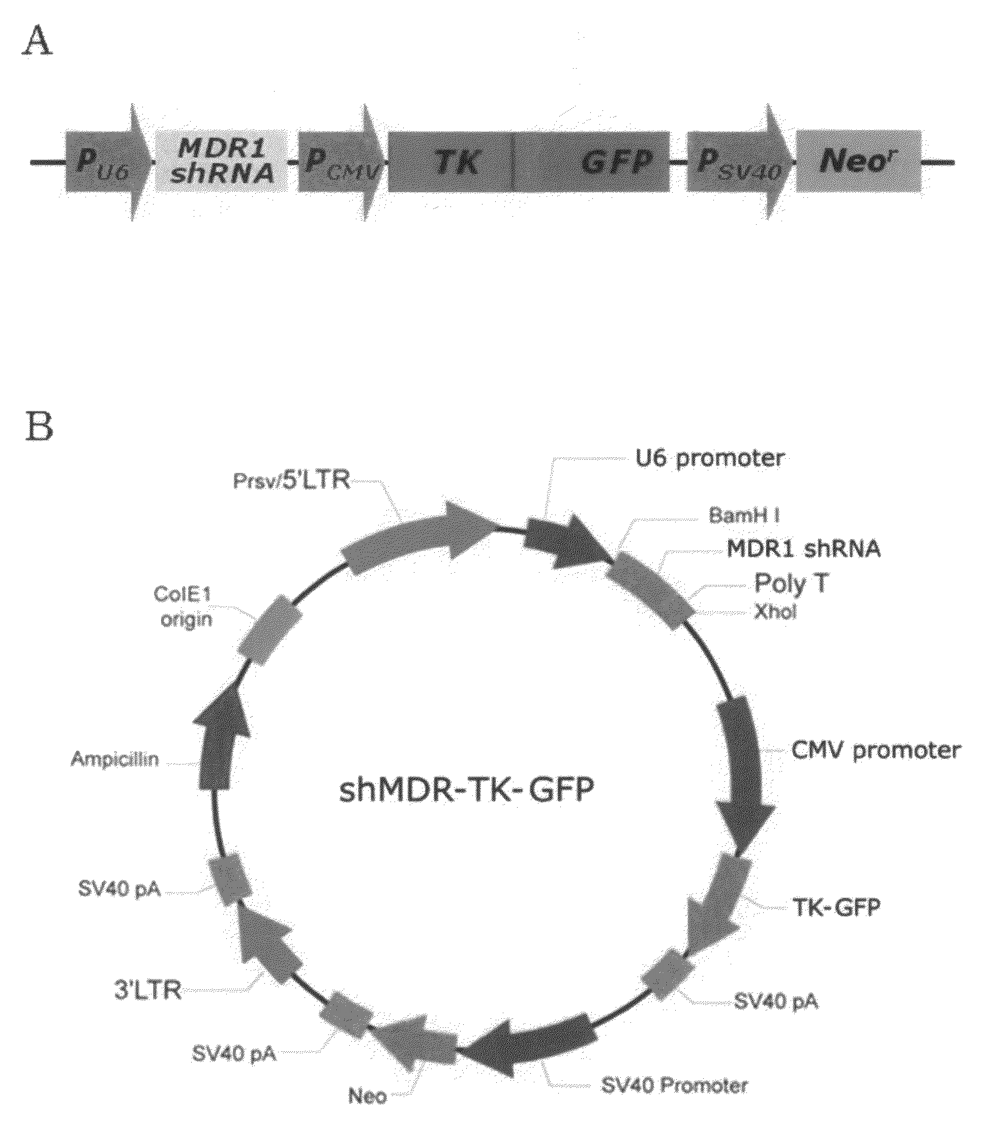
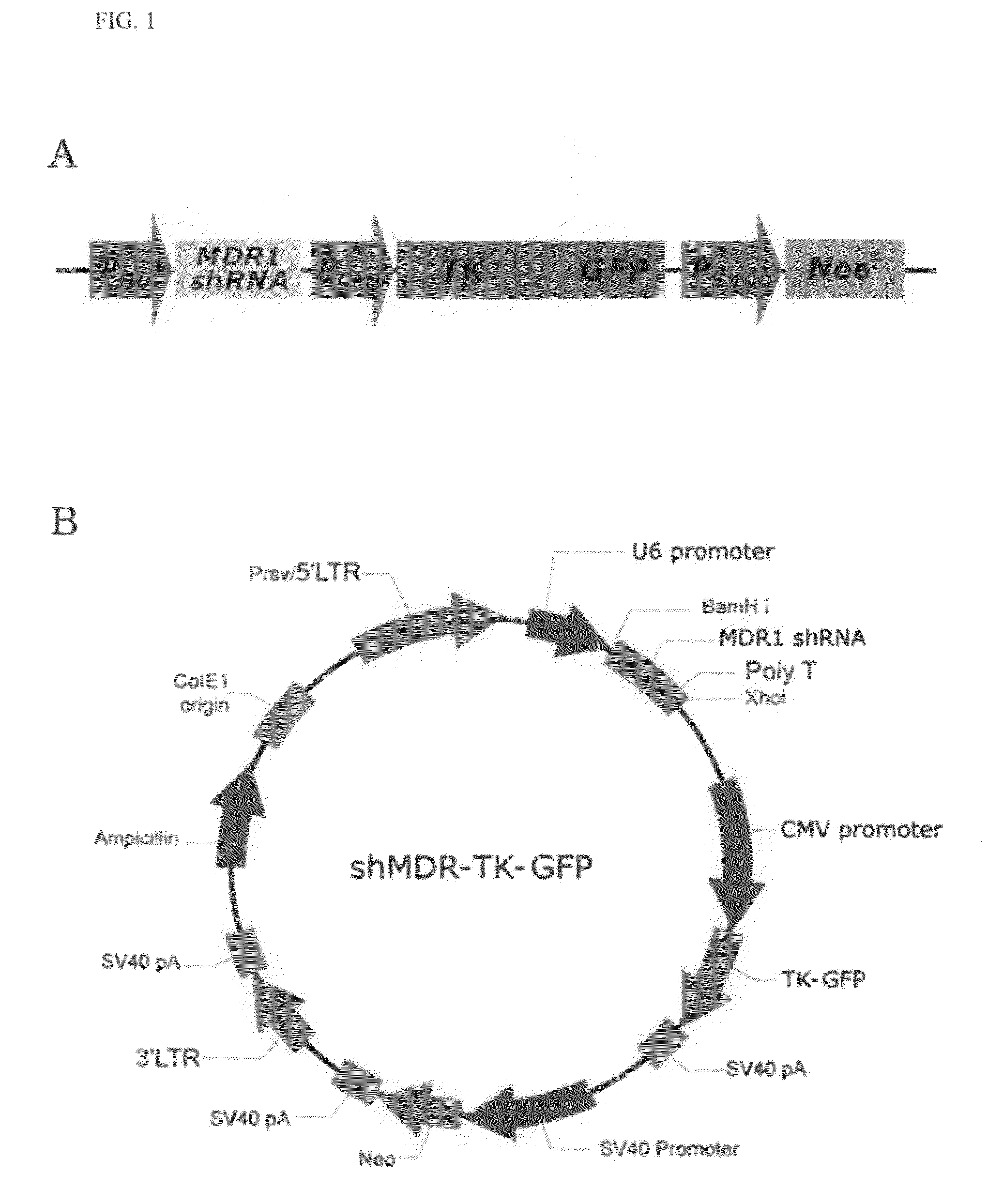
Cloning of Vector with Co-Expression of MDR1 shRNA and Thymidine Kinase Gene

[0067]1-1. Construction of HSV-Thymidine Kinase / GFP Fusion Gene

[0068]For construction of a fusion gene of a Herpes simplex virus-thymidine kinase (HSV-tk) gene and a GFP (green fluorescent protein) gene (hereinafter, referred to as “TK-GFP gene”), PCR amplification was carried out using HSV-tk cDNA (by courtesy of Dr. Jae Yong Park, Department of Internal Medicine, Division of Pulmonary, Medicine College of Kyungpook National University, Korea) as a template, and primers having sequences as set forth in SEQ ID NOS: 7 and 8. PCR was carried out as follows: initial denaturation of template cDNA at 94° C. for 2 min, followed by 30 cycles of denaturation at 94° C. for 30 seconds, annealing at 60° C. for 30 seconds and extension at 72° C. for 1 min. The amplified PCR product was cleaved with Nhe I and BamH I restriction endonucleases and ligated into the same restriction sites (Nhe I and BamH I recognition sites)...

example 2

Expression of MDR1 shRNA in Transfected Cells

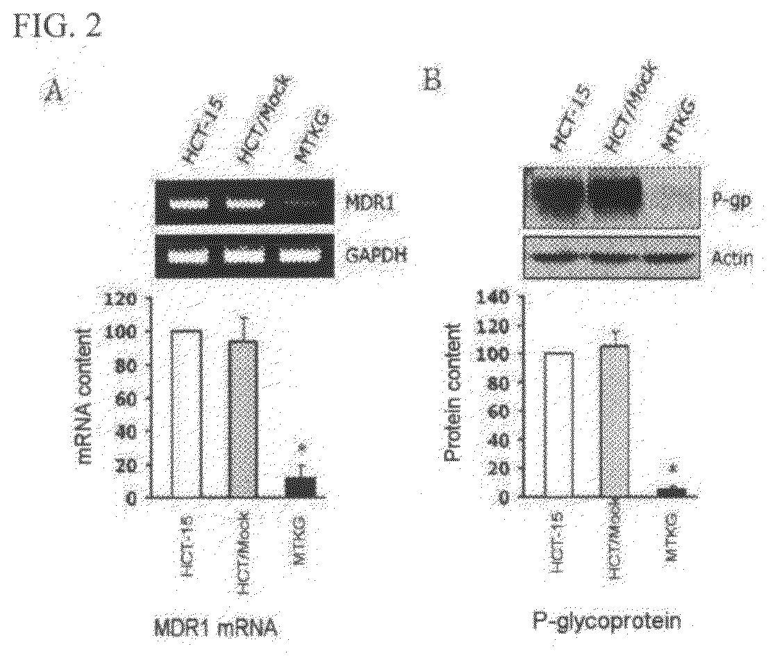
[0077]2-1. Analysis of mRNA Expression

[0078]Expression of MDR1 shRNA in MTKG cells of Section 1-3 of Example 1 was examined by RT-PCR For this purpose, total RNA was isolated from HCT-15, HCT / Mock, and MTKG cells, using Trizol reagent (Invitrogen) according to the manufacturer's instructions. Then, cDNA was prepared using 2 μg of the isolated total RNA as a template, and Oligo(dT)15 primer (Bionics, Seoul, Korea) and reverse transcriptase (Promega). PCR amplification for MDR1 mRNA was carried out using the prepared cDNA as a template and a pair of primers (SEQ ID NOS: 12 and 13). PCR was carried out as follows: initial denaturation of template cDNA at 94° C. for 2 min, followed by 30 cycles of denaturation at 94° C. for 30 seconds, annealing at 60° C. for 30 seconds and extension at 72° C. for 1 min.

TABLE 2Primers for MDR1 amplificationSEQ IDPrimersSequencesNOMDR1 sense5′-gga gtg tcc gtg gat cac12a-3′MDR1 antisense5′-aat aca tca ttg cct g...

example 3

Expression of TK-GFP Gene in Transfected Cells

[0083]3-1. Analysis of Protein Expression

[0084]In order to examine expression of an intracellularly incorporated TK-GFP gene, Western blot analysis was carried out in the same manner as in Section 2-2 of Example 2, except that anti-GFP antibodies (clone B-2, Santa Cruz, USA) were used for proteins isolated from HCT-15, HCT / Mock, and MTKG cells.

[0085]From the experimental results shown in FIG. 3A, it can be seen that the MTKG cells exhibit significant expression of the TK-GFP fusion protein, as compared to a control group.

[0086]3-2. Fluorescence Analysis

[0087]In order to further examine expression of an intracellularly incorporated TK-GFP gene, MTKG cells were observed under a light microscope and a fluorescence microscope, respectively.

[0088]From the experimental results shown in FIG. 3B, it can be seen that all the cells observed under the light microscope exhibited the fluorescence of the GFP protein upon examination of the cells under...


PUM

PropertyMeasurementUnit
pHaaaaaaaaaa
pHaaaaaaaaaa
resistanceaaaaaaaaaa

Abstract

A recombinant vector capable of expressing MDR1 shRNA and thymidine kinase, and a use thereof. More specifically, provided is a recombinant vector capable of efficiently expressing MDR1 shRNA and thymidine kinase in a host cell, a transfectant cell comprising the same recombinant vector, a composition for treating neoplastic diseases, comprising the same recombinant vector, and a method for imaging of neoplastic lesions using the same recombinant vector. The recombinant vector of the present invention is capable of achieving efficient intracellular expression of MDR1 shRNA and a thymidine kinase-GFP fusion protein within the host cell and is therefore highly effective for combined therapy of anticancer drugs. Further, the recombinant vector of the present invention enables imaging of neoplastic lesions. Therefore, the recombinant vector of the present invention can be used in combination with other anticancer drugs for treatment of neoplastic diseases.

Description

BACKGROUND OF THE INVENTION[0001]1. Field of the Invention[0002]The present invention relates to a recombinant vector capable of expressing MDR1 shRNA and thymidine kinase, and a use thereof. More specifically, the present invention relates to a recombinant vector capable of efficiently expressing MDR1 shRNA and thymidine kinase in a host cell, a transfectant cell comprising the same recombinant vector, a composition for treating neoplastic diseases, comprising the same recombinant vector, and a method for imaging of neoplastic lesions using the same recombinant vector.[0003]2. Description of the Related Art[0004]Cancer is a complex disease characterized by uncontrolled division and abnormal growth of malignant cells. Most of cancers result from various pathogenic factors including the genetic and epigenetic alterations of oncogenes and tumor suppressor genes. Cancer cells initially proliferate, then invade and destroy adjacent tissues, and finally spread into the circulatory system...

Claims


Application Information

Patent Timeline
Patent Type & Authority Applications(United States)
IPC IPC(8): A61K31/7088C12N15/00C12N5/08C12Q1/02
CPCA61K31/337A61K31/522C12N15/85C12N9/1211A61K49/0045A61K45/06A61K31/7088A61K31/704A61K2300/00C12N15/66C12N15/10C12N15/09
Inventor KIM, IN SANPARK, SEUNG YOONLEE, JAE TAEAHN, BYEONG CHEOL
Owner KYUNGPOOK NAT UNIV IND ACADEMIC COOP FOUND
Why Eureka
Try Eureka