
Systems and methods for cardiac remodeling

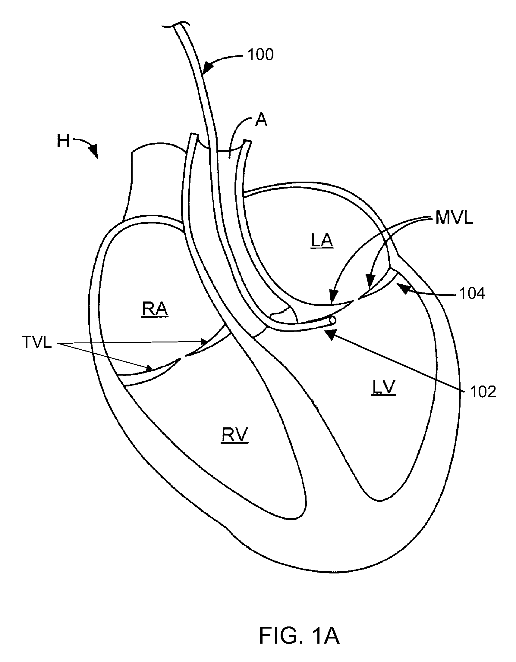
a system and cardiac technology, applied in the field of systems and methods for cardiac remodeling, can solve the problems of loss of cardiac valve competence, valve dysfunction, and loss of leaflet coaptation, and achieve the effect of improving the hemodynamic function of patients

Active Publication Date: 2009-09-17
ANCORA HEART INC
View PDF99 Cites 180 Cited by
  • Summary
  • Abstract
  • Description
  • Claims
  • Application Information

AI Technical Summary

Benefits of technology

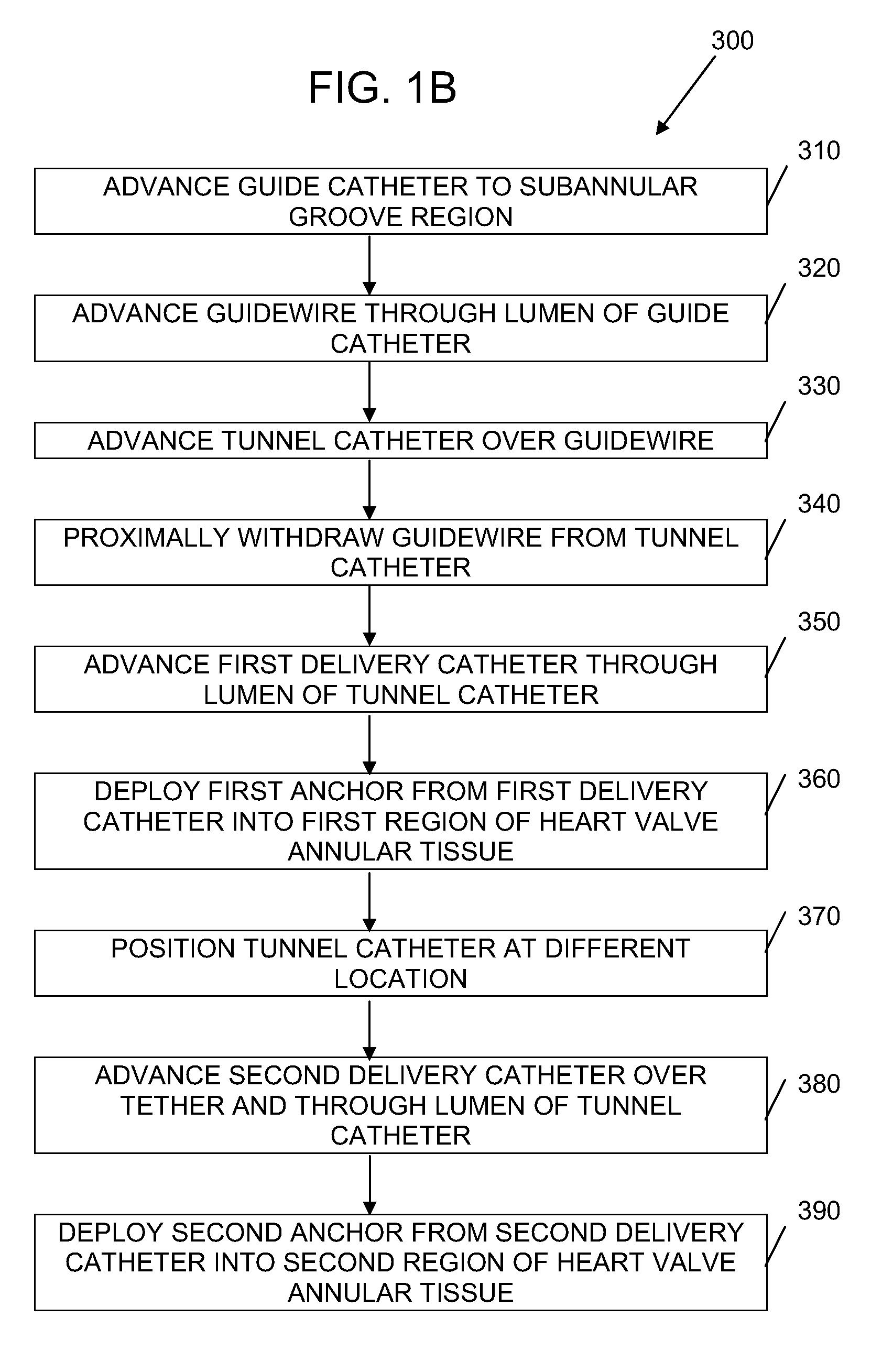
[0006]In one embodiment, a method for reshaping a heart is provided. The method comprises accessing a first cardiac tissue at a subvalvular space of a ventricle, positioning a first therapy device adjacent the first cardiac tissue using a first delivery tool, reconfiguring the first cardiac tissue using the first therapy device and reconfiguring a second cardiac tissue at a different location from the first cardiac tissue using a second therapy device. Thus, more than one therapy device may be used. In some embodiments, a septolateral dimension of a heart chamber is reduced.
[0010]In another embodiment, a method for reducing valve regurgitation is provided. The method comprises accessing a ventricle in a patient with a pre-existing annuloplasty implant, positioning a therapy device adjacent a wall of the ventricle, and reconfiguring the wall of the ventricle using the therapy device. The therapy device may comprise a plurality of tissue anchors movably coupled to a tether. At least one tissue anchor may be self-attaching or self-securing. The method may be performed to reduce a distance between a first papillary muscle and a second papillary muscle in the ventricle, or reduce a distance between a valve leaflet and a papillary muscle. The papillary muscle may be attached to the valve leaflet by a chordae tendineae, or may be an unassociated papillary muscle.

Problems solved by technology

Disease of the valvular apparatus can result in valve dysfunction, where some fraction of the ventricular blood regurgitates back into the atrial chambers.
Structural abnormalities at one or more of these anatomical sites may eventually lead to loss of coaptation of the leaflets, loss of competence of the valve and decreased efficiency of the heart as a one-way pumping mechanism.

Method used


Image

  • Systems and methods for cardiac remodeling
  • Systems and methods for cardiac remodeling
  • Systems and methods for cardiac remodeling

Examples


Embodiment Construction

[0042]While existing treatment options, such as the implantation of an annuloplasty ring or edge-to-edge leaflet repair, have been developed to treat structural abnormalities of the disease process, these treatments may fail to return the patient to a normal hemodynamic profile. Furthermore, atrio-ventricular valve regurgitation itself can also cause secondary changes to the cardiac function. For example, compensatory volume overload of the left ventricle may occur over time to maintain the net forward flow from the ventricle. This in turn will cause ventricular dilation, and further worsen mitral valve regurgitation by reducing valve coaptation. Ventricular dilation may also cause non-structural changes to the heart that can cause arrhythmias or electrophysiological conduction delays.

[0043]Devices, systems and methods are generally described herein for reshaping or remodeling atrio-ventricular valves. In some variations, procedural efficiencies may be gained by facilitating the del...


PUM


Abstract

Described herein are devices and methods for improving the hemodynamic function of a patient. In particular, a first device adapted to reshape an atrio-ventricular valve is used in combination with a second device configured to further alter the blood flow through the valve. The first device is typically an implant positioned in the subvalvular space of a ventricle. The second device may be an annuloplasty implant, a non-annulus valve apparatus implant, a ventriculoplasty implant, or a cardiac rhythm management device.

Description

CROSS-REFERENCE TO RELATED APPLICATIONS[0001]The present application claims priority under 35 U.S.C. 119(e) to U.S. Provisional Application No. 60 / 981,423 filed on Oct. 19, 2007, which is hereby incorporated by reference in its entirety.BACKGROUND OF THE INVENTION[0002]Blood returning to the heart from the peripheral circulation and the lungs generally flows into the atrial chambers of the heart and then to the ventricular chambers, which pump the blood back out of the heart. During ventricular contraction, the atrio-ventricular valves between the atria and ventricles, i.e. the tricuspid and mitral valves, close to prevent backflow or regurgitation of blood from the ventricles back to the atria. The closure of these valves, along with the aortic and pulmonary valves, maintains the unidirectional flow of blood through the cardiovascular system. Disease of the valvular apparatus can result in valve dysfunction, where some fraction of the ventricular blood regurgitates back into the at...

Claims


Application Information

Patent Timeline
Patent Type & Authority Applications(United States)
IPC IPC(8): A61M31/00A61F2/24
CPCA61B17/00234A61B17/0401A61B17/064A61B17/0644A61B2017/00243A61F2/2487A61B2017/0414A61B2017/0496A61F2/2445A61F2/2481A61B2017/00783
Inventor LOULMET, DIDIERSTARKSEN, NIEL F.FABRO, MARIELSERINA, EUGENE
Owner ANCORA HEART INC
Learn More