
Systems and Methods for Monitoring and Controlling Internal Pressure of an Eye or Body Part

a technology of system and pressure sensor, applied in the field of living system systems for monitoring and controlling pressure in body cavities, tissues and organs, can solve the problems of glaucoma not being directly treated, iop being considered a primary risk factor, drug and surgically implanted passive valves have their own limitations, etc., to avoid undesirable pressure loss and prevent device clogging

Active Publication Date: 2009-11-05
EASTERN VIRGINIA MEDICAL SCHOOL
View PDF112 Cites 119 Cited by
  • Summary
  • Abstract
  • Description
  • Claims
  • Application Information

AI Technical Summary

Benefits of technology

[0012]The present invention has a number of advantages compared to conventional therapies. There is no concern about patient compliance, drug side effects or drug interactions. Control of pressure is maintainable and not affected by normal physiological changes, or, for that matter, by changes that may occur due, for example, to drug administration or changes in pathology. Preferred embodiments of the present invention are capable of monitoring their own performance and adjusting for changes in liquid flow in a body part (e.g., aqueous humor flow in an eye or cerebrospinal fluid in the brain) and other variables that might affect pressure in the body part. The devices of the present invention can, in preferred embodiments, inform the care-giver of any problems related to pressure immediately, with the result that any acute or chronic pressure-mediated damage that might occur with other devices or drugs may be reduced or prevented. In addition, the apparatus and systems of the present invention can accommodate the use of larger orifices, to prevent device clogging, while avoiding undesirable loss of pressure (e.g., IOP) inside a body part. Preferred systems of the present invention may also have a fail-safe mode for adjusting valve function.

Problems solved by technology

Although the mechanism for glaucomatous optic neuropathy is uncertain, elevation of IOP is considered a primary risk factor.
These therapeutic interventions do not treat glaucoma directly.
However, drugs and surgically implanted passive valves have their own limitations.
Since patients cannot detect an immediate benefit, their motivation to administer the drugs is lacking and patient compliance is often poor.
In addition, drugs, even those applied topically, often cause adverse effects, are uncomfortable to apply in the eye, and present a significant, continuing expense.
Although the reduction of IOP by surgical methods also effectively reduces progression of visual defects, long-term control is rarely achieved.
Although patient compliance is not a factor here, the surgical approach is often of limited effectiveness due to formation of fibrous scar tissue and may predispose patients to cataracts.
In addition, when compared to medical therapy, surgical patients reported a slightly higher incidence of long term ocular burning, redness and dry eye.
A major limitation of existing approaches to treatment is that neither drugs nor surgical procedures are capable of automatically adjusting their actions based on the patient's IOP.
This drawback is of particular concern because maintenance of IOP in POAG below 18 mm Hg is generally considered essential to decrease the onset of glaucomatous optic neuropathy (or, once established, prevent its progression).
In addition, the lack of adjustment further limits the utilization of any other drugs that may elevate or decrease IOP as one of their primary or side effects.
However, existing valve-regulated devices are limited.
Conventional valves are passive devices that often allow too little or occasionally too much drainage of aqueous humor.
Another disadvantage is the inability to actively monitor IOP on a long-term basis.
In addition, conventional devices, even when working optimally, cannot be adjusted once in place, which leaves the patient and the physician at the mercy of pre-existing valve parameters.
Furthermore, these valves may fail and, therefore, must be removed due to poor control in terms of proper pressure regulation.

Method used


Image

  • Systems and Methods for Monitoring and Controlling Internal Pressure of an Eye or Body Part
  • Systems and Methods for Monitoring and Controlling Internal Pressure of an Eye or Body Part
  • Systems and Methods for Monitoring and Controlling Internal Pressure of an Eye or Body Part

Examples


Embodiment Construction

, including the description of various embodiments of the invention, will be best understood when read in reference to the accompanying figures wherein:

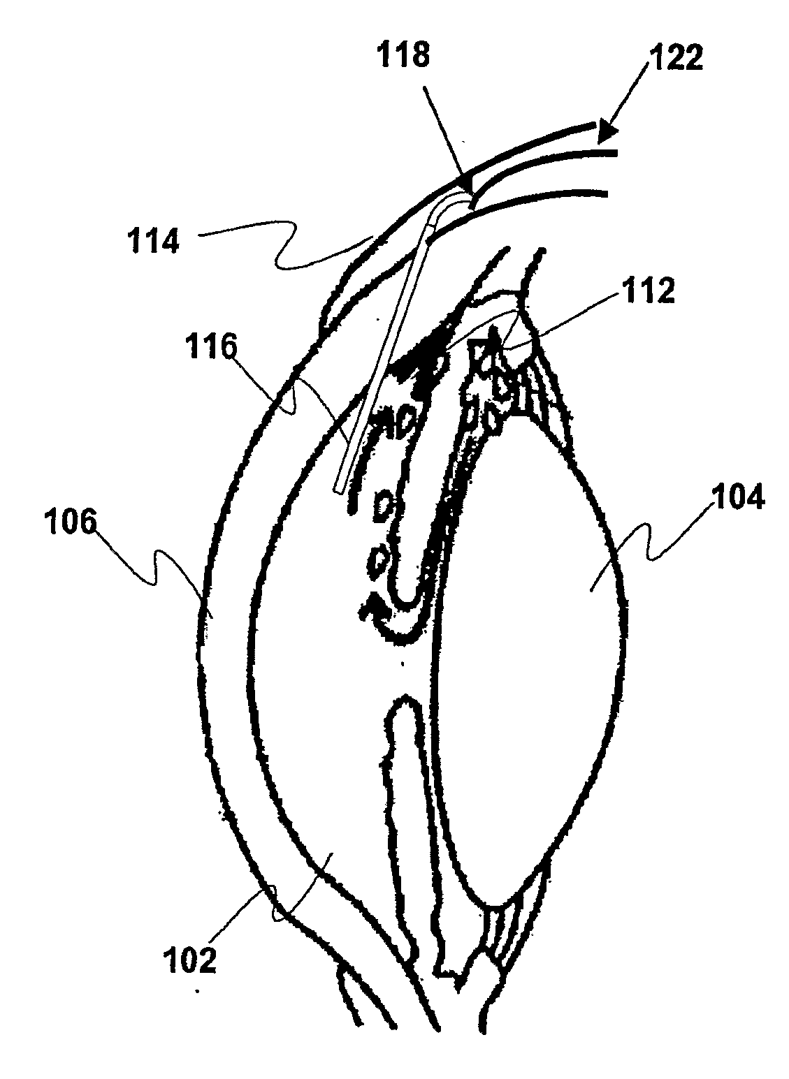
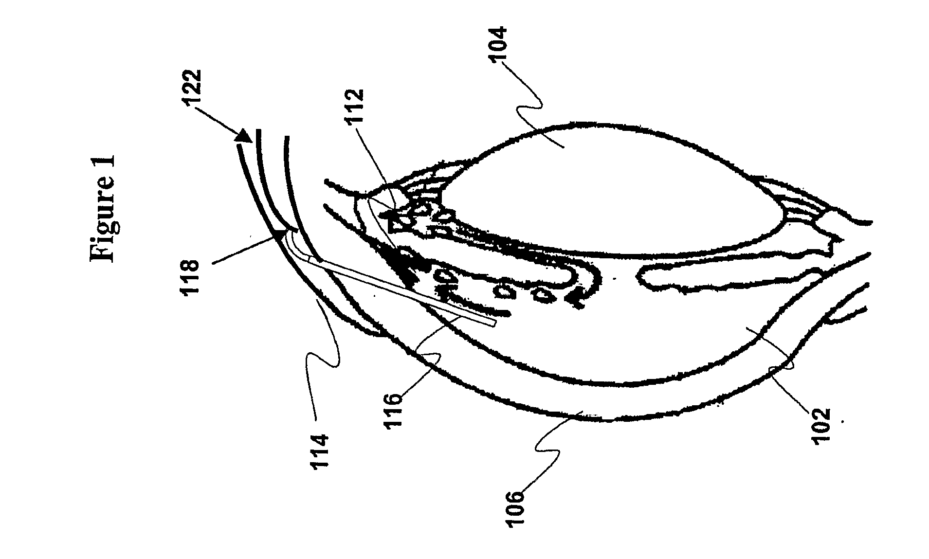
[0021]FIG. 1 is a cross-sectional view depicting a portion of a human eye and a pressure control system according to various embodiments of the present invention;

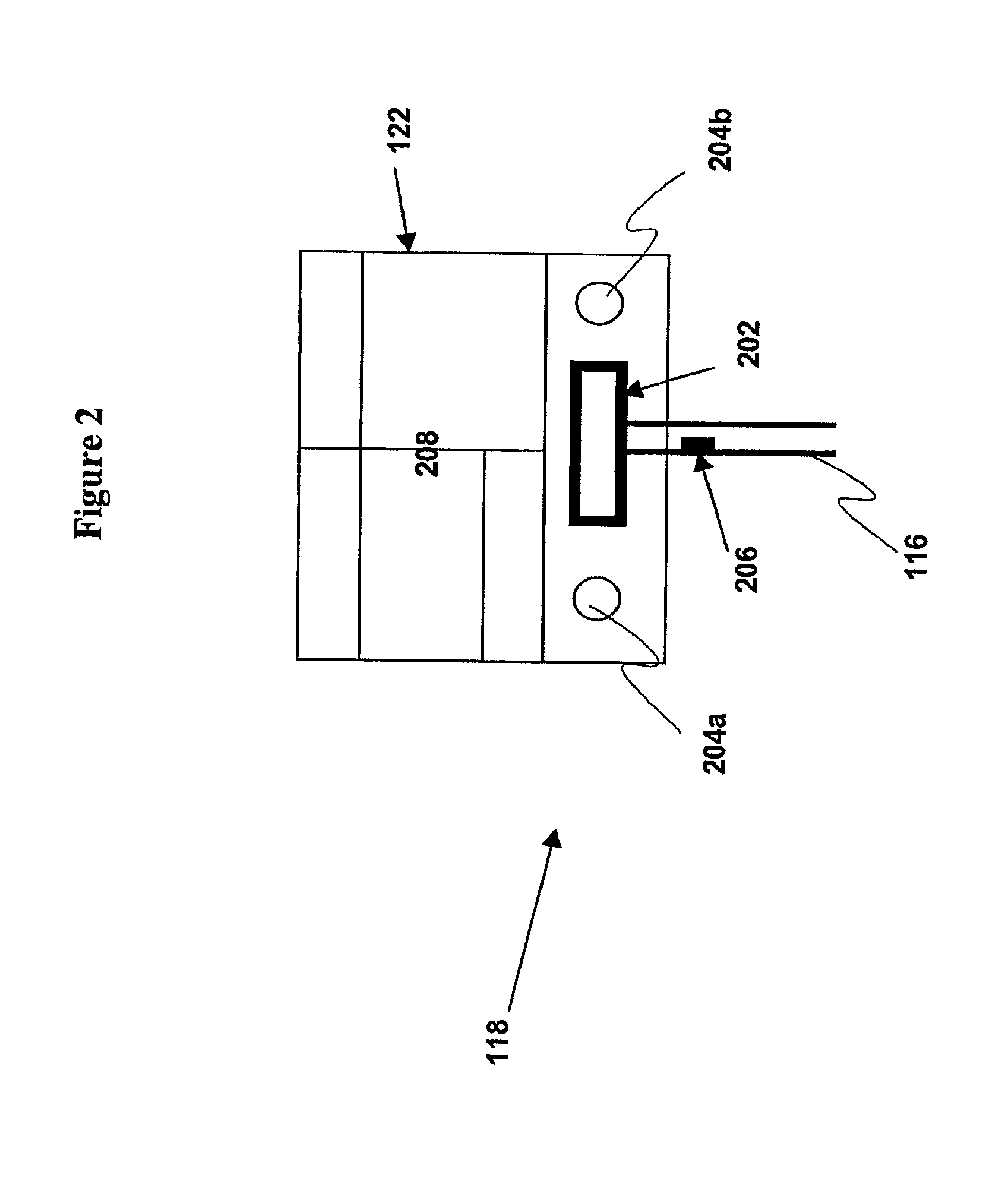
[0022]FIG. 2 is a diagram depicting a pressure control system according to various embodiments of the present invention;

[0023]FIG. 3 is a cross-sectional view depicting a pressure sensor, according to various embodiments of the present invention;

[0024]FIG. 4 is a top view depicting a pressure sensor, according to various embodiments of the present invention;

[0025]FIG. 5a is a perspective view depicting an electromagnetic valve, according to various embodiments of the present invention;

[0026]FIG. 5b is a top view depicting an electromagnetic valve, according to various embodiments of the present invention;

[0027]FIG. 6 is a block diagram illustrating the layout of an electroni...


PUM


Abstract

Systems and methods for automatically monitoring and controlling pressure in a body part are disclosed. The systems include an implantable tube with one open end of the tube implanted in the body part, an implantable valve coupled with the tube having at least one open state and a closed state, an implantable sensor for measuring pressure, and an implantable control device coupled with the sensor and the valve. The control device switches the valve between the at least one open state and the closed state, based on pressure information received from the sensor. When the valve is in the at least one open state, the tube drains fluids from the body part due to a difference of pressure between the open ends of the tube. Methods for using the systems to administer drugs and monitor and control fluid pressures in various biological systems are also disclosed.

Description

FIELD OF THE INVENTION[0001]Embodiments of the present invention relates to new and useful apparatus, systems and methods for monitoring and controlling pressure in body cavities, tissues and organs in living systems and, more particularly, to intraocular pressure sensor and valve apparatus, systems and methods of using them. Embodiments of the present invention may also be used to administer drugs and / or other agents to living or non-living systems by controlling fluid access to an implanted or remote reservoir.BACKGROUND OF THE INVENTION[0002]Primary open-angle glaucoma (POAG), a widespread, chronic disease, is classified by the World Health Organization as the third leading cause of blindness. As many as 67 million people suffer from glaucoma and 10% will lose vision in both eyes as a result. Even in developed countries with advanced medical care, fewer than half of those with glaucoma associated with clinically elevated intraocular pressure (IOP) are even aware of their conditio...

Claims


Application Information

Patent Timeline
IPC IPC(8): A61F9/00
CPCA61B3/16A61B5/076A61B5/4839A61M39/28A61F9/00781A61M5/16804A61M39/22A61B2560/045
Inventor LATTANZIO, FRANK A.DELAHANTY, THOMAS C.
Owner EASTERN VIRGINIA MEDICAL SCHOOL
Learn More