Organ-specific gene, method for identifying the same and use thereof

a technology of organs and genes, applied in the field of organ-specific genes and methods for identifying the same, can solve the problems of large amount of compound required, inability to reflect toxicity in humans, and high cost of purchasing and maintaining laboratory animals

Inactive Publication Date: 2009-12-17
TAKEDA PHARMA CO LTD
View PDF2 Cites 1 Cited by
  • Summary
  • Abstract
  • Description
  • Claims
  • Application Information

AI Technical Summary

Benefits of technology

[0014]According to the method of extraction of the present invention, it is possible to extract a set of genes that are highly expressed truly specifically in an organ or tissue. The set of genes highly expressed specifically in an organ (tissue) thus obtained are useful as an organ or tissue differentiation marker. Furthermore, according to the method of identification of the present invention, it is possible to identify an organ or tissue for which no specific highly expressed gene has been obtained.

Problems solved by technology

Meanwhile, an evaluation of the toxicity of a drug to various organs is normally performed by an in vivo toxicity study by compound administration to laboratory animals such as rats; however, such studies are faulty in that (1) several days to several months are taken before onset of the toxicity, (2) a large amount of compound is required, (3) much costs are taken to purchase and maintain laboratory animals, and the like, and, in addition, the radical problem arises that the toxicity in humans is not always reflected.
In in vitro screening, it is common practice to use a primary culture of a tissue isolated from various organs or a cell line established therefrom; however, primary culture involves the above-described drawback (3) as with in vivo studies; meanwhile, when a cell line is used, problems arise, including the disappearance of the characteristics of the organ or tissue from which the cell line is derived during repeated passage, and the inability to fully reflect the pathology of the organ or tissue as an organic integration thereof on the basis of individual cells.
For these reasons, use of a human cell does not always accurately reflect the toxicity in human individuals.
However, differentiation into albumin-producing cells or insulin-producing cells, if confirmed, does not immediately lead to the conclusion that they are hepatocytes or pancreatic β cells.
This method of extraction, as a means for comprehensively identifying tissue / organ-specific genes, was better than conventionally known methods; however, even so, the specific highly expressed genes extracted by the method sometimes included those that cannot be said to be truly specific for the tissue (organ).

Method used


Image

  • Organ-specific gene, method for identifying the same and use thereof
  • Organ-specific gene, method for identifying the same and use thereof
  • Organ-specific gene, method for identifying the same and use thereof

Examples


example 1

Materials and Methods

1. Database Used and Method of Data Extraction

[0081]Gene expression data on male C57BL / 6 mice were extracted from BioExpress of Gene Logic on Apr. 21, 2006. The parameters used in the extraction are shown in Table 1. Mouse Genome 430 2.0 Array includes 45101 probe sets.

TABLE 1Parameter settings at time of extractionof C57BL / 6 mouse gene expression dataParameter setting inBioExpressParameterDonor SpeciesM. musculusStrainC57BL / 6GenderMaleChipsetMouse430_2(Affymetrix, Mouse Genome 4302.0 Array)AlgorithmMAS5.0 (Affymetrix)NormalizationAffymetrixGeneral Pathologic CategoryNormalIVT Labeling Protocol-Double BiotinMouse430_2

2. Method of Calculating Log-Ratio Values of the Gene Expression Data

[0082]The gene expression data extracted were subjected to a logarithmic conversion with the base 2 using the Avadis version 3.3 software program (manufactured by Strand Genomics). The mean of AFFX-GapdhMur / M32599—5_at, AFFX-GapdhMur / M32599_M_at, and AFFX-GapdhMur / M32599—3_at was s...

example 2

Comparison of Organ / Tissue Specificity with Specific Genes Obtained by Other Method of Extraction

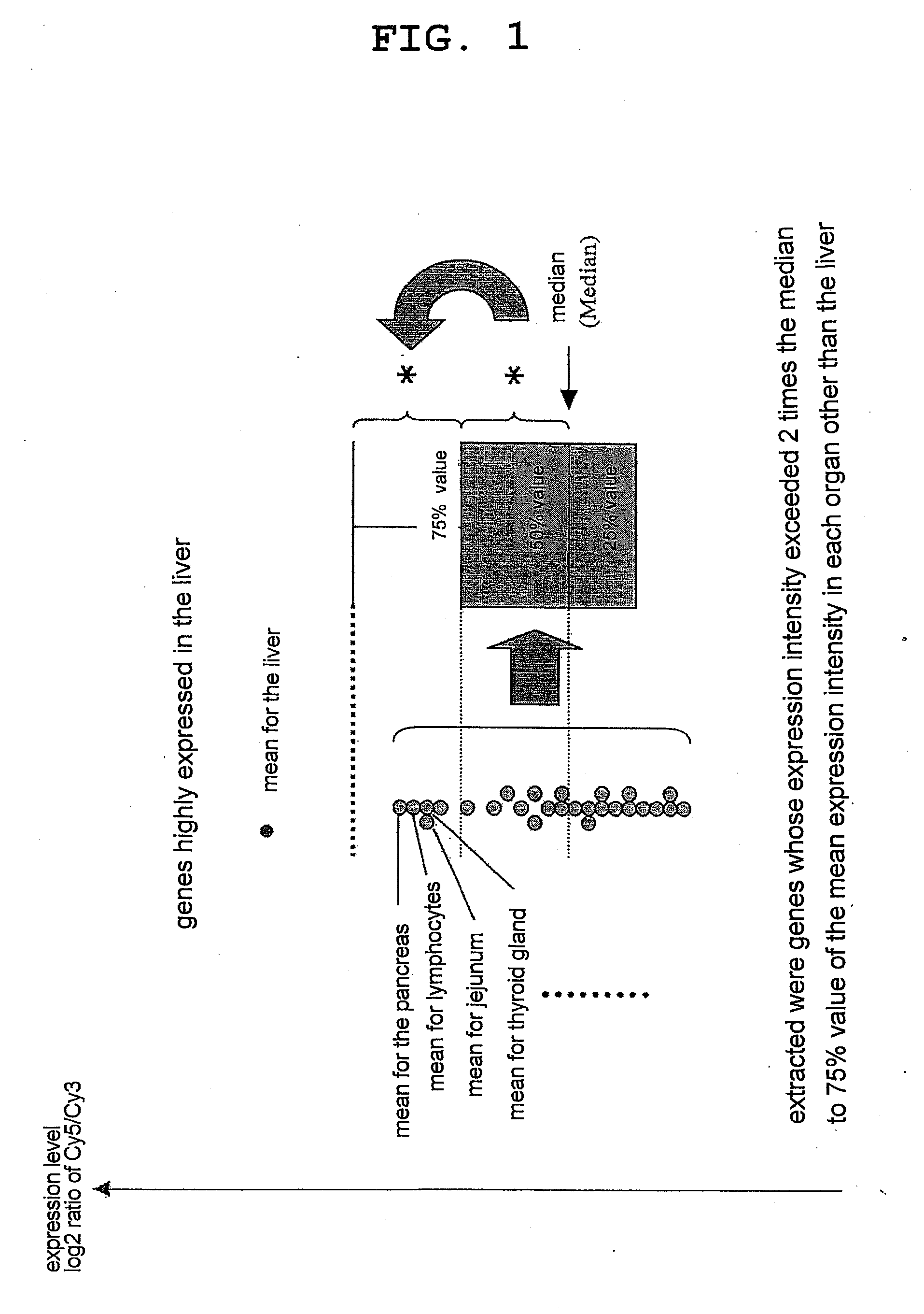
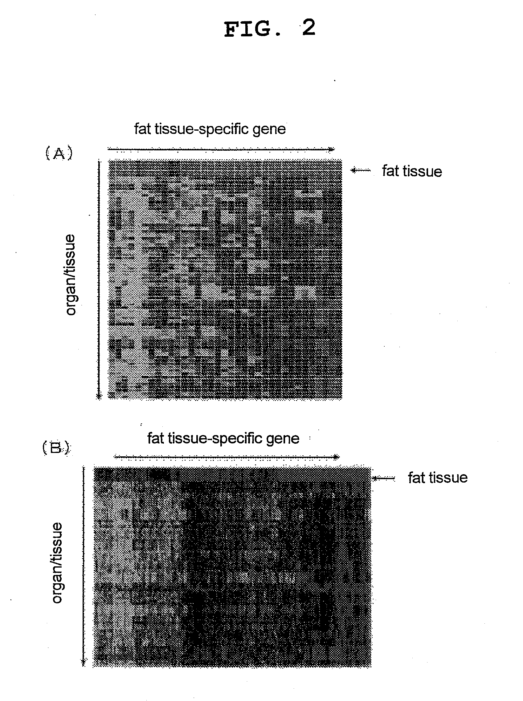
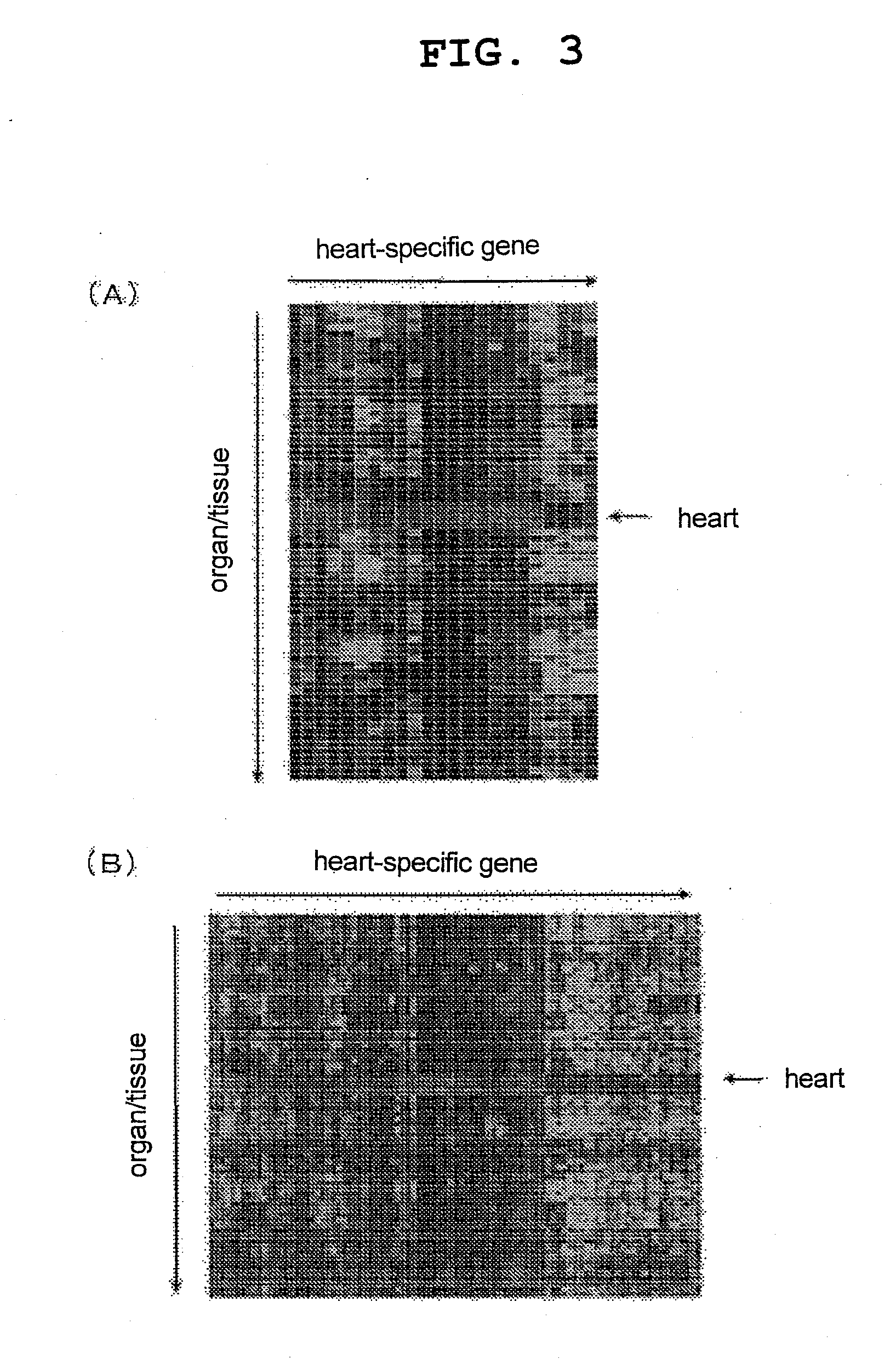
[0086]The expression of the genes specific for various organs / tissues obtained in Example 1 and the expression of the organ / tissue-specific genes obtained by a method previously published by the present inventors (see the non-patent document 1 and FIG. 1) (Comparative Example) in various mouse organs / tissues were compared by generating heat maps. FIG. 2 shows a comparison of both for a fat tissue-specific gene group, FIG. 3 shows a comparison for a heart-specific gene group, FIG. 4 shows a comparison for a liver-specific gene group, FIG. 5 shows a comparison for a thymus-specific gene group, and FIG. 6 shows a comparison for a stomach-specific gene group (in all graphs, A represents the results from the gene groups obtained in Example 1, and B represents the results from the gene groups obtained in Comparative Example). For all organ / tissue-specific gene groups, almost all of the genes o...

example 3

Identification of Genes Highly Expressed Specifically in Combinations of Organs / Tissues

[0087]For the kidney (left), kidney (right), prostate and skeletal muscle, for which no specific highly expressed gene was identifiable in Example 1, an attempt was made to identify specific highly expressed genes using combinations with other organs / tissues. As a result, it was found that when the kidney (left) and the kidney (right) were combined, 66 probe sets were expressed specifically in the kidneys (left and right). For the prostate, it was found that when it was combined with the pancreas, 1 probe set was highly expressed specifically in the prostate / pancreas (Table 3). For skeletal muscle, an analysis was attempted after it was combined with the heart, which mainly consists of muscular tissue, or with the prostate, from which no specific gene was acquired when examined alone, but no specific highly expressed gene was acquired.

TABLE 3Numbers of genes highly expressed specificallyin combina...


PUM

PropertyMeasurementUnit
Center for Biotechnology Information Basic Local Alignment Search Toolaaaaaaaaaa
lengthaaaaaaaaaa
fluorescentaaaaaaaaaa

Abstract

The present invention provides a method of extracting an organ- or tissue-specific highly expressed gene, including:
(1) a step for measuring expression level of a specified gene group for each organ or tissue in 2 or more individuals,
(2) a step for acquiring (a) a minimum value of expression levels in a particular organ or tissue in all individuals, and (b) a maximum value of expression levels in other organs and tissues in all individuals, for each gene, and
(3) a step for extracting the gene as a gene highly expressed specifically in the particular organ or tissue if the above-described (a)/(b) ratio is larger than 1. By the present invention, truly organ- or tissue-specific genes are extracted.

Description

TECHNICAL FIELD[0001]The present invention relates to a method of identifying organ- or tissue-specific highly expressed genes of various organisms, a set of mouse organ- or tissue-specific highly expressed genes obtained by the method, and a method of identifying an organ or tissue for which no specific highly expressed gene has been obtained, by combining organ- or tissue-specific highly expressed genes identified by the method.BACKGROUND OF THE INVENTION[0002]In recent years, there has been remarkable progress in regenerative medicine (medical practice). Since a method of establishing human embryonic stem cells (ES cells) was developed, it has been a realistic goal to produce cells / tissues, and even organs themselves, for transplantation from ES cells, as well as from somatic stem cells, including mesenchymal stem cells (MSCs), on an industrial scale using approaches based on cell engineering / embryological engineering. In fact, for the blood, nerves, heart muscle, liver, pancreas...

Claims


Application Information

Patent Timeline
Patent Type & Authority Applications(United States)
IPC IPC(8): C12Q1/68C07H21/04G16B25/10
CPCC12Q1/6881C12Q2600/158G16B25/00G16B25/10C12Q1/6876
Inventor MIYAMOTO, SAKUTAKAMI, KENJIHORINOUCHI, AKIRA
Owner TAKEDA PHARMA CO LTD
Why Eureka
Try Eureka