

Method for Detecting Autoantibodies Formed in Rheumatoid Arthritis
a technology for rheumatoid arthritis and autoantibodies, which is applied in the field of autoantibodies detection, can solve the problems of not making any observation of telopeptides, not disclosing studies of the role of collagen telopeptides, etc., and achieves the effect of preventing their broad us
- Summary
- Abstract
- Description
- Claims
- Application Information
AI Technical Summary
Benefits of technology
Problems solved by technology
Method used
Image

Examples
example 1
[0054]ELISA Assay
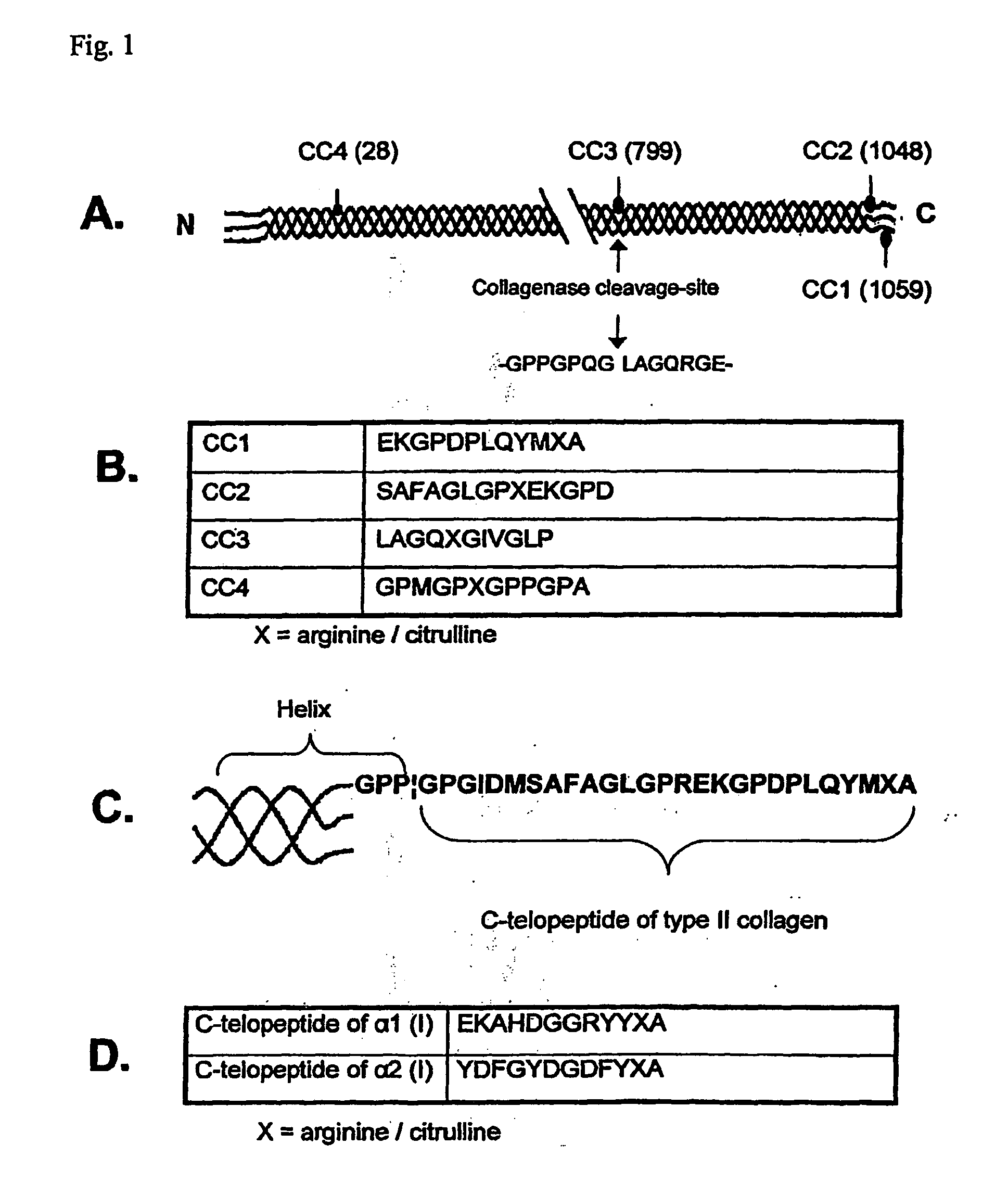
[0055]The serum samples from 120 patients with RA were obtained from the Division of Rheumatology of Oulu University Hospital. The controls consisted of 81 sera from healthy people matched for age and sex. Six pairs of biotinylated peptides were synthesised by NeoMPS (Strasbourg, France). FIG. 1 shows the sequences and locations of the chosen peptides in the primary structures of human type I and II collagens. FIGS. 1A-C show the localization of the peptides CC1-CC4 in type II collagen and their sequences; the numbers in brackets refer to arginine residues: CC1 (1059), CC2 (1048), CC3 (799) and CC4 (28). Collagenase cleavage site is shown by arrow in FIG. 1A -GPPGPQG|LAGQRGE- (SEQ ID NO:9). FIG. 1B shows the sequences CC1 EKGPDPLQYMXA (SEQ ID MO:3), CC2 SAFAGLGPXEKGPD (SEQ ID NO:6),CC3 LAGQXGIVGLP (SEQ ID NO:7) and CC4 GPMGPXGPPGPA (SEQ ID NO:8); X is arginine / citrulline. FIG. 1C shows detailed structure of the carboxy terminal telopeptide of type II collagen (GPP b...
example 2
[0061]Chemiluminesence Assay
[0062]The measurements were done with two-site chemiluminescence immunoassays, which detect IgG antibodies against the synthetic C-telopeptides of the α1 chains of human type I and II collagens (EKAHDGGRYYRA (SEQ ID NO:4), and EKGPDPLQYMRA (SEQ ID NO:5), or EKAHDGGRYYXA (SEQ ID NO:1), and EKGPDPLQYMXA (SEQ ID NO:3), respectively, where X stands for citrulline, from NeoMPS, Strasbourg, France). The serum samples were first diluted 1:10 in a buffer containing 10 mM Tris-HCl, 350 mM NaCl, 1% BSA, 1% [vol / vol] Triton X-100, 0.5% [wt / vol] Na-deoxycholate, 0.1% SDS; pH 7.6 and incubated with suitable concentrations of one of the above peptides (in biotinylated form) and streptavidin-coated magnetic particles for 10 minutes at 37° C. The unbound biotinylated antigen and antibodies were separated from the complex bound to the magnetic particles by aspiration of the reaction mixture and subsequent washing. Thereafter, acridinium-labeled anti-human IgG antibodies w...
example 3
[0065]The Specificity of Autoantibodies In sera of Rheumatoid Arthritis Patients
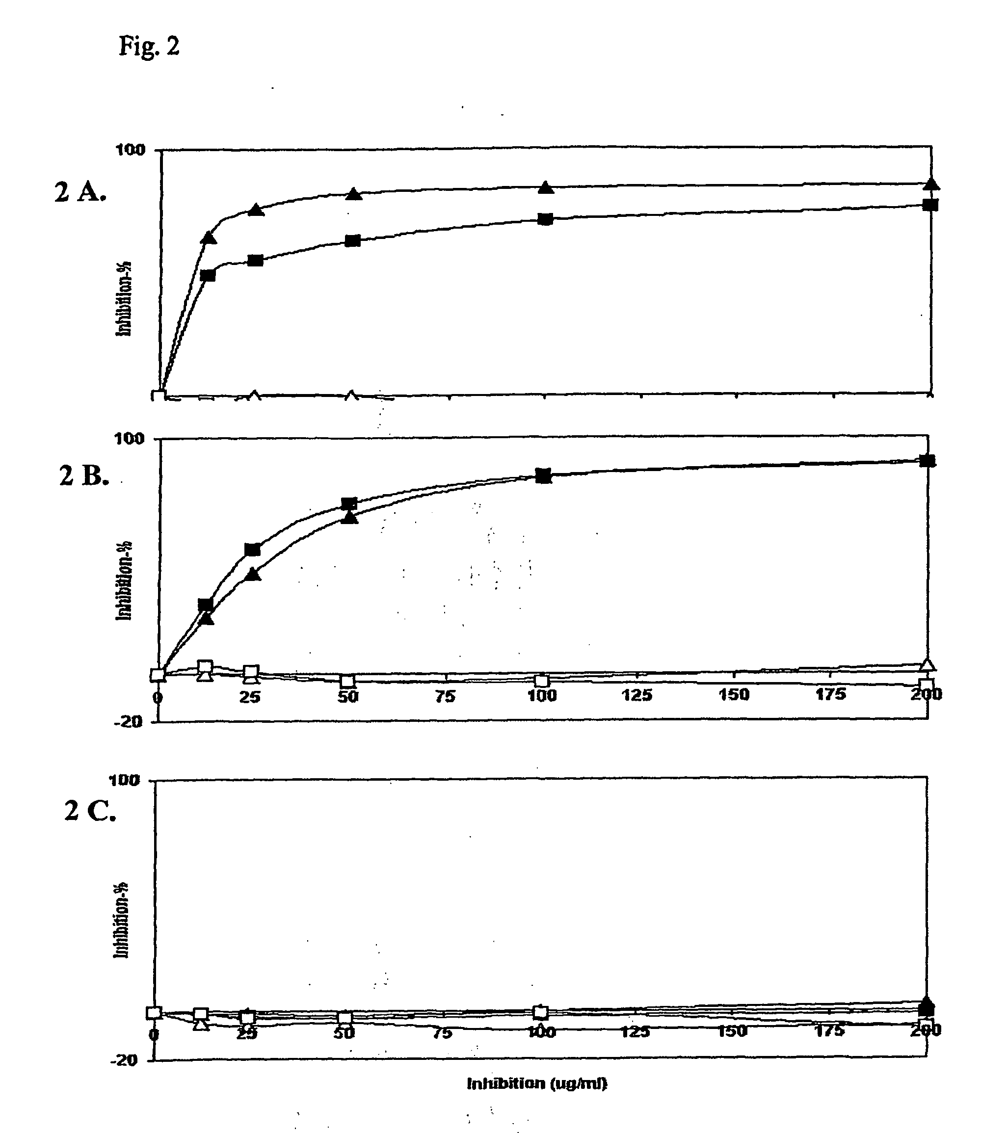
[0066]For detecting the antibody specificity we tested both type I and II collagen telopeptide assays and an anti-CCP Mark2 assay kit. Patient sera were diluted in assay buffer so that in each assay the initial binding could be noticeably inhibited with the respective peptides. Serial dilutions of competing peptides (arginine and citrulline forms of collagen C-telopeptides of α1(I) and α1(II)) were added (FIG. 2). Inhibition by the soluble form of the same peptide as that coupled to the plate served as a specificity control in both telopeptide assays. For anti-CCP assay no such peptide was available for specificity testing. The percentage inhibition was plotted against the soluble peptide concentration. The signal (wavelength of 450 nm) obtained with the human serum only (initial binding) was defined as 0% inhibition, and the signal of the blank (no serum sample) was defined as 100% inhibition. We inhibi...
PUM
| Property | Measurement | Unit |
|---|---|---|
| Mass flow rate | aaaaa | aaaaa |
| Mass flow rate | aaaaa | aaaaa |
| Volume | aaaaa | aaaaa |
Abstract
Description
Claims
Application Information