Obtaining optical tissue properties

a tissue and optical technology, applied in the field of medical tissue examinations, can solve the problems of inability to know whether a specimen is taken from the correct part of the tissue, inability to obtain optical tissue properties, and inability to achieve optimal guidance methods, so as to achieve the effect of improving the accuracy of the biopsy procedure and minimizing the number of needle biopsies

Inactive Publication Date: 2009-12-31
KONINKLIJKE PHILIPS ELECTRONICS NV
View PDF15 Cites 32 Cited by
  • Summary
  • Abstract
  • Description
  • Claims
  • Application Information

AI Technical Summary

Benefits of technology

[0045]In other words, Raman spectroscopy merely allows for inspecting the tissue locally before taking the actual specimen. Therefore, the number of needle biopsies can be minimized while the accuracy of the biopsy procedure is actually improved.
[0046]According to a further embodiment of the invention the second fiber end provides an interior lateral field of view, which is directed from the shaft wall towards the central longitudinal axis of the hollow shaft. This may provide the advantage that a tissue specimen, which has been removed from the pat

Problems solved by technology

Although helpful, these methods of guidance are far from optimal.
The spatial resolution is limited and, furthermore, these imaging modalities can in most cases not discriminate between benign and malignant tissue.
As a result, during a biopsy procedure one does not know whether a specimen is taken from the cor

Method used


Image

  • Obtaining optical tissue properties
  • Obtaining optical tissue properties
  • Obtaining optical tissue properties

Examples


first embodiment

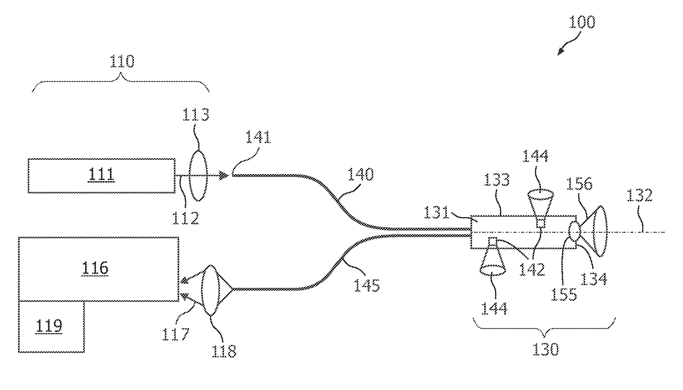
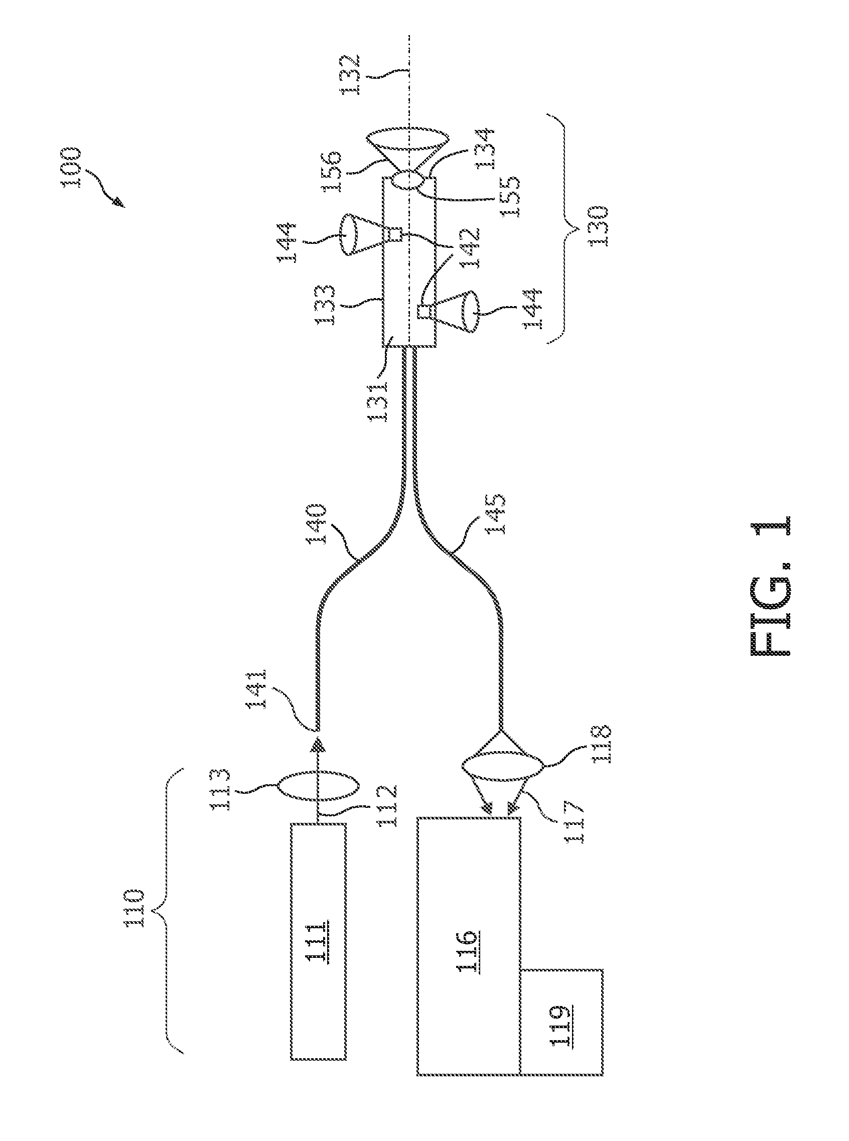
[0074]FIG. 1 shows a medical apparatus 100 according to the present invention. The medical apparatus 100 comprises an optical instrument 110 and a medical device 130. According to the embodiment described here the medical device is an optical needle 130. The medical apparatus 100 is in particular suitable for optically investigating tissue material being surrounded laterally with respect to the medical device 130.

[0075]The optical instrument 110 comprises a light source 111, which is adapted to generate illumination light 112. According to the embodiment described here, the light source is a laser 111, which emits a monochromatic radiation beam 111. The radiation beam is directed via an optic 113 onto a first fiber end 141 of an optical fiber 140.

[0076]The optical instrument 110 further comprises a spectrometer device 116, which is optically coupled to an optical fiber 145 by means of an optic 118. The spectrometer device 116 is used for spectrally analyzing measurement light 117, w...

second embodiment

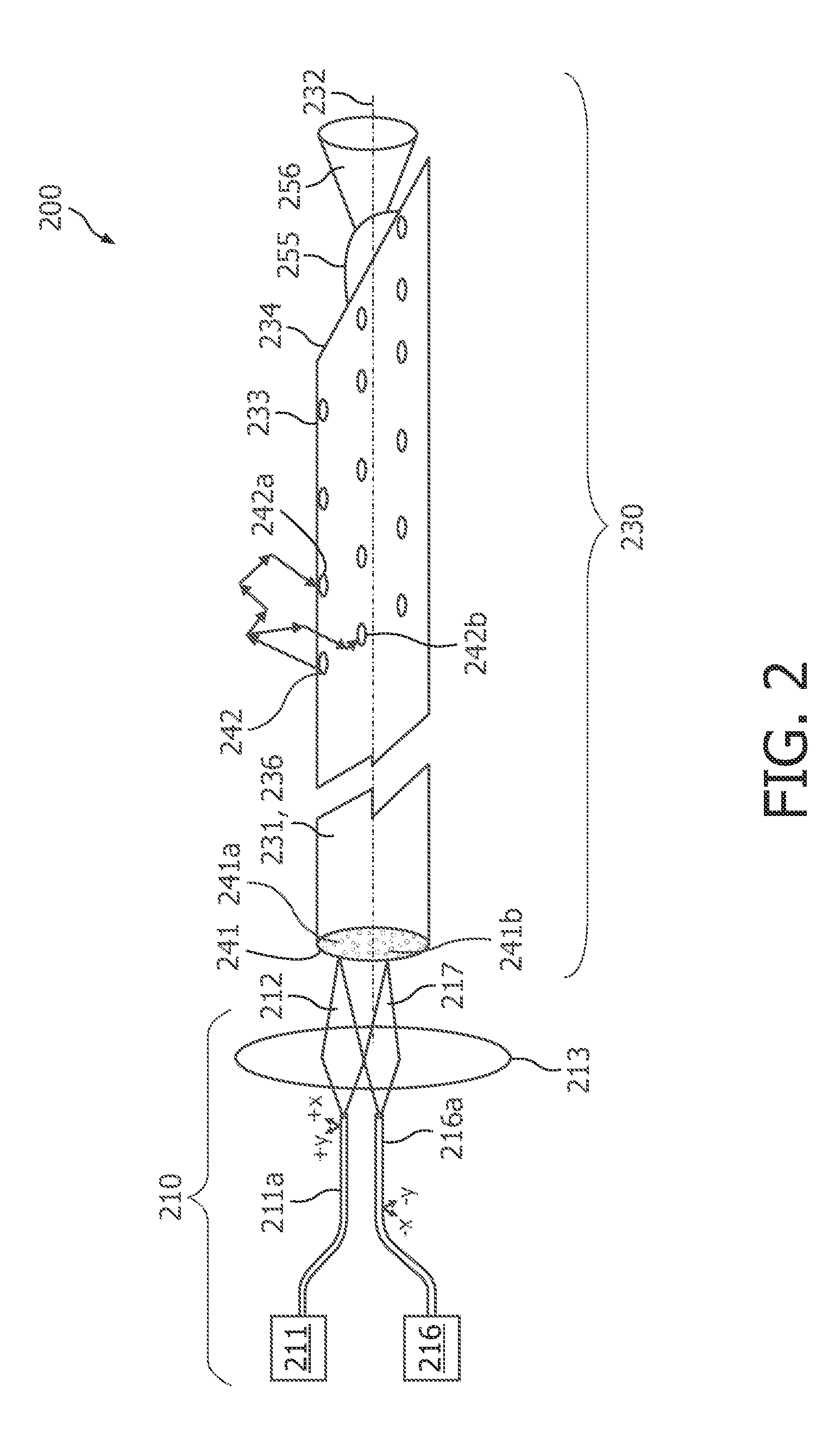
[0081]FIG. 2 shows a medical apparatus 200 according to the present invention. The medical apparatus 200 comprises an optical instrument 210 and a medical device 230. According to the embodiment described here the medical device is a solid optical needle 130.

[0082]The optical instrument 210 comprises a light source 211, which is adapted to generate illumination light 212. The illumination light is guided by an optical fiber 211a, which may also be denominated an illumination fiber 211a. The optical instrument 210 further comprises a spectrometer device 216, which is adapted to receive a measurement light 217 by means of a measurement fiber 216a. An optic 213 is provided in order to optically couple the optical fiber 211a respectively the measurement fiber 216a with selected optical fibers being accommodated within the medical device 230.

[0083]The spectrometer device 216 may also be replaced with an optical detector 216 solely measuring the light intensity. The detector 216 may be eq...


PUM


Abstract

This application describes a medical device (230) for obtaining optical tissue properties of a target material. The medical device (230) comprises an elongated body (231) having a longitudinal axis (232) and an optical fiber being integrated within the elongated body (231). The optical fiber has a second fiber end (242, 242a, 242b), which is arranged at a side wall (233) of the elongated body (231) and which provides a lateral field of view with respect to the longitudinal axis (232). According to an embodiment many optical fibers are integrated each having an optical outlet (242, 242a, 242b) around the elongated body (231). Using the outlets (242, 242a, 242b) to do diffuse optical tomography and also use optical fibers to do an optical inspection, one can get information on the presence of tumors in a volume around the medical device (230) and a tissue characterization in the vicinity of the medical device (230). Thereby, an optical biopsy may be carried out, wherein no real tissue is removed. According to another embodiment an optical detection system is integrated into a real biopsy needle (330) allowing inspection and taking real biopsy simultaneously.

Description

FIELD OF INVENTION[0001]The present invention relates to the field of medical tissue examinations. In particular, the present invention relates to a medical device such as a needle for obtaining optical tissue properties of a human or animal body. The medical device, which is insertable into tissue to be probed, comprises at least one optical fiber for directing illumination light to the tissue and for receiving measurement light having interacted with the tissue. The present invention further relates to a medical apparatus and to a method for obtaining optical tissue properties of a human or animal body. Both the medical apparatus and the method take benefit from the described medical device.ART BACKGROUND[0002]For correct diagnosis of various cancer diseases biopsies are taken. This can either be via a lumen of an endoscope or via needle biopsies. Biopsies may be taken for instance from the prostate via the rectum. In order to find the correct position to take the biopsy, various ...

Claims


Application Information

Patent Timeline
IPC IPC(8): A61B6/00
CPCA61B5/0066A61B5/0071A61B5/6848A61B5/0084A61B5/0075
Inventor HENDRIKS, BERNARDUS HENDRIKUS WILHELMUSKUIPER, STEINDE BOER, RUTH WILHELMINE IREENBRAUN, AUGUSTINUS LAURENTIUSVAN BEEK, MICHAEL CORNELISRENSEN, WOUTER HARRY JACINTH
Owner KONINKLIJKE PHILIPS ELECTRONICS NV
Why Eureka
Try Eureka