Method and apparatus for diagnostic and treatment using hard tissue or material microperforation

a micro-perforation and hard tissue technology, applied in the field of minimally invasive diagnostic procedures and treatment, can solve the problems of inconvenient tissue removal, limited drilling capabilities of known techniques and devices, undesirable side effects, etc., and achieve the effect of reliable and minimally invasive fastening of a prosthesis

Inactive Publication Date: 2010-01-21
REJUVEDENT
View PDF12 Cites 66 Cited by
  • Summary
  • Abstract
  • Description
  • Claims
  • Application Information

AI Technical Summary

Benefits of technology

[0009]It is another object of the present invention to control diffusion in the hard tissue of a substance delivered into the incision. It is yet another object of the invention to provide a method and device for reliable and minimally invasive fastening of a prosthesis onto the hard tissue or hard material.

Problems solved by technology

However, the drilling capabilities of the known techniques and devices are limited to a relatively large diameter (>0.5 mm).
This is appropriate when tissue removal is required for caries treatment or for restoration, but not desirable in other instances, when the orifice or incision is only needed to gain access to the tissue or for the delivery of drugs or other compounds.
In such circumstances, therefore, tissue removal is an undesirable side effect.
However laser heads of the industrial lasers are typically bulky and impractical for use in clinical applications.
Delivery of a therapeutic or cosmetic compound in or through hard tissue is limited due low permeability of hard tissue.
Due to the low permeability of the enamel, high concentrations of bleaching agent are used, increasing the risk of gum damage and hypersensitivity.

Method used


Image

  • Method and apparatus for diagnostic and treatment using hard tissue or material microperforation
  • Method and apparatus for diagnostic and treatment using hard tissue or material microperforation
  • Method and apparatus for diagnostic and treatment using hard tissue or material microperforation

Examples


Embodiment Construction

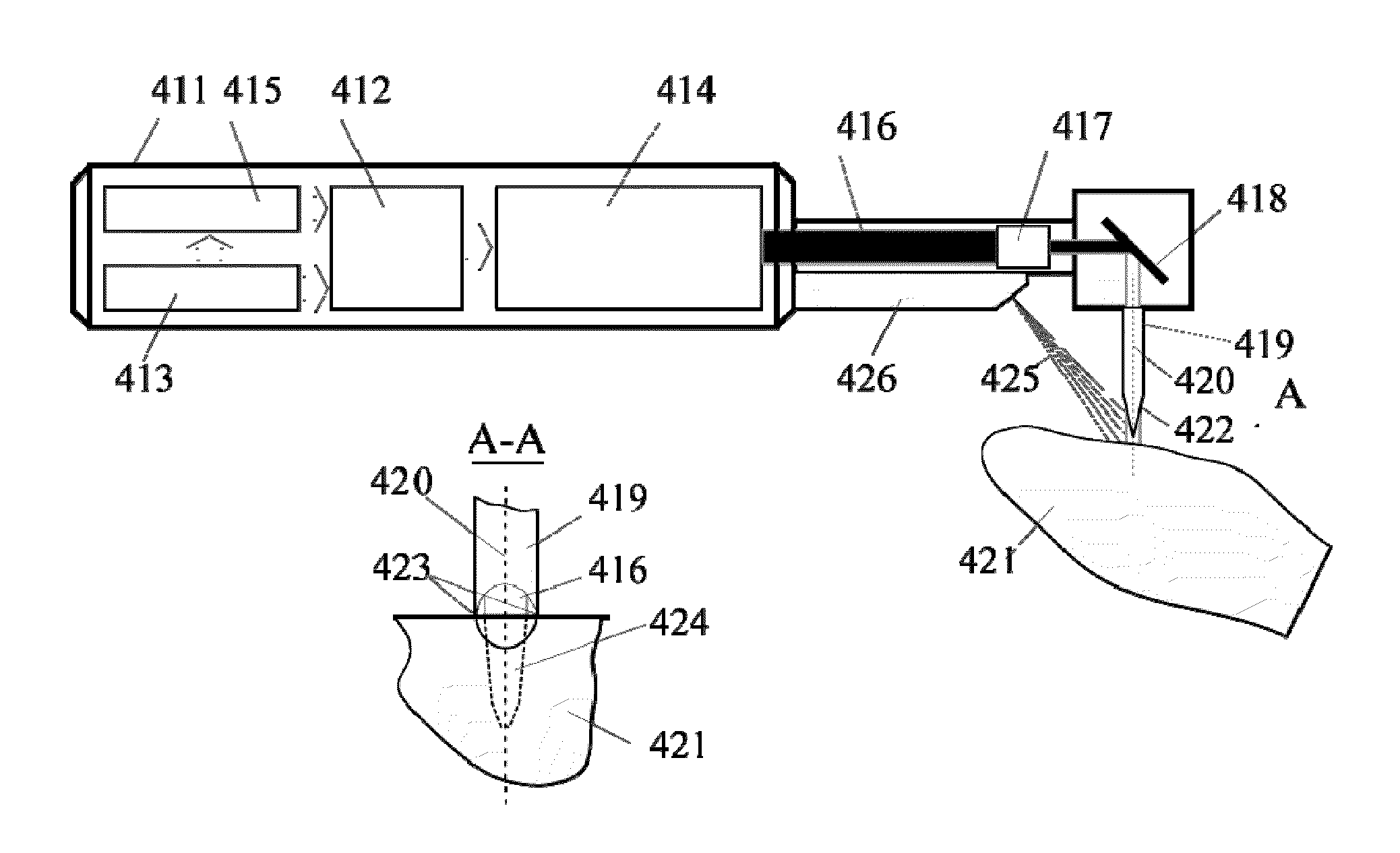
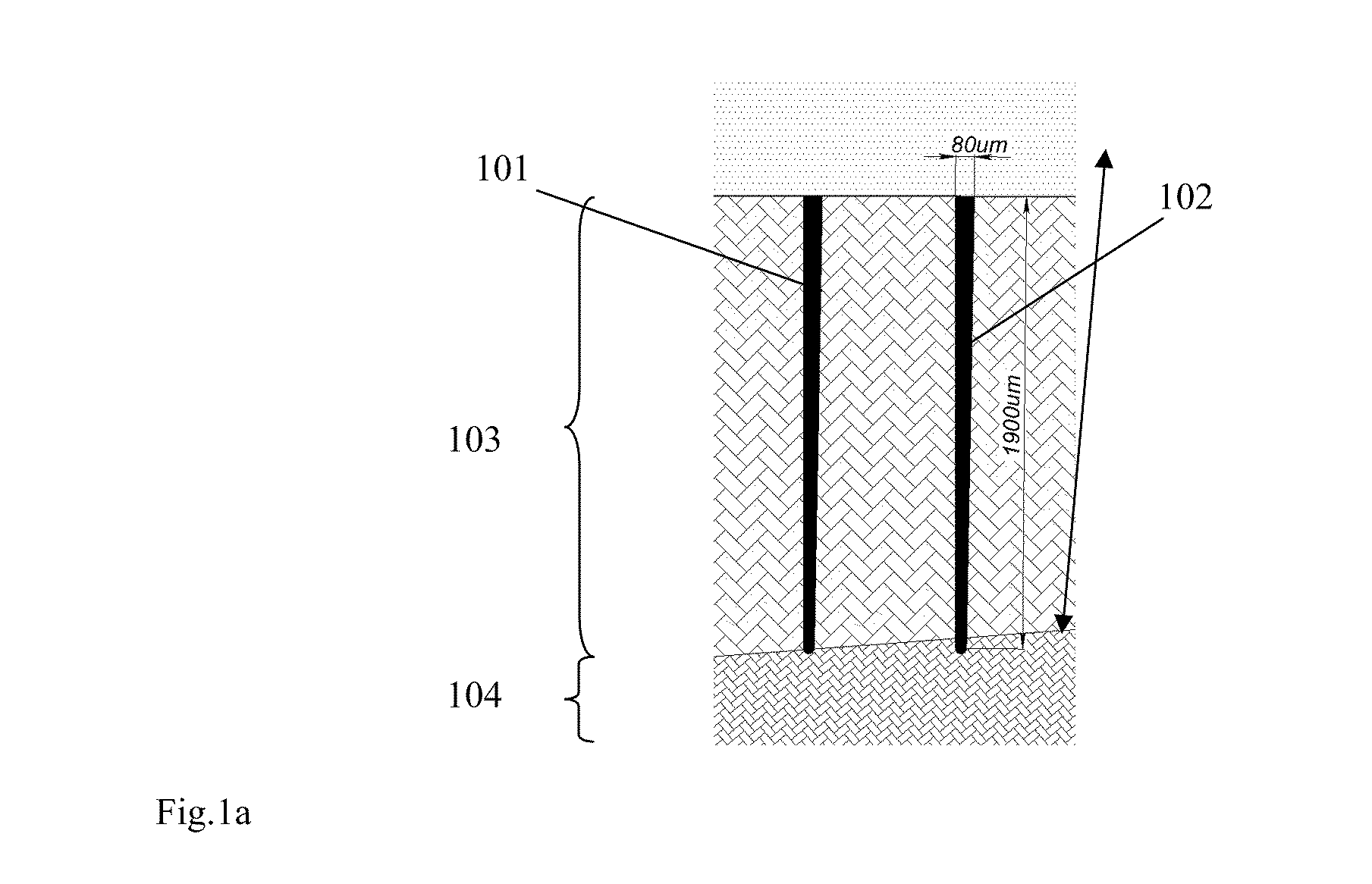
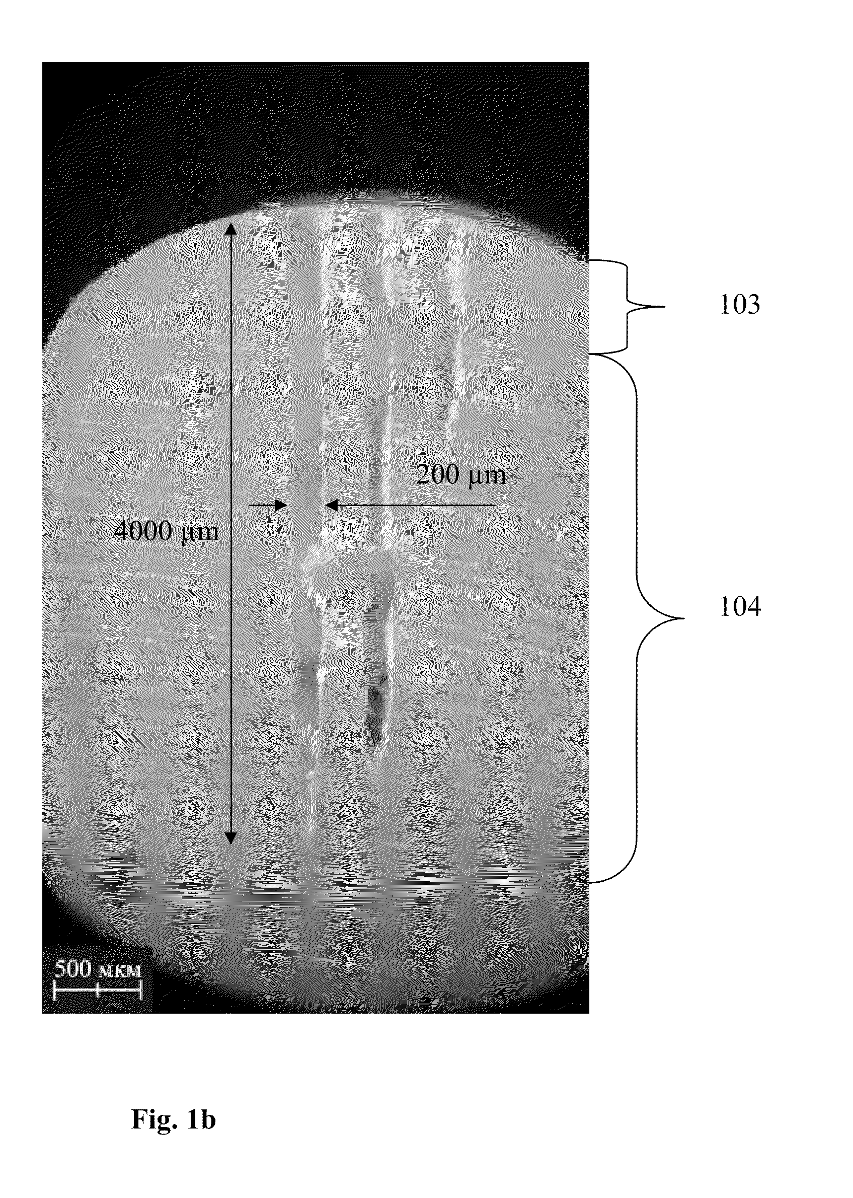
[0028]According to the invention, one or more micro-incisions are first drilled in hard tissue, including, but not limited to, dental enamel, cementum, dentine, bone, or oral soft tissues, including but not limited to, oral mucosa, muscle, glands, tongue, pulp, frenum, palate, uvula, and tonsil. The microperforations have a cylinder-like or cone-like shape and are characterized by a certain aspect ratio (AR). The aspect ration can be defined as the ratio of a length of a cylinder-like structure to its diameter. For the referenced microperforations the AR is in the range from 1 to 100, preferably from 5 to 30. For the incisions themselves, the diameter can vary in the range from 1 to 500 μm and the length can very in the range from 100 to 10000 μm. These microincisions can be used for a variety of dental and oral procedures, such as, for example, teeth whitening, delivering therapeutic compounds through the incision to the treatment target, using this incision for dental and oral dia...


PUM


Abstract

A method of modifying or treating biological tissue by microperforating hard tissue is disclosed. The method comprises identifying a target area associated with the hard tissue, using a laser beam to perforate at least one incision in the hard tissue, wherein at least one incision has a diameter from a range of 0.001 mm to 0.5 mm and an aspect ratio from a range of 1 to 100 times, introducing a treatment substance into the incision, and causing the treatment substance to interact with the target area. Also a device for microperforating hard biological tissue is disclosed, comprising a laser pump system and a laser head coupled to the laser pump system for generating a pulsed laser having ranges of wavelengths, a pulse duration, pulse energy from a selected range, a beam divergence factor less than 5, a repetition rate higher than 50 Hz; and a beam delivery system comprised of a focusing system for creating a beam having a diameter from a range of 0.001 mm to 0.5 mm.

Description

CROSS REFERENCE TO RELATED APPLICATION[0001]This application claims priority to an earlier filed US provisional patent application number 60 / 885,009, filed on Jan. 16, 2007, which is incorporated herein by reference in its entirety.FIELD OF THE INVENTION[0002]The invention relates to the minimally invasive diagnostic procedures and treatment of hard biotissues or soft biotissues obscured by hard tissue or hard material. More particularly, the invention relates to the method and apparatus for perforating small diameter incisions in the hard tissue or material to gain minimally invasive access to the biotissue for diagnosis or treatment.BACKGROUND OF THE INVENTION[0003]Traditionally, mechanical drilling or sawing is performed to gain access into or through hard tissues, such as dental enamel, dentin or bone. This access may be needed for drug delivery, visualization and diagnostics, dental whitening, to name a few examples.[0004]In the field of dentistry, several methods of gaining ac...

Claims


Application Information

Patent Timeline
Patent Type & Authority Applications(United States)
IPC IPC(8): A61C5/08A61C19/00
CPCA61B5/0066A61C1/0046A61B5/415A61B5/0088
Inventor ALTSHULER, GREGORY B.BELIKOV, ANDREY V.FELDCHTEIN, FELIX I.TUCHIN, VALERY V.VYBORNOV, ALEXANDER G.
Owner REJUVEDENT
Why Eureka
Try Eureka