Method for preparing biological scaffold material

a biological scaffold and material technology, applied in the field of regenerative medical technology, can solve the problems of limited durability, restenosis and other adverse effects, and the inability to use biodegradable vascular grafts in the arterial system,

Inactive Publication Date: 2010-01-28
HEALTH SCI TECH TRANSFER CENT JAPAN HEALTH SCI FOUND
View PDF2 Cites 29 Cited by
  • Summary
  • Abstract
  • Description
  • Claims
  • Application Information

AI Technical Summary

Problems solved by technology

In case of glutaraldehyde-fixed xenogenic vascular grafts, there exists, in addition to the limited durability due to calcification, another disadvantage.
However, restenosis and other adverse effects have been reported.
In addition, the biodegradable vascular grafts cannot be used in the arterial system because of possible puncture due to the hydrolysis of the polymer.
However, the decellularized tissues as such do not have a strength sufficient to withstand relatively high pressures required as, for example, the aortic valve.

Method used


Image

  • Method for preparing biological scaffold material
  • Method for preparing biological scaffold material
  • Method for preparing biological scaffold material

Examples


example 1

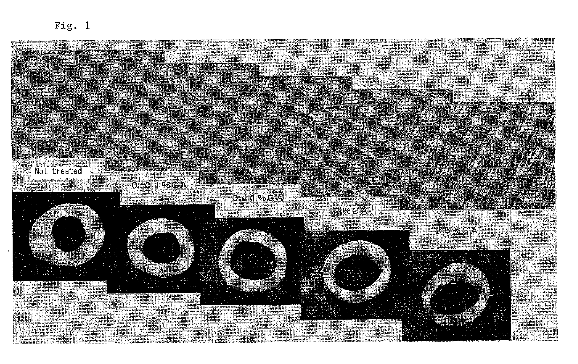
[0020]Porcine aorta tissue segments were treated with 0.01% GA, 0.1% GA, 1% GA and 25% GA, respectively, at room temperature for 1 hour, and then subjected to enzymatic degradation of elastin under the above constant condition. As a control, a tissue segment not treated with GA was also subjected to the enzymatic degradiation of elastin under the same conditions. FIG. 1 shows in the photographs in the lower row appearance of annularly cut aorta segments and histographic pictures of the tissue segment stained with hemotoxilin-eosin in the upper row. As shown in FIG. 1, untreated tissue and treated tissue with 0.01% GA were excessively loosened by the enzymatic removal of elastin and did not retain the original shape. In contrast, the tissues treated with 1% GA and 25% GA, respectively, were observed to contain an amount of elastin sufficient to retain the original shape after the treatment with the enzyme due to partial cross-linking with GA. Essentially complete removal of elastin w...

example 2

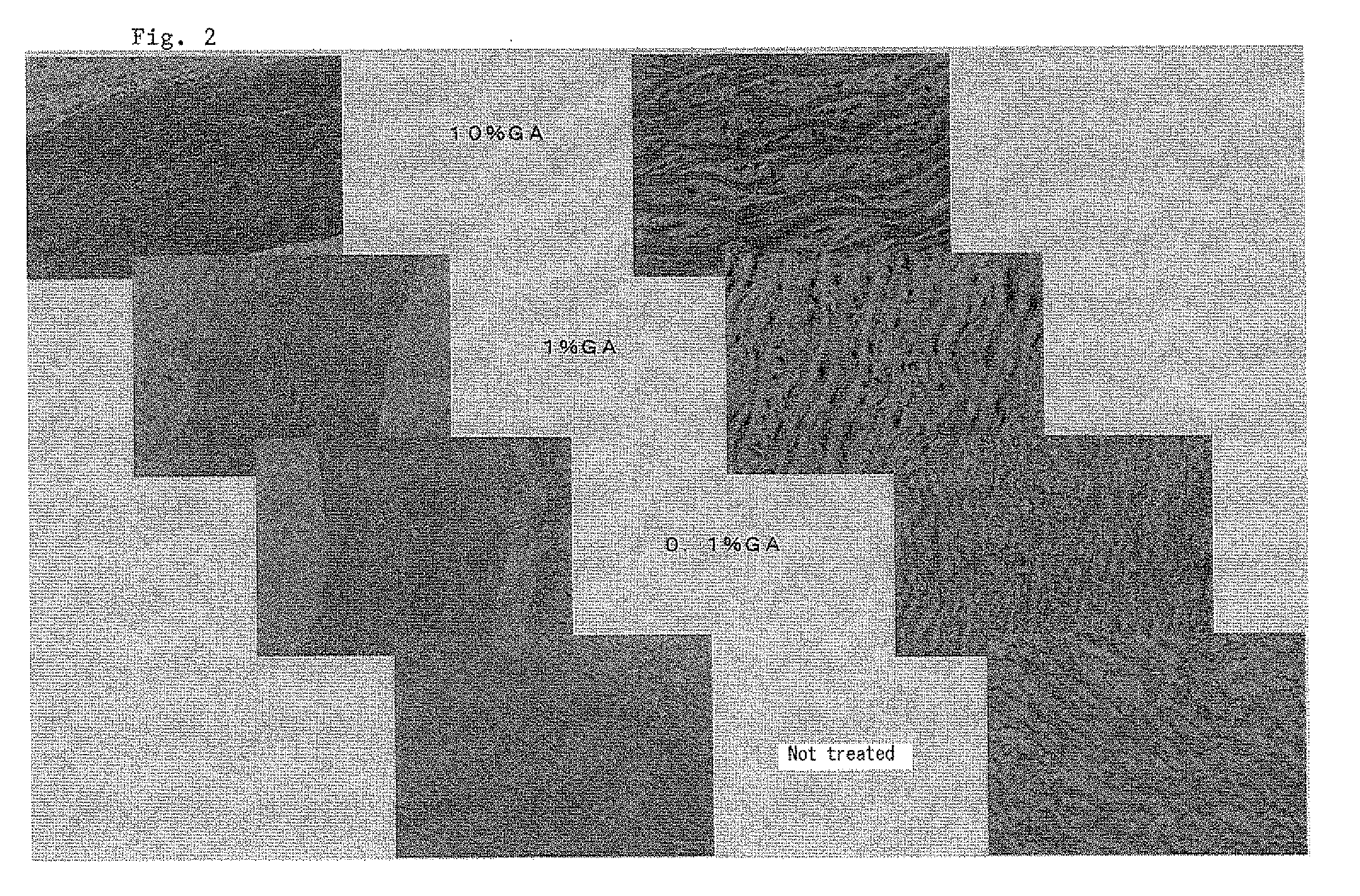
[0021]As in Example 1, porcine aorta tissue segments were treated with GA at varying concentrations from 0% to 25%, and then subjected to enzymatic degradation of elastin. Cytographic pictures were taken after staining hemotoxylin-eosin at different enlargement scales. As shown in FIG. 2, excessive loosening of the tissue was observed in the tissue not treated with GA. In the tissue treated with 1% GA or 10% GA, many elastin fibers and cell nuclei were observed.

example 3

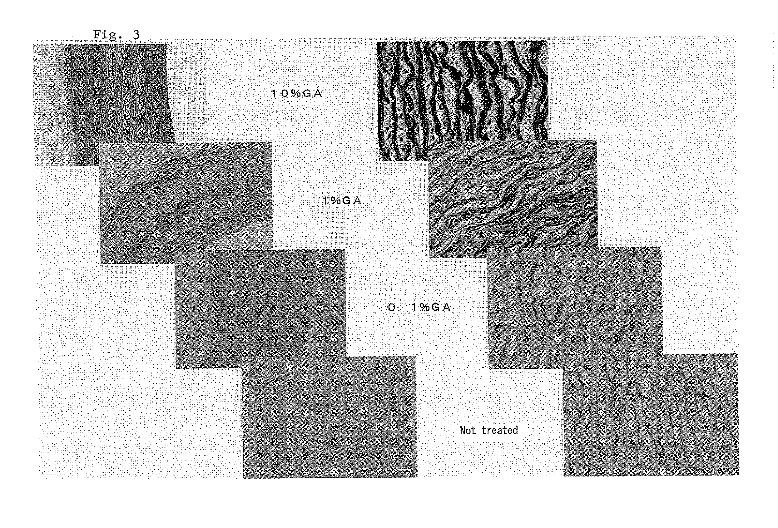
[0022]Porcine aorta tissue segments were treated with GA and elastase under the same conditions as in Example 2, and the stained by the Elastica von Gieson method, FIG. 3 shows enlarged histographic pictures of tissues thus treated. According to the above staining method, collagen fibers appear significantly darker than elastin fibers in monochromatic photographs. As observed from the pictures, residual elastin fibers increased with increase in GA concentration.

[0023]The tissue segment treated with 0.1% GA in Example 2 was digested with collagenase (228 U / mg, Wako Pure Chemical) in tris-buffer and the digested solution was analyzed spectroscopically. An absorption near 280 nm responsible for aromatic amino acids was observed indicating the biodegradability of the scaffold produced by the method according to the present invention.

[0024]As a result of the foregoing experiments, the effect of the treatment of a native tissue with glutaraldehyde under appropriate conditions before remov...


PUM

PropertyMeasurementUnit
pHaaaaaaaaaa
incubation timeaaaaaaaaaa

Abstract

Disclosed is a method for production of a bio-derived scaffold for use in regenerative medicine, which has a bio-compatibility and biodegradability and can self-organize and grow without causing calcification. The method comprises the steps of partially fixing a biological soft tissue with glutaraldehyde by cross-linking and incubating the partially fixed tissue together with an elastase.

Description

FIELD OF THE INVENTION[0001]The present invention relates to regenerative medical technology. Particularly, it relates to a method for preparing biological scaffold materials made of soft tissues of mammalian origine.BACKGROUND ART[0002]Prosthetic surgery is aimed to repair impaired organs by replacing with artificial devices or by grafting artificial or native scaffolds for developing into autogenic tissues. Currently more than 10,000 patients undergo heart valve replacement surgery in Japan. Mechanical valves are used in about 7% of the patients and xenogenic valves made from glutaraldehyde-fixed porcine or bovine tissues are used in the balance of patients. In case of mechanical valves, continuous administration of an antithrombotic agent is essential after replacement while affecting patient's quality of life. The xenogenic valves are durable in vivo only for about 15 years due to calcification and requires repeated replacement surgery for young generation.[0003]In case of gluta...

Claims


Application Information

Patent Timeline
Patent Type & Authority Applications(United States)
IPC IPC(8): C12P1/00
CPCA61L27/3604A61L27/56A61L27/3687
Inventor FUJISATO, TOSHIYATERADA, DOHIKOSAWADA, KAZUYANAKATANI, TAKESHI
Owner HEALTH SCI TECH TRANSFER CENT JAPAN HEALTH SCI FOUND
Why Eureka
Try Eureka