
Synaptotagmin and Collapsin Response Mediator Protein as Biomarkers for Traumatic Brain Injury

Active Publication Date: 2010-02-25
UNIV OF FLORIDA RES FOUNDATION INC +1
View PDF6 Cites 1 Cited by
  • Summary
  • Abstract
  • Description
  • Claims
  • Application Information

AI Technical Summary

Benefits of technology

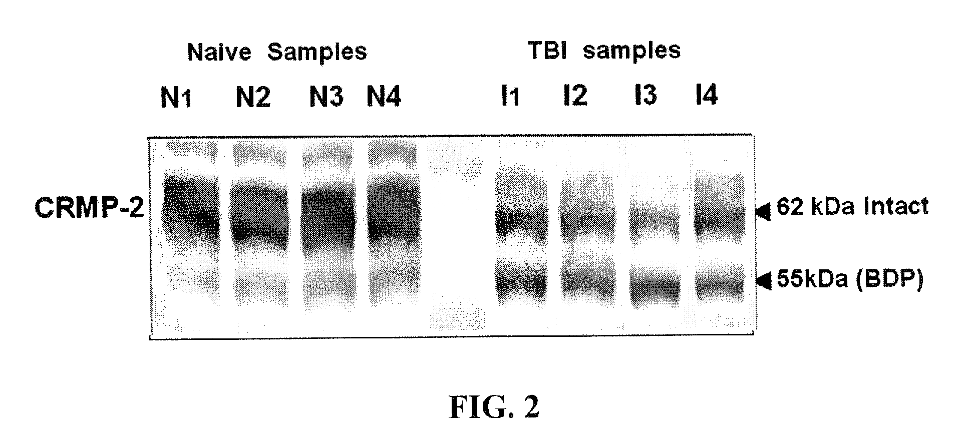
[0014]The present invention provides evidence that neuronal protein markers are differentially present in brain tissue after neural injury due to traumatic brain injury as compared with normal subjects. The measurement of these markers, alone or in combination, provides information useful for correlation with extent of injury and a means for assessing recovery as levels of the markers return to normal levels.

Problems solved by technology

However, despite dramatically improved emergency triage systems based on these outcome measures, most TBI patients suffer long term impairment and a large number of TBI survivors are severely affected despite predictions of “good recovery” on the GOS.
In addition, CT and MRI are expensive and cannot be rapidly employed in an emergency room environment.
The neural pathways of a mammal are particularly at risk if neurons are subjected to mechanical or chemical trauma or to neuropathic degeneration sufficient to put the neurons that define the pathway at risk of dying.

Method used


Image

  • Synaptotagmin and Collapsin Response Mediator Protein as Biomarkers for Traumatic Brain Injury
  • Synaptotagmin and Collapsin Response Mediator Protein as Biomarkers for Traumatic Brain Injury
  • Synaptotagmin and Collapsin Response Mediator Protein as Biomarkers for Traumatic Brain Injury

Examples


example 1

Proteolysis of CRMP2

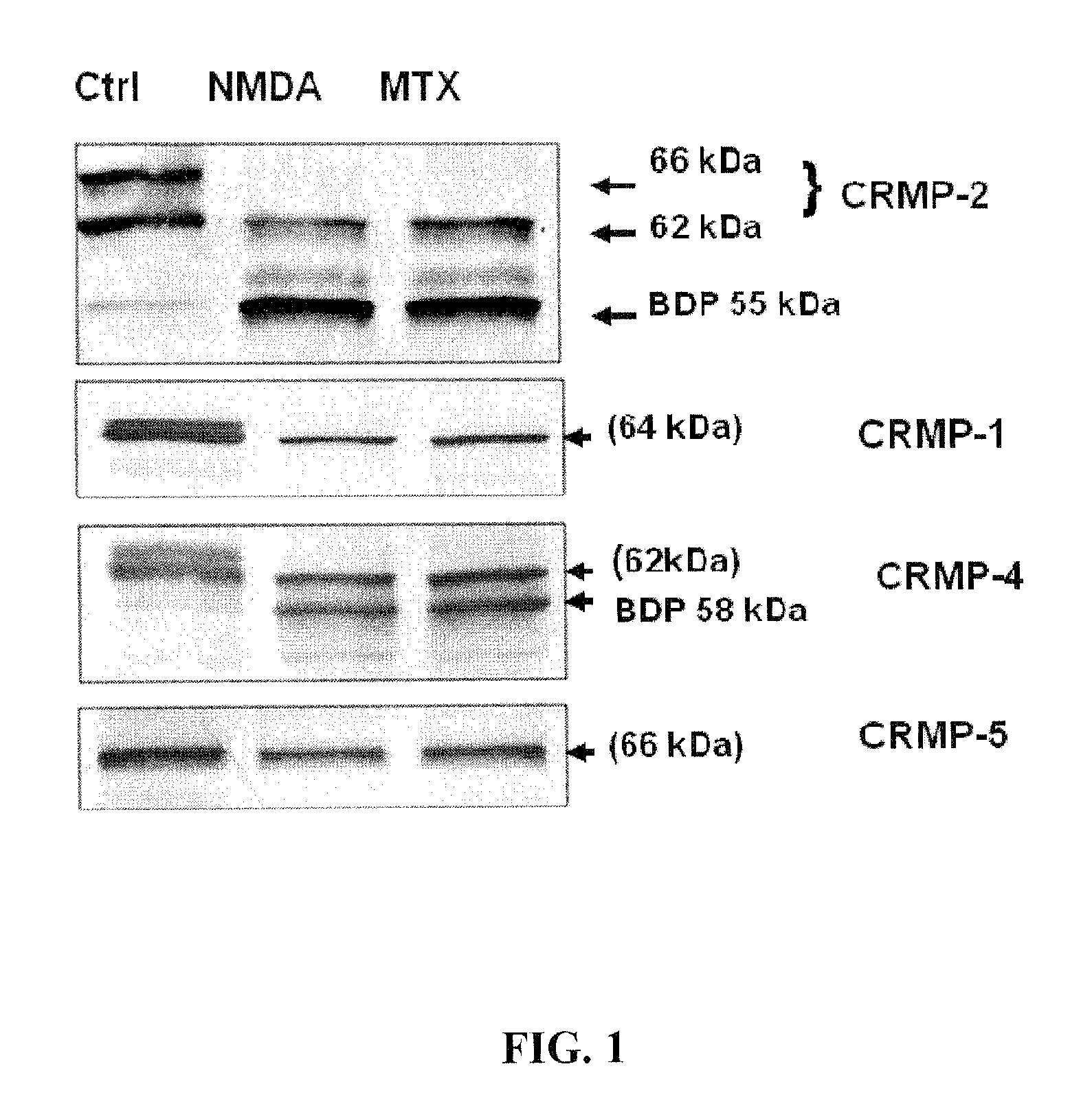
[0093]The integrity of CRMP2 following NMDA and MTX neurotoxin induction in primary cortical neurons was examined. A marked reduction in the intact CRMP-2 (66 kDa and 62 kDa) was noticed along with the appearance of a 55 kDa band after excitotoxic injury (200 μM NMDA) in rat primary cortical neuron culture (FIG. 1). Similar results were observed after MTX treatment.

[0094]Two anti-phsopho-CRMP2 specific antibodies (3F4 and C-terminal Phospho-CRMP-2) were used to rule out the possibility that the 55 kDa band was due to de-phosphorylation. The altered profile of the 66 kDa and 62 kDa CRMP2 was not observed after NMDA and MTX treatment (data not shown). Thus, the 55 kDa fragment was likely a breakdown product of CRMP2.

[0095]The integrity of CRMP1, 4 and 5 was then examined under identical conditions. Decreases in intact CRMP1 and CRMP4 were observed, as well as the increase of a 58 kDa CRMP4 band; however, CRMP5 levels remained unchanged following neurotoxic treatmen...

example 2

Inhibition of CRMP2 Proteolysis

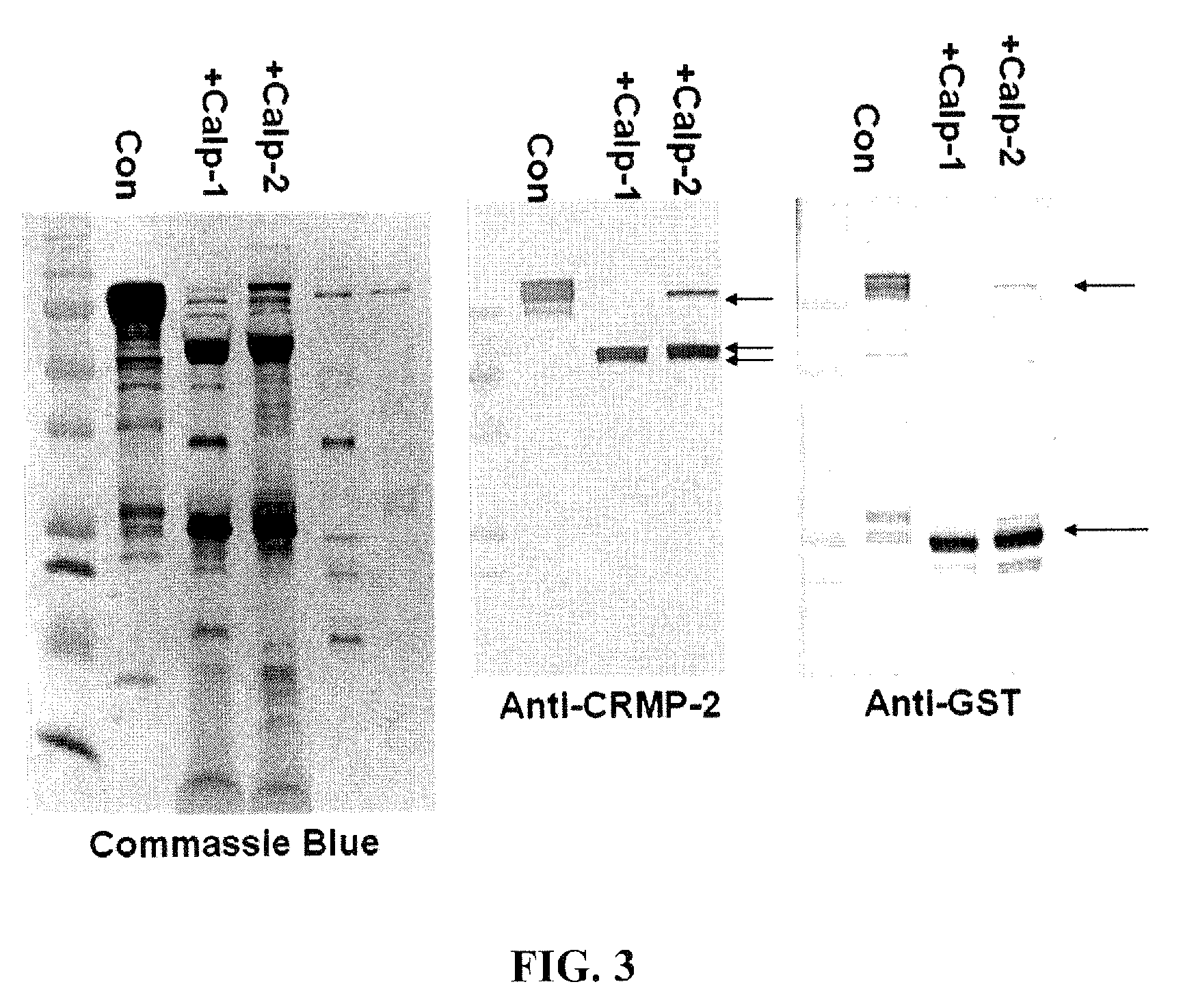
[0097]The apoptosis inducer staurosporine (STS, 0.5 μM), a calpain and caspase mixed challenge, and the calcium chelator EDTA (5 mM), a caspase-dominant challenge, were used in primary cortical neurons. Results showed that intact 62 kDa CRMP2 was rapidly degraded to the 55 kDa BDP upon STS treatment, but not upon the caspase-activating EDTA treatment. STS-mediated generation of the 55 kDa CRMP2 BDP was also effectively blocked by SJA6017, while Z-VAD offered no protection. The production of the 55 kDa CRMP2 BDP strikingly paralleled the production of the 150 and 145 kDa αII-spectrin breakdown products, which were monitored as markers for calpain activity in NMDA and STS treatment.

example 3

Blocking of CRMP2 Redistribution

[0098]LDH release assays were performed to determine the role of calpain and caspase inhibition on NMDA induced neuronal cell injury, and to draw a link with CRMP2 degradation. The release of LDH, normally present in the cytoplasm of neurons, into the cell culture media can be used as a measure of dying cells. Results showed that NMDA treatment induced CRMP2 proteolysis in a time-dependent manner. NMDA treatment induced significant neuron death after a 3 hour induction, peaking at 24 hours, which is consistent with the producing of the 55 kDa CRMP2 BDP. Moreover, the calpain inhibitor (SJA6017) provides significant protection within 6 hours, while the caspase inhibitor (Z-VAD) has no protection throughout NMDA treatment.

[0099]The distribution of CRMP2 following 6 hours' NMDA treatment with or without calpain and caspase inhibitor was examined to further explore the association of CRMP2 and NMDA induced neurite damage. In a normal healthy state, neuron...


PUM


Abstract

Collapsin response mediator proteins (CRMPs) decreased in tissue and increased in biological fluids after neural injury from traumatic brain injury (TBI). Significant decreases of CRMP1, CRMP2, CRMP4 and CRMP5 were accompanied by the appearance of distinct 58 kDa (CRMP-2) or 55 kDa (CRMP-4) breakdown products from proteolytic cleavage by calpain. Synaptotagmin breakdown products were also associated with TBI and could be detected along with intact protein in human cerebral spinal fluid (CSF). Both biomarkers were detected in human biofluid and related to recovery from traumatic brain injury.

Description

CROSS-REFERENCE TO RELATED APPLICATIONS[0001]This application is a continuation-in-part of International Application No. PCT / US2008 / 001644, filed Feb. 6, 2008, which claims priority from U.S. Provisional Application Ser. No. 60 / 888,432, filed Feb. 6, 2007, now abandoned.BACKGROUND OF THE INVENTION[0002]1. Field of the Invention[0003]The invention concerns neuroprotein changes associated with neurological damage and particularly to assays and diagnosis relating to traumatic brain injury.[0004]2. Description of Background Art[0005]The incidence of traumatic brain injury (BI) in the United States is conservatively estimated to be more than 2 million persons annually with approximately 500,000 hospitalizations. Of these, about 70,000 to 90,000 head injury survivors are permanently disabled. The annual economic cost to society for care of head-injured patients is estimated at $25 billion. Assessment of pathology and neurological impairment immediately after TBI is crucial for determinati...

Claims


Application Information

Patent Timeline
IPC IPC(8): G01N33/53
CPCG01N2800/28G01N33/6896
Inventor OTTENS, ANDREW K.KOBAISSY, FIRAS H.HAYES, RONALD L.ZHANG, ZHIQUNLIU, MING CHENOLI, MONIKA
Owner UNIV OF FLORIDA RES FOUNDATION INC
Learn More