Porous bioabsorbable implant

a bioabsorbable, porous technology, applied in the field of porous bioabsorbable implants, can solve the problems of few successful commercialization, few implants have met with significant commercial success, and the effect of easing dosage determination and simplifying irradiation patterns

Active Publication Date: 2010-04-01
SENORX
View PDF101 Cites 28 Cited by
  • Summary
  • Abstract
  • Description
  • Claims
  • Application Information

AI Technical Summary

Benefits of technology

[0007]The implant is sized and shaped so as to fit within the body cavity and to conform tissue lining the cavity about the implant. Generally, the implant will be spherical or oval in shape, although other shapes may be employed. It is preferred that the implant expand somewhat after deployment within the cavity, e.g. the implant materials swell (by taking up water or hydrating) upon contact with aqueous based fluids such as body fluids and other fluids which may be at the cavity site to ensure that tissue lining the cavity conform to the exter

Problems solved by technology

Biopsy and other tissue removal procedures in soft tissue can frequently lead to dimpling and other disfigurements unless a prostheses or implant is deployed within the cavity from which tissue

Method used


Image

  • Porous bioabsorbable implant
  • Porous bioabsorbable implant
  • Porous bioabsorbable implant

Examples


example i

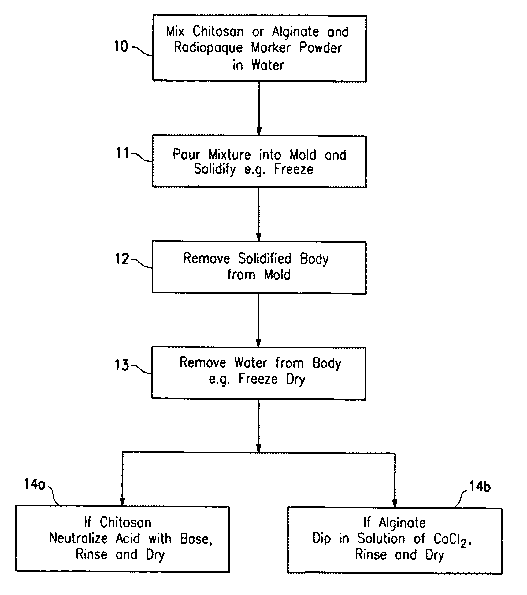
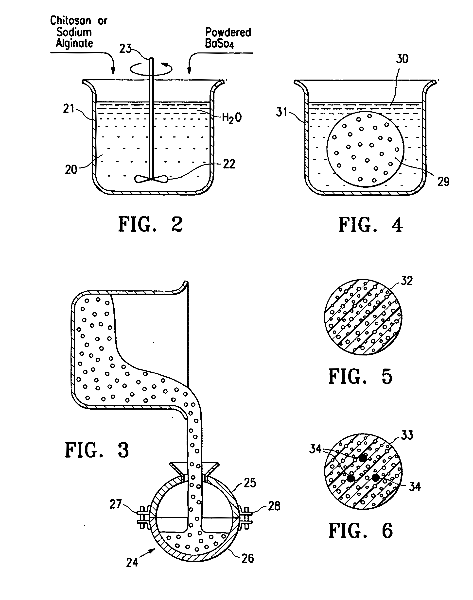
[0026]An acidic aqueous solution (12.5% acetic acid) was prepared containing 4% by weight chitosan and 2% by weight barium sulfate. The solution was placed in a spherical mold and was then frozen in the mold at −30° C. for 16 hours. The frozen body was removed from the mold and lyophilized for 3 days to remove water. The lyophilized body was neutralized in a 10% solution of ammonium hydroxide for one hour and then rinsed free of the ammonium hydroxide with deionized water. The body was vacuum dried for 16 hours. The body had the spongy consistency approximating breast tissue and had sufficient compressive strength to support breast tissue surrounding a lumpectomy cavity. It comprised 67% chitosan and 33% barium sulfate. An SEM micrograph (30×) of the surface porosity is shown in FIG. 6 and an SEM micrograph (30×) of the central porosity is shown in FIG. 7. The implant had the spongy consistency approximating breast tissue. The implant could be made harder by increasing the amount of...

example ii

[0027]A quantity of sodium alginate (0.5 to about 4% (wt.)) is dissolved in water to form a paste, viscous fluid or gel and air or other biocompatible gas is introduced into the mixture. The mixture is placed in a mold of a desired implant shape and then freeze dried or air dried in the desired shape. The formed implant structure of sodium alginate is introduced into a solution of calcium chloride (0.5 to about 4% (wt.)) where at least part of the sodium alginate is converted to calcium alginate which precipitates. The precipitated porous structure of the implant is introduced into a body cavity from which tissue has been removed. The implant remains at the site for sufficient period of time so as to act as scaffolding to facilitate tissue in-growth within the body cavity. Starch, such as corn starch in finely divided particulate form, can be incorporated into the sodium alginate-water mixture so that when the calcium alginate is formed, it precipitates about the starch particles to...

example iii

[0028]This example is similar to Example II except 30 grams of salt (NaCl) granules are mixed with about 30 ml of 3% (wt.) sodium alginate aqueous solution. The solution placed in a spherical mold and then is frozen for 4 hours. The frozen implant was removed from the mold and placed in a 2% (wt.) calcium chloride solution, forming calcium alginate gel and dissolving at least some of the incorporated salt granules to form a porous structure. The implant had the spongy consistency approximating breast tissue. The implant could be made harder by increasing the amount of sodium alginate in solution, decreasing the amount of salt or decreasing the size of the salt granules.


PUM


Abstract

The bioabsorbable implant described has a porous body formed of bioabsorbable materials that have an in vivo life span of at least 2 weeks, preferably at least three weeks and not greater than 20 weeks, preferably no greater than ten weeks. The implant has a scaffolding structure which facilitates tissue in-growth and ultimately tissue replacement of the scaffolding structure. The implant has a radiopaque imaging agent at least at the exterior margins and an orientation plurality of radiopaque elements in the interior of implant. The implant preferably has three radiopaque elements within the interior that form a plane within the implant interior.

Description

RELATED APPLICATION[0001]This application relates to and claims priority from Provisional Application Ser. No. 61 / 192,896, filed on Sep. 23, 2008, which is incorporated herein in its entirety.FIELD OF THE INVENTION[0002]The invention is generally directed to porous bioabsorbable implants, for cavities in soft tissue such as in breast tissue after biopsy or lumpectomy procedures. Implants embodying features of the invention are particularly suitable for supporting such cavities and are imagable to facilitate conformal three dimensional irradiation.BACKGROUND OF THE INVENTION[0003]Biopsy and other tissue removal procedures in soft tissue can frequently lead to dimpling and other disfigurements unless a prostheses or implant is deployed within the cavity from which tissue has been removed. See for example U.S. Pat. No. 6,214,045, U.S. Pat. No. 6,638,308 and U.S. Pat. No. 6,881,226 (Corbitt et al.). Moreover, after tissue removing procedures involving cancer, such as lumpectomies, it is...

Claims


Application Information

Patent Timeline
IPC IPC(8): A61F2/02
CPCA61L27/54A61L27/56A61L27/58A61L31/146A61L2300/44A61L31/18A61L2300/102A61L2300/104A61L31/148A61F2/0077A61F2/02
Inventor GOVIL, AMITJONES, MICHAEL L.LUBOCK, PAUL
Owner SENORX
Why Eureka
Try Eureka