Universal Cell for Medical Imaging of a Small Animal Under Anesthesia

a technology of anesthesia and medical imaging, applied in the field of universal cells for medical imaging of small animals under anesthesia, can solve the problems of affecting the treatment effect of patients, requiring the animal to be prevented from moving, and requiring considerable overall treatment time, so as to maximize the effect of suction of excess polluted gas

Inactive Publication Date: 2010-04-29
EQUIP VETERINAIRE MINERVE
View PDF14 Cites 16 Cited by
  • Summary
  • Abstract
  • Description
  • Claims
  • Application Information

AI Technical Summary

Benefits of technology

[0036]The isolation cell is constituted by a main body and by a moving portion with an isolating bubble. The assembly rests on a stationary structure that provides mechanical stability to the apparatus. When separated from its stationary structure, the isolation cell containing the animal can be transferred manually or by means of a robotic system to a unit for acquiring medical imaging data.
[0037]The main body is provided with two independent channels, one serving to pass cold gas extracted from the isolation cell and the other to pass said gas in return, which gas is reintroduced after being treated by the heater system. These two independent channels are connected at one end to pipes of a bed made of an extruded section member and at the other end to a heat exchanger integrated in the main body. The heater system operates in a closed loop and is made up of a gas extractor pump that is connected to a unit for regulating temperature and to the heat exchanger of the body, thereby serving to heat the extracted cold gas so that it is reintroduced into the cell at a temperature that is sufficient to enable the animal contained on the bed to conserve or recover the desired body temperature. In this main body there is engaged a holder tunnel with a service channel, with or without a perforatable membrane, enabling catheters, probes, electrodes, breathing circuits, pneumatic control circuits, etc. to be passed through. The moving portion is constituted by a support bed and by an outer transparent container. The bed is made of extruded section member having longitudinal channels for passing various fluids that need to be conveyed (induction/extraction of anesthetic gas, heat-conveying fluid, . . . ). The bed engages in the holder tunnel (itself engaged in the main body). The moving portion as a whole enables the animal to be restrained ...

Problems solved by technology

In order to ensure pre-anesthesia of an animal, there exist induction cages or chambers that have in common:they require the available volume to be filled with anesthetic gas and then the same volume to be emptied prior to extracting the animal for continued processing;they are heavy consumers of anesthetic agents, vector gas, and other consumables for collecting the extracted polluting vapors;they are awkward to handle, requiring the animal to be prevented from moving for a relatively long time and they involve considerable overall treatment time (for given induction parameters, depending on the model of cage an...

Method used


Image

  • Universal Cell for Medical Imaging of a Small Animal Under Anesthesia
  • Universal Cell for Medical Imaging of a Small Animal Under Anesthesia
  • Universal Cell for Medical Imaging of a Small Animal Under Anesthesia

Examples


Embodiment Construction

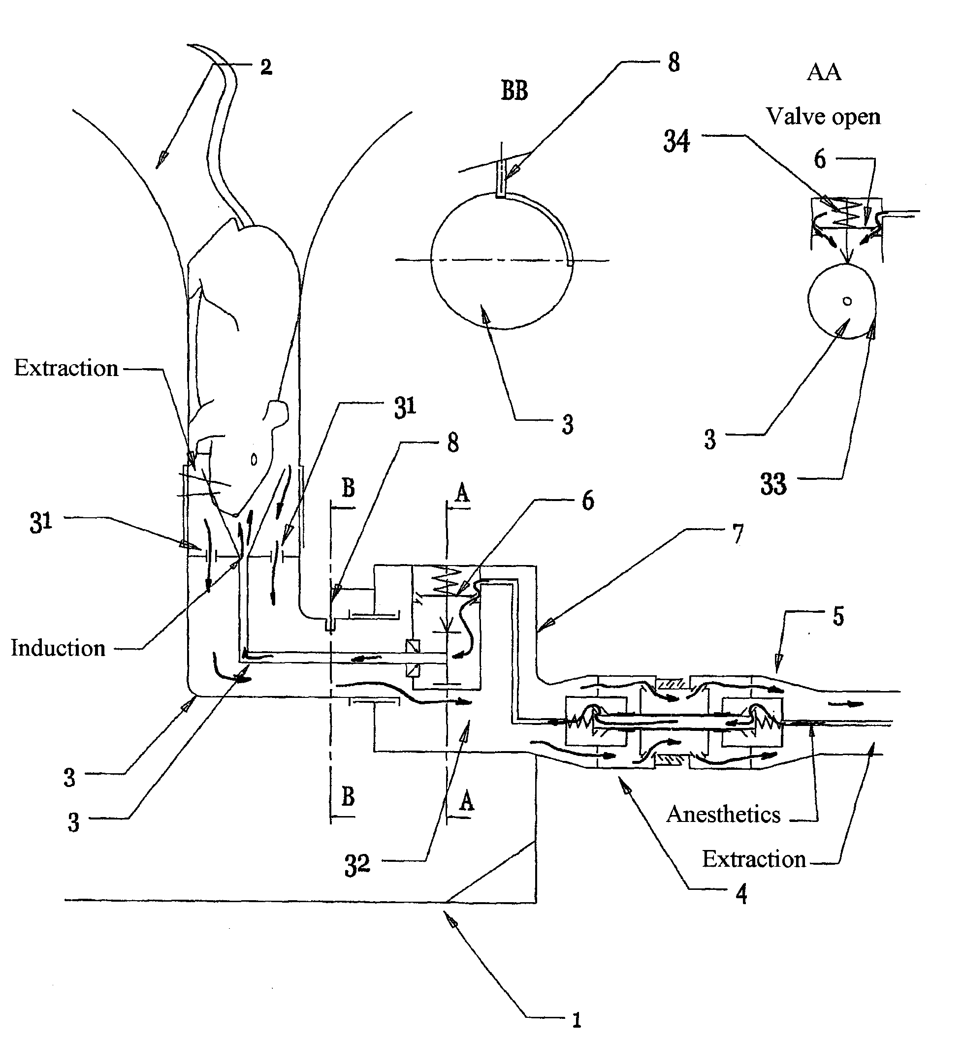
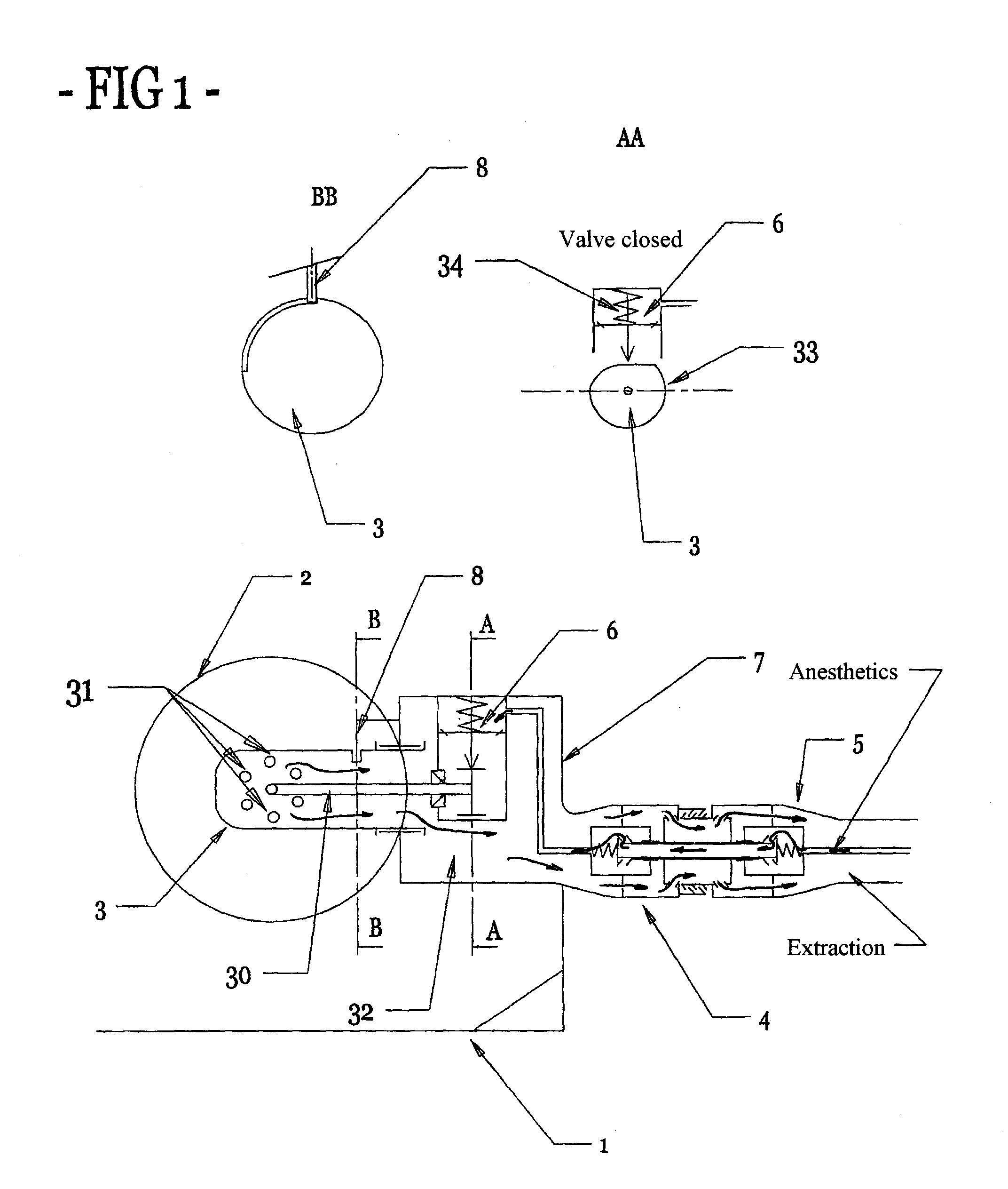
with reference to the diagrams of FIGS. 1 to 8

[0056]FIGS. 1 and 2: the induction device is permanently connected to a stationary structure (1) placed on a mattress or on a work surface. This structure that provides stability to the mechanical assembly is of dimensions, shape, and weight that ensure it remains stable during the induction stage. The induction device comprises a main body (7) fastened to the structure, having a double-closure female coaxial connector (4) integrated therein and two moving portions that can be secured to each other. The first portion is constituted by the induction and restraint cylinder (2), and the second is constituted by the moving body (3). The female coaxial connector (4) fitted to the main body (7) needs to be connected (FIG. 3) and screwed to the double-closure male coaxial connector (5) of the coaxial circuit connecting the anesthetic gas generator to the induction device in order to enable two free and independent flows of gas to take place. Th...


PUM


Abstract

The invention relates to equipment for a small laboratory animal designed to keep it under volatile anesthesia then to insert it into an imaging system for medical imaging investigations. It makes sure the vigilant animal has pre-anesthesia by means of a direct induction device of anesthetizing gas, its anesthesia prolonged by means of the usable device for keeping it in the open or closed position. This device makes direct access possible by the blood or respiratory pathway. The temperature of the animal is controlled and modulated by means of a nonmagnetic heat exchanger. The invention takes into account the safety of the users in regard to their exposure to halogens thanks to the double sealing connection system and thanks to the induction device that does not impregnate the coat of the animal that they have to handle.

Description

[0001]The invention lies in the field of research activities associated with techniques involving medical imaging investigation of small laboratory animals such as magnetic resonance imaging, X-ray micro-tomodensitometry, techniques making use of scintigraphy by single-photon and two-photon emitters, and optical imaging. Image acquisition can be spread over several hours, and the possibly contaminated animals need to be kept calm, insensitive to pain, and unmoving. This physiological state is characterized by good chemical or volatile general anesthesia, good ventilation, and good maintenance of the body temperature of the animal.[0002]The invention, made of non-magnetic materials, is constituted by two devices, one of which performs pre-anesthesia and the other of which maintains the laboratory animal under prolonged volatile anesthesia and isolates it from ambient air while enabling it to be subjected to medical treatment via the blood or respiration. Furthermore, the latter devic...

Claims


Application Information

Patent Timeline
IPC IPC(8): A61D7/04
CPCA61B5/0555A61D7/04A61B6/045A61B5/055
Inventor SANNIE, VINCENTSANNIE, SEBASTIEN
Owner EQUIP VETERINAIRE MINERVE
Why Eureka
Try Eureka