

Method for diagnosis of severity and prediction of recurrence in eosinophilic inflammatory disease
a technology of eosinophilic inflammation and diagnosis method, which is applied in the direction of instruments, peptides/protein ingredients, peptides, etc., can solve the problems of limited chronic administration, and achieve the effect of preventing the progress of eosinophilic inflammatory diseases and preventing recurren
- Summary
- Abstract
- Description
- Claims
- Application Information
AI Technical Summary
Benefits of technology
Problems solved by technology
Method used
Image

Examples
example 1
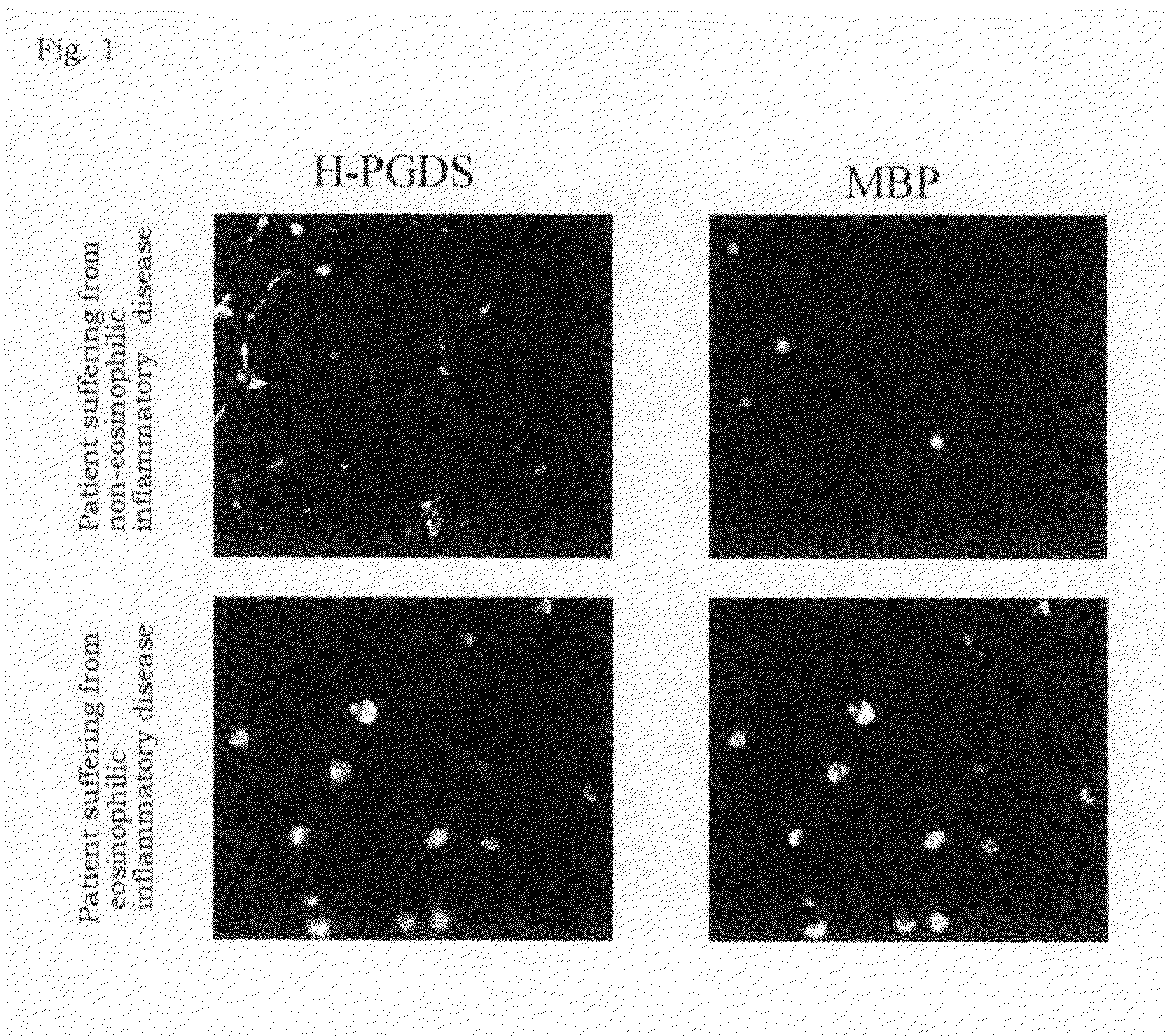
[0046]As a treatment for a patient suffering from chronic sinusitis, an excision operation of nasal polyp was conducted. For the purpose of analyzing the infiltration of the eosinophils in the excised nasal polyp, a histochemical analysis was carried out using the nasal polyp tissue. At first, the excised nasal polyp was fixed with 10% neutralized formalin and then a paraffin section was prepared. The slice was stained with hematoxylin-eosin (HE) by a conventional method and observed under a microscope to measure the numbers of eosinophils per infiltrated cell and the case where the ratios were 20% or more and 10% or less were classified as examples of high eosinophil infiltration and non-eosinophilic nasal polyp, respectively.
[0047]Backgrounds of the patients are shown in Table 1. In nasal polyps used for the experiments, the seven cases were severe degree and the another seven cases were mild degree shown in Table 1. In seven cases among the above, specific IgE antibody against to...
example 2
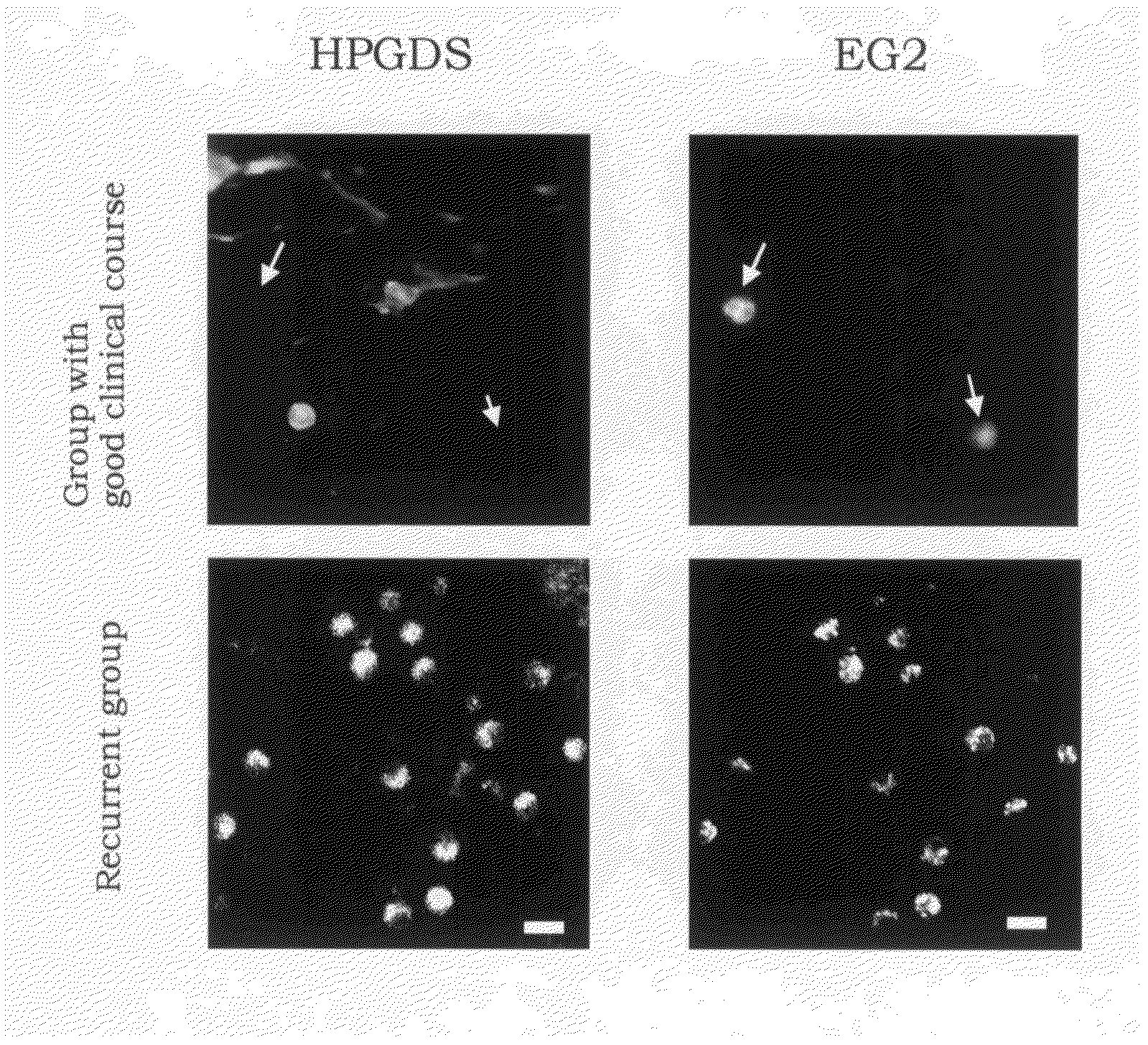
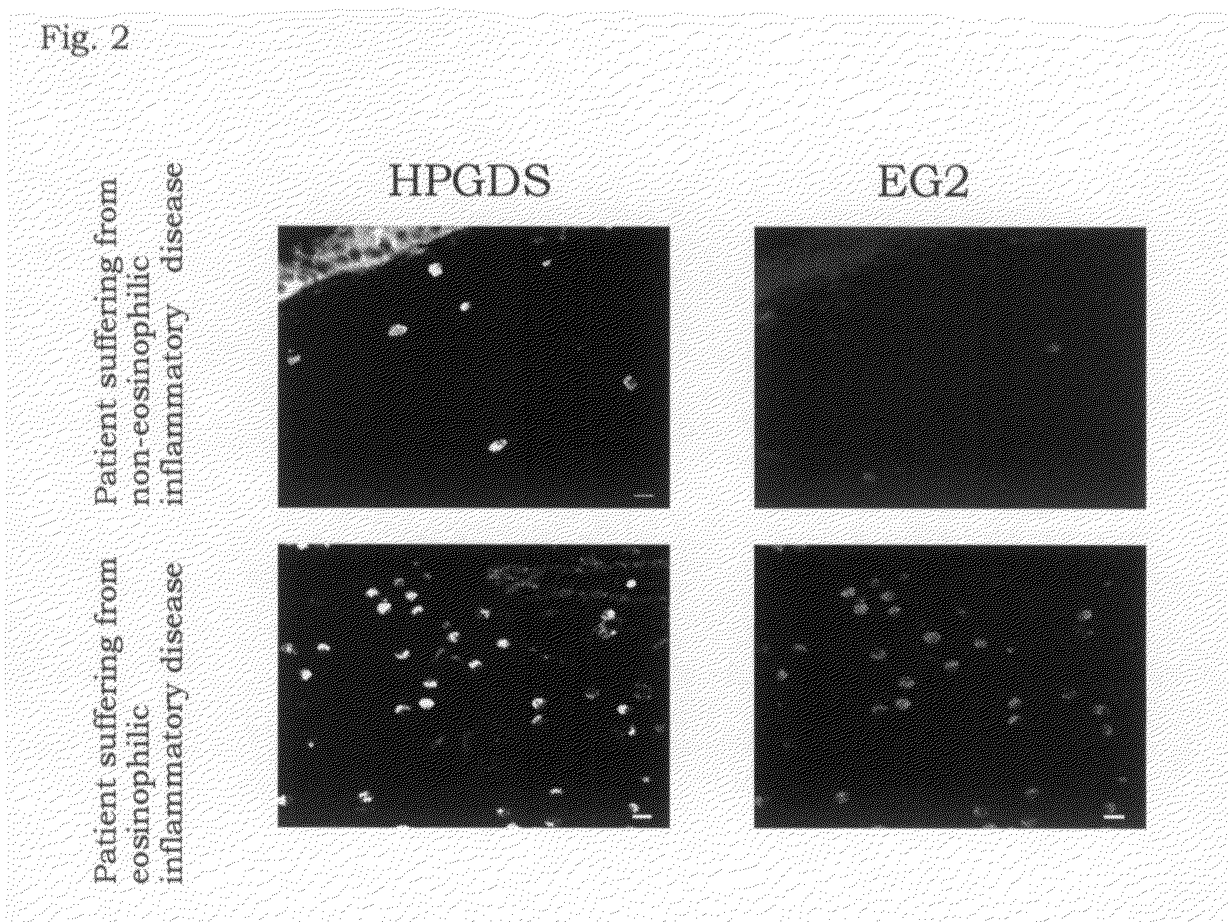
Expression of H-PGDS in EG2-Positive Activated Eosinophils
[0052]For a purpose of investigating the distribution of H-PGDS in nasal polyp in the activated eosinophils, expression of H-PGDS and the antibody (Bentley A M, Jacobson M R, Cumberworth V, Barkans J R, Moqbel R, et al., Immunohistology of the nasal mucosa in seasonal allergic rhinitis: increases in activated eosinophils and epithelia mast cells, J. Allergy Clin. Immunol., 1992; 89(4):877-83) which specifically recognizes EG2 antigen (injury protein recognized in eosinophils activated by eosinophil cationic protein (ECP)) which is an index for activated eosinophils was compared by an immunohistochemical staining method.
[0053]At first, a paraffin section of nasal polyp fixed with 10% neutralized formalin was dipped in 100% xylene to remove paraffin and then antigenicity was activated by a 0.3% pepsin solution. After that, a blocking was carried out using a PBS containing 10% normal goat serum and 0.1% Triton X, the primary ant...
example 3
Western Blotting of Nasal Polyp Tissue Specimens Collected from Patients Suffering from Chronic Sinusitis (1)
[0055]For a detection of the expression of H-PGDS protein in nasal polyp, an SDS gel electrophoresis was conducted using an extract of nasal polyp tissue and a Western blotting by using an antibody against H-PGDS was conducted. After the electrophoresis, the proteins were transferred to a nylon membrane. A western blotting was conducted by using an antibody against H-PGDS.
[0056]At first, about 50 mg of the excised specimen was homogenized in 0.5 ml of PBS, centrifuged at 7,000 g (at 4° C. for 20 minutes) and then centrifuged at 100,000 g (at 4° C. for 1 hour) to separate into a supernatant liquid and a microsome fraction. The supernatant obtained by 7,000 g centrifugation and the microsome obtained by 100,000 g centrifugation were subjected to electrophoresis to 10 / 20% SDS-PAGE gradient gel and 4 / 20% SDS-PAGE gradient gel (both manufactured by Daiichi Kagaku), respectively an...
PUM
| Property | Measurement | Unit |
|---|---|---|
| Volume | aaaaa | aaaaa |
| Volume | aaaaa | aaaaa |
| Volume | aaaaa | aaaaa |
Abstract
Description
Claims
Application Information