

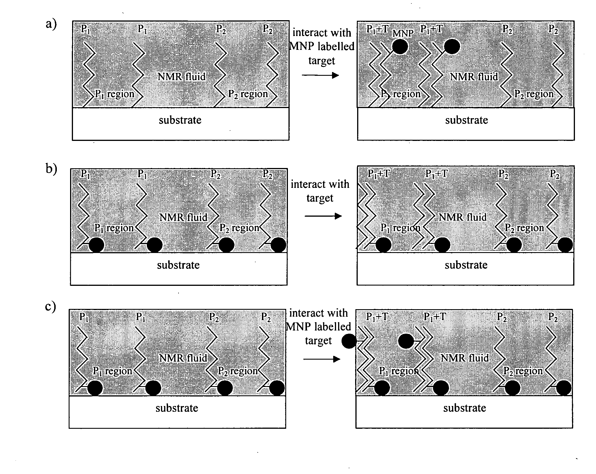
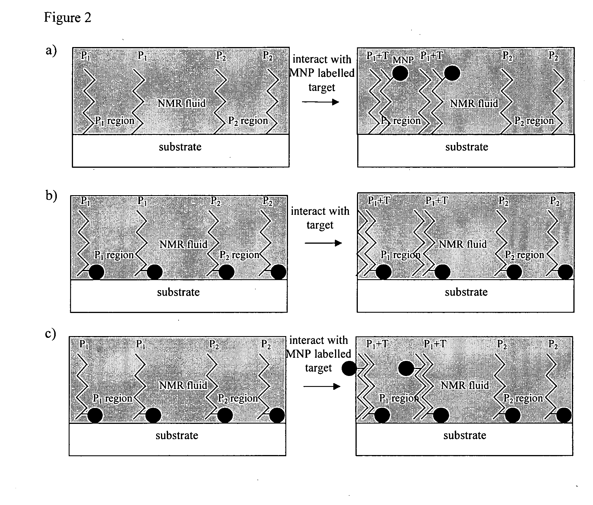
Method of detecting interactions on a microarray using nuclear magnetic resonance
a microarray and nuclear magnetic resonance technology, applied in the field of nuclear magnetic resonance-based interactions detection on microarrays, can solve the problems of non-uniform immobilisation of dna and non-uniform fluorescence, and achieve the effects of improving signal-to-noise ratio, improving sensitivity, and rapid collection of signals
- Summary
- Abstract
- Description
- Claims
- Application Information
AI Technical Summary
Benefits of technology
Problems solved by technology
Method used
Image

Examples
examples
Overview
[0097]Two types of NMR system have been used to confirm the ability of NMR and MRI to image magnetic nanoparticle based microarrays. In the first set of experiments a unilateral NMR profiling instrument was used to detect immobilization of a chemical species on glass surfaces. In the second set of experiments a non-unilateral, standard, cylindrical, horizontal small bore MRI instrument was used to image chemical arrays and oligonucleotide arrays.
a) Experiments Using a Unilateral NMR Instrument
[0098]In these experiments a profile NMR MOUSE® [Perlo, J, Casanova, F., and Blumich, B. Profiles with microscopic resolution by single-sided NMR. J. Mag. Res. 2005. 176 (1): p 64-70] that collects the NMR signal coming from a thin and flat-volume of sensitivity (approximately 200 μm×20 mm×20 mm) at 5 to 10 mm above the instrument was used. A strong (11.4 T / m) magnetic field gradient resides permanently across the selected slice. The presence of this gradient in conjun...
example 3.1
Imaging of a Single Spot Using SPIO Particles and a Droplet of Water as the Imaging Fluid
[0112]Droplets of water were deposited as the imaging fluid onto the glass slide which had previously been prepared with a SPIO labelled region and a non-SPIO labelled region (see section 2d). A repetition time of TR=100 ms was used to reveal the presence of SPIOs as a positive contrast. Five hundred averages were acquired, resulting in a 50 second duration experiment.
[0113]In FIG. 7, the upper / red curve shows the signal acquired from a droplet deposited on area of the glass slide that had SPIO labelled region and the lower / blue curve is the NMR signal acquired using a droplet deposited on the side without a SPIO labelled region. The z-axis location of the peak in the upper curve corresponds to the plane of the surface of the glass slide at which the SPIO labelled region resides. The results show that a measurable difference as a positive contrast in SPIO labelled and non-SPIO labelled regions c...
example 3.2
Imaging a Multiple Spots Using SPIO Particles and Immersion in Water as the Imaging Fluid
[0115]The glass dish prepared for example 3.1 with multiple SPIO-labelled and non-SPIO labelled spots was imaged by immersing the slide in water as the imaging fluid. Four different repetition times, TR, were used, 0.1 s, 0.5 s, 0.9 s and 1.3 s and one minute duration experiments were performed for each repetition time.
[0116]FIG. 8 shows a comparison between NMR signal for spot 2, created using a 0.01 ml concentration of SPIO's, and spot 6, created using a 1 ml concentration of SPIO's. Spot 6 demonstrates a much higher signal, and the difference between the two signals decreases for increasing TR value, as expected. On the long TR value, the water content can be seen. This demonstrates that NMR signals related to the concentration of SPIO's within a region can be obtained.
[0117]By dividing the signal-to-noise ratio (SNR) of the short TR measurement by the SNR of the long TR measurement, an estim...
PUM
| Property | Measurement | Unit |
|---|---|---|
| size | aaaaa | aaaaa |
| size | aaaaa | aaaaa |
| diameter | aaaaa | aaaaa |
Abstract
Description
Claims
Application Information