Generation of brachial, thoracic and lumbar spinal motor neurons from embryonic stem cells in the absence of all-trans retinoic acid supplement

a technology of embryonic stem cells and retinoic acid, which is applied in the field of generation of brachial, thoracic and lumbar spinal motor neurons from embryonic stem cells in the absence of alltrans retinoic acid, can solve the problems of unaddressed basic study of cell differentiation into mns, and achieve the effects of reducing retinoid conditions, increasing the number of hoxd9+ mns, and blocking endogenous fg

Inactive Publication Date: 2010-08-05
THE TRUSTEES OF COLUMBIA UNIV IN THE CITY OF NEW YORK
View PDF35 Cites 39 Cited by
  • Summary
  • Abstract
  • Description
  • Claims
  • Application Information

AI Technical Summary

Benefits of technology

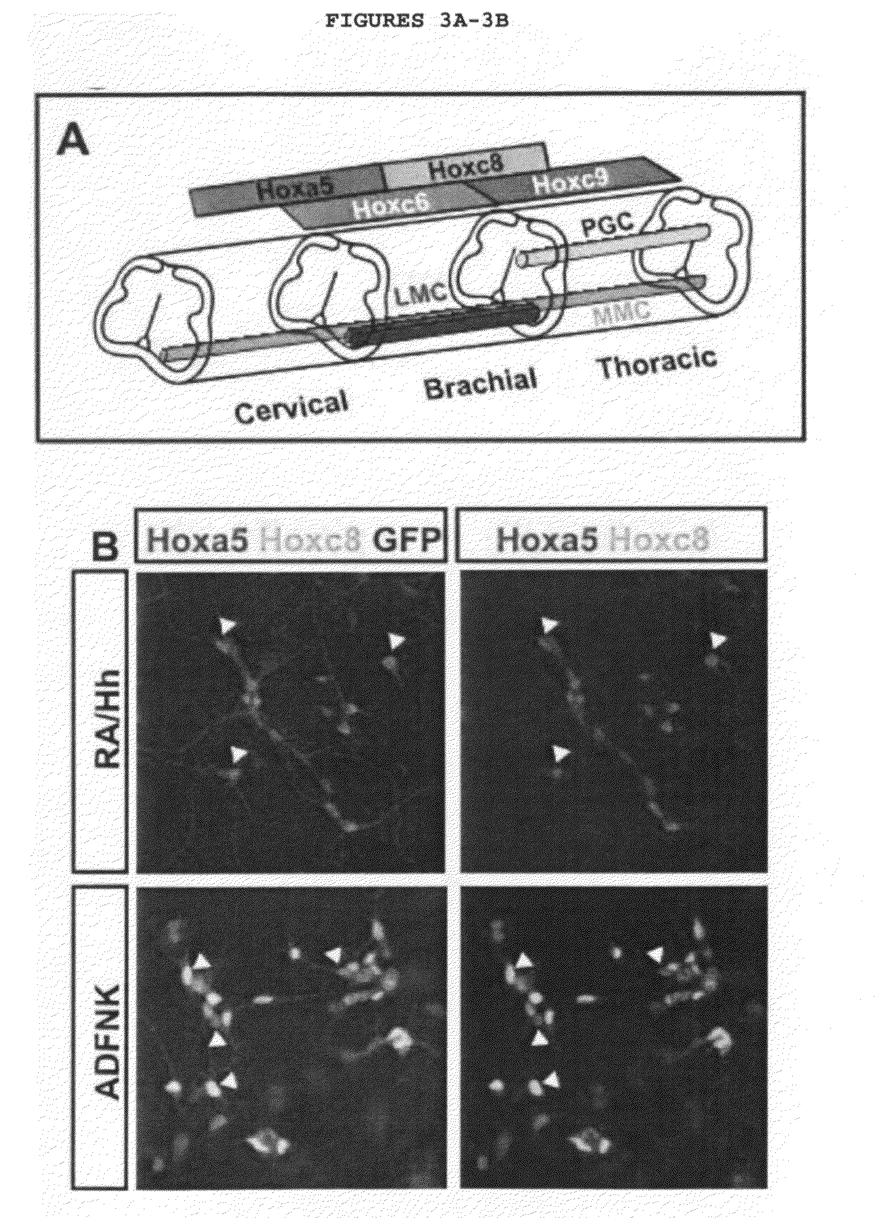
[0041]FIG. 21: Titration of Retinoic Acid in the Presence and Absence of Hh Agonist: A) To determine whether decreasing concentrations of RA may result in the specification of caudal brachial ES-MNs EBs were treated with different RA concentrations (100 nM-0 nM) in the presence and absence of 1 μM Hh agonist and expression of Hoxa5, Hoxc8, and Hb9-GFP was examined in the resulting day 7 EBs. While Hoxa5+ MNs are detected in all conditions, Hoxc8+ MNs are efficiently specified only in the absence or low concentrations of exogenous RA and Hh. B) To quantify ES-MN induction efficiency in conditions resulting in the specification of Hoxc8+ ES-MNs (0 RA/Hh, 1 nM RA, 1 μM Hh, and, 1 nMRA/1 μM Hh), EBs were dissociated into single cells on day 7 of dissociation and the number of Hb9-GFP+ and total cells were determined. In general all four conditions resulted in the similar levels of MN induction suggesting that these pharmacological treatments did not significantly affect the fraction of ES-MNs generated during differentiation. Graph represents three independent differentiation experiments (mean±SEM). C) To quantify the fraction of MNs that express Hoxc8 EBs grown in Hoxc8+ ES-MN generating conditions (0 RA/Hh, 1 nM RA, 1 μM Hh, and 1 nMRA/1 μM Hh) were fixed and immunostainings were performed for Hoxc8 and Hb9. 0 RA/0 Hh condition resulted in the highest number of Hoxc8+ ES-MNs. Graph represents two independent experiments (mean±SEM).
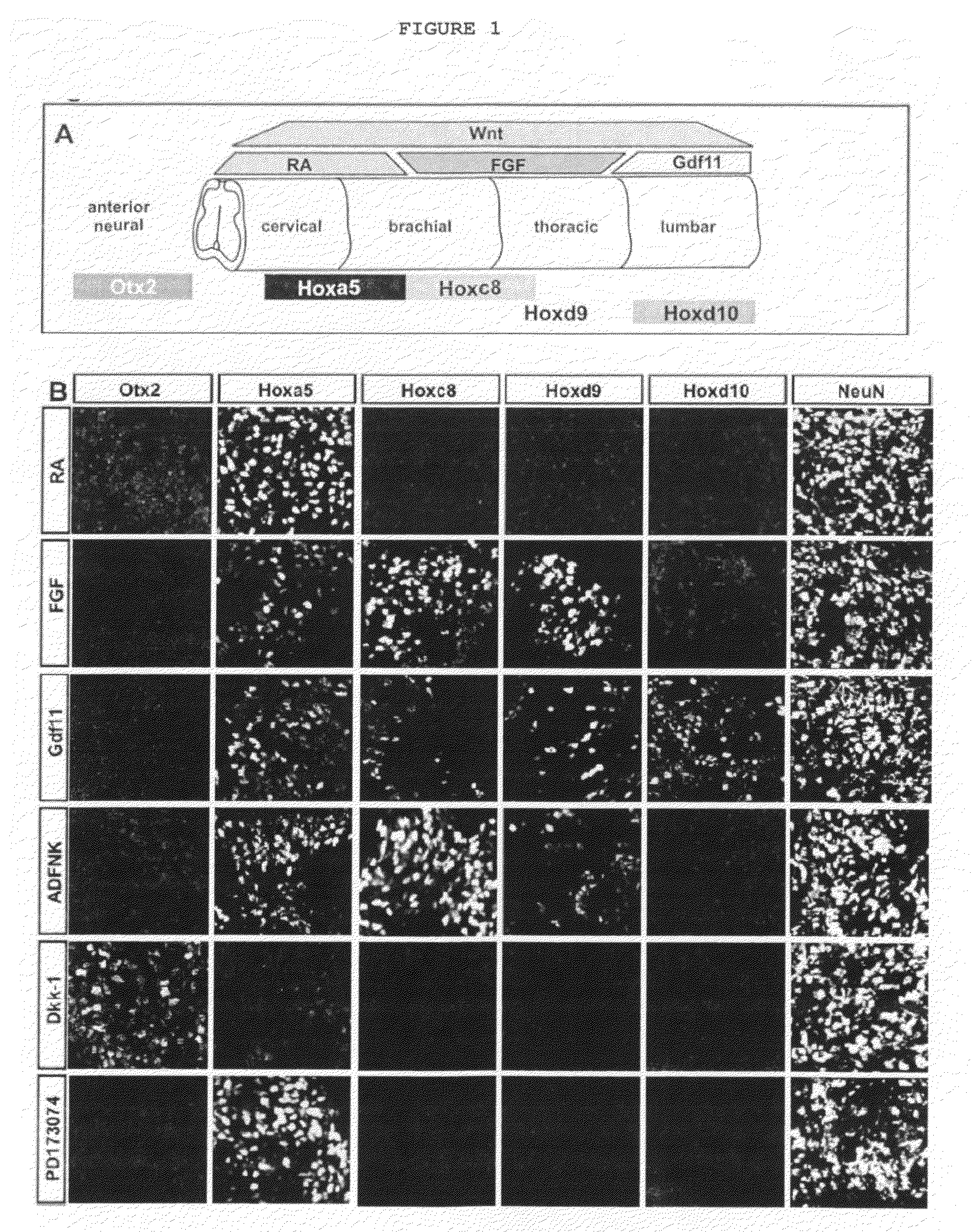
[0042]FIG. 22: Rostro-caudal Patterning of Differentiating ES Cells in Response to Endogenous and Exogenous Signals: A) Schematic of the developing spinal cord with principal signals that control the rostro-caudal patterning and Hox expression profile in spinal cord MNs. B-H) To determine if FGF/Hh (control) differentiation condition could be used for patterning ES-MNs along the rostro-caudal axis EBs were grown under low-density FGF/Hh conditions (control) and treated with RA, FGF, RA/FGF/Hh, Gdf11, and PD173074 on day 3 of differentiation and Dkk-1 on day 2 of differentiation. Sections of EBs were immunostained on day 7 of differentiation, except Otx2 on day 5 of differentiation. All differentiation conditions resulted in the specification of NeuN+ neural cells and Hb9-GFP expressing MNs. C) Addition of 10 nM-1 μM RA on day 3 of differentiation resulted in the generation of Hoxa5+ cervical MNs lacking expression of more posterior Hox genes. D) Addition of FGF2 (100 ng/ml) resulted in an increase in the number of Hoxd9+ MNs. E) Treatment of cells with 100 nM RA, 100 ng/ml FGF2, and 100 nM Hh agonist resulted in the specification of Hoxc8+ MNs. F) Treatment of differentiating cells with 20 ng/ml Gdf11 resulted in specification of MNs expressing Hoxd10. G) Blocking Wnt signaling by addition of 1 μg/ml Dkk1 on day 2 of differentiation prevented specification of spinal cells (absence of Hoxa5 and Hoxc8 expression) and resulted in the appearance of Otx2+ forebrain/midbrain neural cells. H) Blocking endogenous FGF signaling by the addition of 50 nM PD173074 on day 3 of differentiation selectively prevented specification of caudal brachial Hoxc8+ ES-MNs. I) Addition of 100 ng/ml of FGF to control condition results in the significant increase in Hoxd9 expressing cells (p=0.007). Graphed are results from three independent experiments (mean±SEM). J) Addition of 20 ng/ml of Gdf11 leads to significant increase in Hoxd10 expressing ES-MNs (p<0.001). Graphed are results from three independent experiments (mean±SEM).
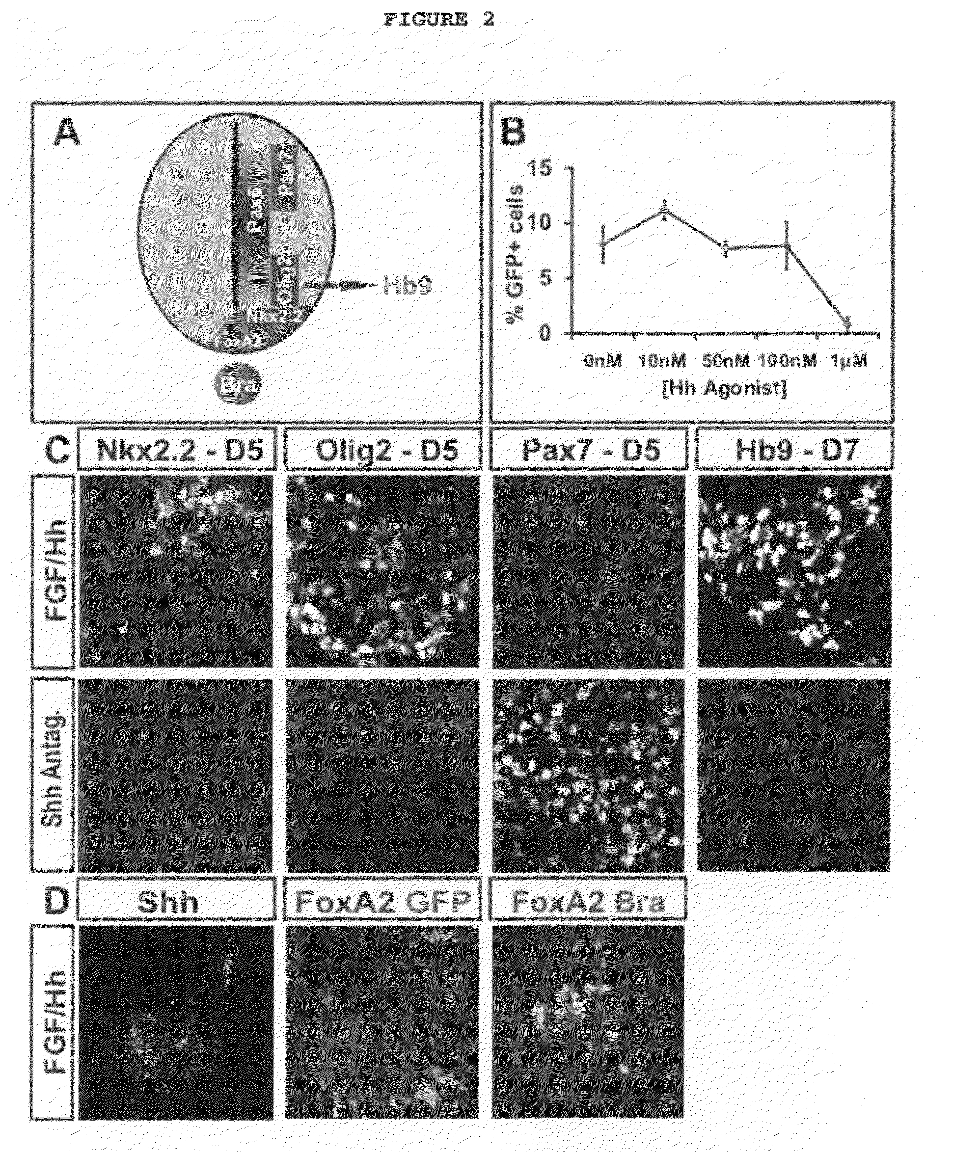
[0043]FIG. 23: Endogenous Shh Signal Controls Ventralization of Spinal Progenitors: A) Schematic of Shh signaling and specification of dorso-ventral progenitor domains in the developing spinal cord. Shh signal secreted from the floor plate and notochord patterns ventral spinal cord and specifies Olig2+ MN progenitor identity. B) To test if Shh signaling is the limiting factor in the efficiency of ES-MN generation in the absence of extrinsic factors, ES cells were differentiated in the presence of increasing concentration of Hh agonist (10 nM, 50 nM, 100 nM, and 1 μM Hh agonist were supplemented on day 2 of differentiation). The resulting EBs were dissociated on day 7 of differentiation and the number of Hb9-GFP expressing and total cells was determined. Addition of increasing concentrations of Hh agonist does not significantly change the percentage of Hb9-GFP+ cells obtained during differentiation suggesting that Shh signaling is not the limiting factor during differentiation. Graphed are results from three independent differentiation experiments (mean±SEM). C) To test if the induction of ES-MNs in the absence of exogenous signals depends on Shh generated by the EBs during differentiation, EBs differentiated in FGF/Hh conditions were treated with 100 nM Shh antagonist on day 2 of differentiation and the expression of Pax7, Olig2, Nkx2.2 (on day 5), and Hb9 (on day 7) was examined. Inhibition of endogenous Shh signaling by addition of Shh antagonist resulted in the loss of Nkx2.2 and Olig2 expression and failure of MN differentiation (lack of Hb9+ cells). When treated with Shh antagonist, the majority of cells acquired expression of dorsal spinal progenitor marker Pax7. These results suggest that ES-MN differentiation in the absence of exogenous factors is dependent on Shh signaling induced in differentiating EBs. D) To determine the sources of Shh signaling in the EBs, immunohistochemistry was performed again using antibodies against Shh, FoxA2 and Bra. on day 5 of differentiation. Endogenous signaling centers express floor-plate/notochord marker FoxA2 and Shh on day 5 of differentiation. A subset of FoxA2 cells co-expressed notochord marker Brachyury. These results indicate that notochord and floor-plate like cells express Shh signal in the differentiating EBs, required for the ventralization of MN progenitors and specification of ES-MNs in the absence of added extrinsic factors.
[0044]FIG. 24: Rostro-caudal Patterning of ES-MNs under Combined RA/FGF exposure: A) To test if RA and FGF signaling together may lead to specification of Hoxc8+ ES-MNs during differentiation, different concentrations of RA and FGF together were tested and the rostro-caudal identity of the resulting cells was determined. Addition of up to 625 ng/ml of FGF2 to 1 μM RA/Hh differentiation condition did not result in the specification of Hoxc8+ ES-MNs. Treatment of differentiating ES cells with 100 nM RA/Hh and 100 ng/ml of FGF2 on day 3 of differentiation resulted in specification of Hoxc8+ ES-MNs, demonstrating that under reduced retinoid conditions differentiating ES cells can acquire brachial identity in response to high concentrations of FGF2. B) To compare the efficiency of MN generation between 1 μM RA/1 μM Hh, 100 nM RA/Hh and 100 ng/ml FGF, and endogenous FGF/Hh conditions, fluorescence activated cell sorting analysis was performed on day 7 dissociated Hb9-GFP+ ES-MNs. Differentiation of ES cells in the presence of 100

Problems solved by technology

There are, however, challenges in the basic study of cell differentiation into MNs which remain unaddressed.
This is in part due to limited understanding of developmental processes and transcriptional program

Method used


Image

  • Generation of brachial, thoracic and lumbar spinal motor neurons from embryonic stem cells in the absence of all-trans retinoic acid supplement
  • Generation of brachial, thoracic and lumbar spinal motor neurons from embryonic stem cells in the absence of all-trans retinoic acid supplement
  • Generation of brachial, thoracic and lumbar spinal motor neurons from embryonic stem cells in the absence of all-trans retinoic acid supplement

Examples


example 1

[0125]Relevant to this application, the current status of knowledge of MN subtypes and spinal AP patterning mechanisms is summarized as follows:

[0126]While generic MN identity is specified by DV patterning mechanisms, MN subtype diversity is manifested primarily along the anteroposterior axis of the spinal cord, as MNs at each spinal segment innervate distinct subsets of muscle targets. Little is currently understood about the mechanisms that underlie acquisition of AP spinal identity and about the genetic cascades that translate the progenitor AP identity to postmitotic MNs.

Columnar Identity

[0127]Traditionally MNs were classified based on two criteria: position within the spinal cord, which defines their columnar identity; and axon trajectory and pattern of muscle innervations, which defines their motor pool identity (Hollyday, 1980; Hollyday and Jacobson, 1990; Landmesser, 1978). Spinal MNs are arranged in longitudinal columns, each innervating a distinct subset of peripheral targ...

example 2

[0171]The generation of diverse neuronal subtypes relies on a precisely orchestrated interplay between extrinsic signaling factors and intrinsic transcriptional programs. This experiment shows 1) that mouse embryonic stem (ES) cells can be induced to differentiate into distinct spinal motor neuron (MN) subtypes by extrinsic signals that pattern the rostro-caudal and dorsoventral axis in vivo; 2) Wnt, FGF, and Hh signals induce brachial and thoracic spinal MN identities, as determined by expression of Hox proteins; 3) Many brachial ES cell-derived motor neurons (ES-MNs) expressed FoxP1, a marker of lateral motor column (LMC) neurons that innervate limb muscles; 4) When introduced by transplantation into the chick developing neural tube, ES-MNs settled in motor columnar territories and projected their axons along nerve branches in a pattern that conformed to their molecular columnar identity; 5) ES-MNs of LMC character acquired molecular properties of flexor carpi ulnaris and cutaneou...


PUM

PropertyMeasurementUnit
Fractionaaaaaaaaaa
Fractionaaaaaaaaaa
Fractionaaaaaaaaaa

Abstract

Disclosed are methods for generating a neuron expressing Hoxc8 transcription factor or a caudal motor neuron comprising culturing an embryonic stem cell in a composition which is essentially free of retinoids and comprises an isotonic salt solution, so as to generate the neuron which expresses Hoxc8 transcription factor or the caudal motor neuron. Disclosed are also methods for generating a caudal brachial motor neuron, a thoracic motor neuron, or a lumbar motor neuron from an embryonic stem cell in a composition essentially free of retinoids and comprising ADFNK medium, an amount of FGF-2, or Gdf11 respectively. Disclosed are also methods of transplanting a motor neuron into a subject comprising generating the motor neuron and transplanting the motor neuron into the subject. Disclosed is also a population of motor neuron cells enriched for motor neuron cells expressing Foxp1 and expressing a gene associated with Spinal Muscular Atrophy (SMA) or Amyotrophic Lateral Sclerosis (ALS).

Description

[0001]The invention disclosed herein was made with government support under National Institute of Neurological Disorders and Stroke (NINDS) 1R01NS058502-01 and National Institutes of Health (NIH) T32 HD055165. Accordingly, the U.S. Government has certain rights in this invention.[0002]Throughout this application, various publications are referred to by first author and year of publication. Full citations for these publications are presented in a References section immediately before the claims. Disclosures of the publications cited in the References section in their entireties are hereby incorporated by reference into this application in order to more fully describe the state of the art as of the date of the invention described herein.BACKGROUND[0003]During the development of the vertebrate central nervous system (CNS), hundreds of distinct neuronal types are generated, establishing a diversity that is essential for the formation of neuronal circuits. The degeneration of specific cl...

Claims


Application Information

Patent Timeline
IPC IPC(8): A61K35/12C12N5/02C12N5/071C12N5/07A61K38/00
CPCC12N5/0619C12N2501/115C12N2501/13C12N2501/19C12N2506/02C12N2501/41C12N2501/155C12N2501/385C12N5/0696C12N5/0606C12N2506/45C12N5/0623A61K35/30
Inventor WICHTERLE, HYNEKJESSELL, THOMAS M.PELJTO, MIRZA
Owner THE TRUSTEES OF COLUMBIA UNIV IN THE CITY OF NEW YORK
Why Eureka
Try Eureka