
Method and Device for Non-Invasive Prenatal Diagnosis

Active Publication Date: 2010-08-05
MENARINI SILICON BIOSYSTEMS SPA
View PDF10 Cites 11 Cited by
  • Summary
  • Abstract
  • Description
  • Claims
  • Application Information

AI Technical Summary

Benefits of technology

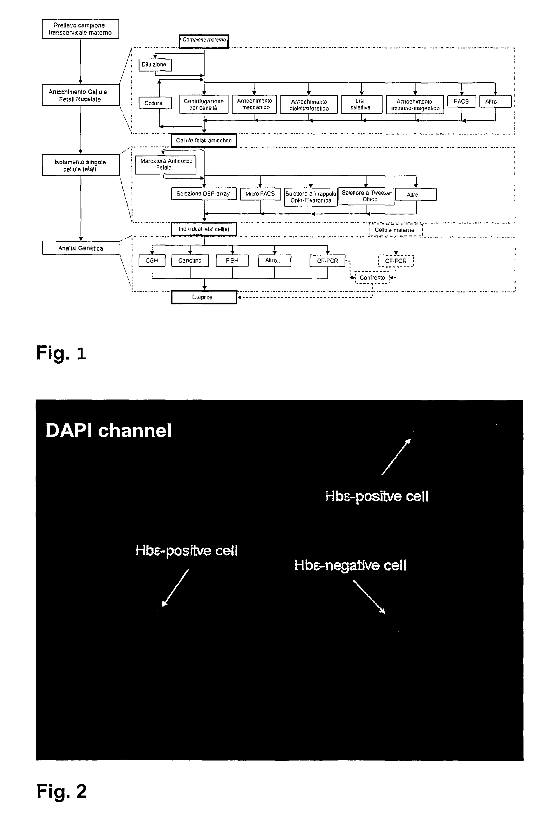
[0017]According to the present invention, the uterine, endocervical or transcervical fluid or the peripheral maternal blood is firstly processed via one or more enrichment stages of the foetal nucleated cells. The method is characterised by the use of a microfluidic system able to individually select, in an easily automatable and repeatable way, single cells from the enriched sample. By means of a microfluidic system of isolation of single cells, it is possible to obtain a set of foetal cells with purity sufficient to perform a genetic diagnosis.
[0022]The use of a microfluidic system offers various advantages including the possibility of having disposable systems, without risks of contamination between the different analyses or the need for thorough washing of the equipment. Furthermore the use of a microfluidic system offers the possibility of having automatic or semiautomatic systems, characterised by a high level of reliability.

Problems solved by technology

All these tests are invasive and involve an increased risk of miscarriage.
The scoring system is very laborious and use of the micromanipulator causes the loss of part of the cells.
The disadvantage of said method is that during the processing for preparation of the slide, part of the transcervical cells are lost, with a no-call due to lack of trophoblasts.
Said method, however, is confined to the selection of foetal cells by means of enrichment of the sample, which does not limit the possibility of having contaminating maternal cells and therefore the consequent risk of an unreliable genetic analysis.
In short, none of the above-mentioned non-invasive methods has so far demonstrated that it can be used as a routine practice for the diagnosis of foetal aneuploidies and / or other chromosome defects.

Method used


Image

  • Method and Device for Non-Invasive Prenatal Diagnosis
  • Method and Device for Non-Invasive Prenatal Diagnosis
  • Method and Device for Non-Invasive Prenatal Diagnosis

Examples


Embodiment Construction

[0034]The subject of the present invention is a method for performing non-invasive prenatal diagnosis.

Collection of Transcervical Samples

[0035]The transcervical samples (TCC) can be taken from different levels of the uterus (external bone, lower part of cervical canal, lower uterine pole, intrauterine cavity) by means of various techniques: aspiration of the cervical mucus, cytobrush or swab, endocervical lavage and intrauterine lavage (IUL).

Enrichment Starting from Peripheral Blood

[0036]The proportion of foetal cells can be enriched using various methods, for example centrifugation on density gradient, consisting of solutions such as Ficoll or Percoll; mechanical enrichment, based on microfabricated filters which select nRBC and empty the sample of RBC; enrichment via dielectrophoretic separation by means of a specific device, the Dielectrophoretic activated cell sorter (DACS); selective lysis, for example selective lysis of the erythrocytes of no interest; immunomagnetic separatio...


PUM

PropertyMeasurementUnit
Densityaaaaaaaaaa
Volumeaaaaaaaaaa
Wavelengthaaaaaaaaaa

Abstract

Method for non-invasive prenatal diagnosis comprising the following steps: a. obtain a sample of an organic fluid having a high probability of containing foetal cells from a pregnant woman; b. enrich said sample of organic fluid in at least one population of cells comprising at least one type of foetal nucleated cells; c. isolate at least one cell from among said at least one type of foetal nucleated cells; d. perform a genetic analysis on said at least one cell isolated from among said at least one type of foetal nucleated cells in order to highlight at least one genetic characteristic of said at least one foetal nucleated cell suitable for permitting said diagnosis; wherein the step of isolating at least one cell from among said at least one type of foetal nucleated cells is performed by individually selecting single cells in a microfluidic device designed for said purpose.

Description

TECHNICAL FIELD[0001]The present invention concerns methods for non-invasive prenatal diagnosis, in particular for the identification of genetic and chromosomic disorders in the foetus.STATE OF THE ART[0002]Prenatal diagnosis for chromosomic disorders was introduced with the aim of highlighting abnormalities of the chromosomes in the foetus.[0003]So far, the diagnostic certainty of a foetus being affected by a chromosome disorder can be obtained only by means of invasive diagnostic tests, examining the embryonic cells in order to determine the karyotype by means of amniocentesis, sampling of the chorionic villi or cordocentesis.[0004]All these tests are invasive and involve an increased risk of miscarriage. They are therefore usually recommended for women over the age of 35, or women who in a previous pregnancy have had a child affected by chromosome disorders or when ultrasound scanning identifies a foetus with a malformation.[0005]The discovery of the existence of foetal cells, al...

Claims


Application Information

Patent Timeline
IPC IPC(8): C12Q1/68
CPCB01L3/5027G01N27/447B01L3/502761
Inventor MANARESI, NICOLFITTIPALDI, ANTONIOGIORGINI, GIUSEPPEMEDORO, GIANNI
Owner MENARINI SILICON BIOSYSTEMS SPA
Learn More