Method and Apparatus for Tomographic Imaging of Absolute Optical Absorption Coefficient in Turbid Media Using Combined Photoacoustic and Diffusing Light Measurements

a technology of optical absorption coefficient and tomography, which is applied in the field of tomography imaging of absolute optical absorption coefficient in turbid media, can solve the problems of difficult to accurately obtain these initial parameters, difficult for such methods to tackle the negative absorbed energy density value, and limit the application of these methods, so as to improve the spatial resolution of dot, improve diagnostic decision-making accuracy, and improve the spatial resolution

Inactive Publication Date: 2010-08-19
UNIV OF FLORIDA RES FOUNDATION INC
View PDF1 Cites 22 Cited by
  • Summary
  • Abstract
  • Description
  • Claims
  • Application Information

AI Technical Summary

Benefits of technology

[0007]Embodiments of the invention pertain to a method and apparatus for imaging a light absorption coefficient distribution. Embodiments of the subject method can be implemented without knowing the strength of incident light in advance and without requiring careful calibrations in the non-scattering medium. Embodiments of the method can combine conventional photoacoustic tomography (PAT) with diffusing light measurements coupled with an optimization procedure based on the photon diffusion equation. Images of absorbing targets as small as 0.5 mm in diameter embedded in a 50 mm diameter background medium can be quantitatively recovered. Small targets with various optical contrast levels relative to the background can be detected well.
[0010]Embodiments of the invention allow the use of PAT for quantitative results, such as obtaining quantitative tissue functional information including oxy-hemoglobin, deoxy-hemoglobin and water and lipid concentrations, for example, which are crucial for accurate diagnostic decision-making. By combining PAT with DOT in accordance with an embodiment of the invention, the spatial resolution of DOT is significantly improved. In a specific embodiment combining PAT with DOT, the spatial resolution of DOT is improved from 5 mm to 1 mm given a large tissue volume. In addition, the hybrid modality can essentially eliminate the crosstalk errors existing in the current DOT techniques.
[0011]Embodiments of the invention can be applied to applications including, but not limited to, breast cancer detection / diagnosis, functional brain imaging, and joint imaging. Embodiments of the invention relate to a medical imaging device / system including both software and hardware, such that considerably higher spatial resolution for tissue functional imaging can be obtained, and much improved accuracy for drastically improved diagnostic decision-making can be achieved.

Problems solved by technology

However, there are several limitations associated with these methods.
First, in these methods, one must know the exact boundary reflection coefficients as well as the exact strength and distribution of an incident light source, which requires careful experimental calibration procedures.
It is often difficult to obtain these initial parameters accurately.
Third, these methods are based on simple data fitting procedures which are highly sensitive to boundary noises.
It is also very difficult for such methods to tackle the negative absorbed energy density values often resulting from conventional PAT.
However, the spatial resolution of DOT is low.
In addition, the functional parameters obtained from DOT are often not accurate because of the strong crosstalk errors contributed from the scattering property of tissue.
However, there are several limitations associated with this simple iteration method.

Method used


Image

  • Method and Apparatus for Tomographic Imaging of Absolute Optical Absorption Coefficient in Turbid Media Using Combined Photoacoustic and Diffusing Light Measurements
  • Method and Apparatus for Tomographic Imaging of Absolute Optical Absorption Coefficient in Turbid Media Using Combined Photoacoustic and Diffusing Light Measurements
  • Method and Apparatus for Tomographic Imaging of Absolute Optical Absorption Coefficient in Turbid Media Using Combined Photoacoustic and Diffusing Light Measurements

Examples


Embodiment Construction

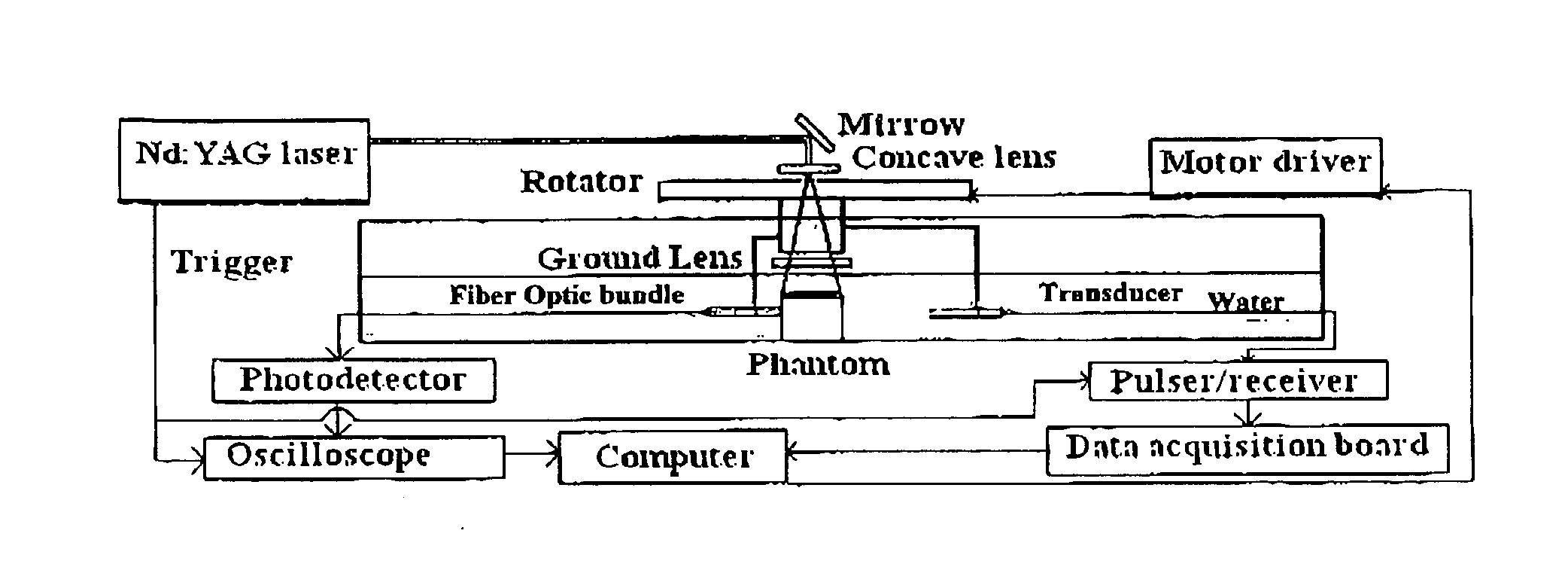
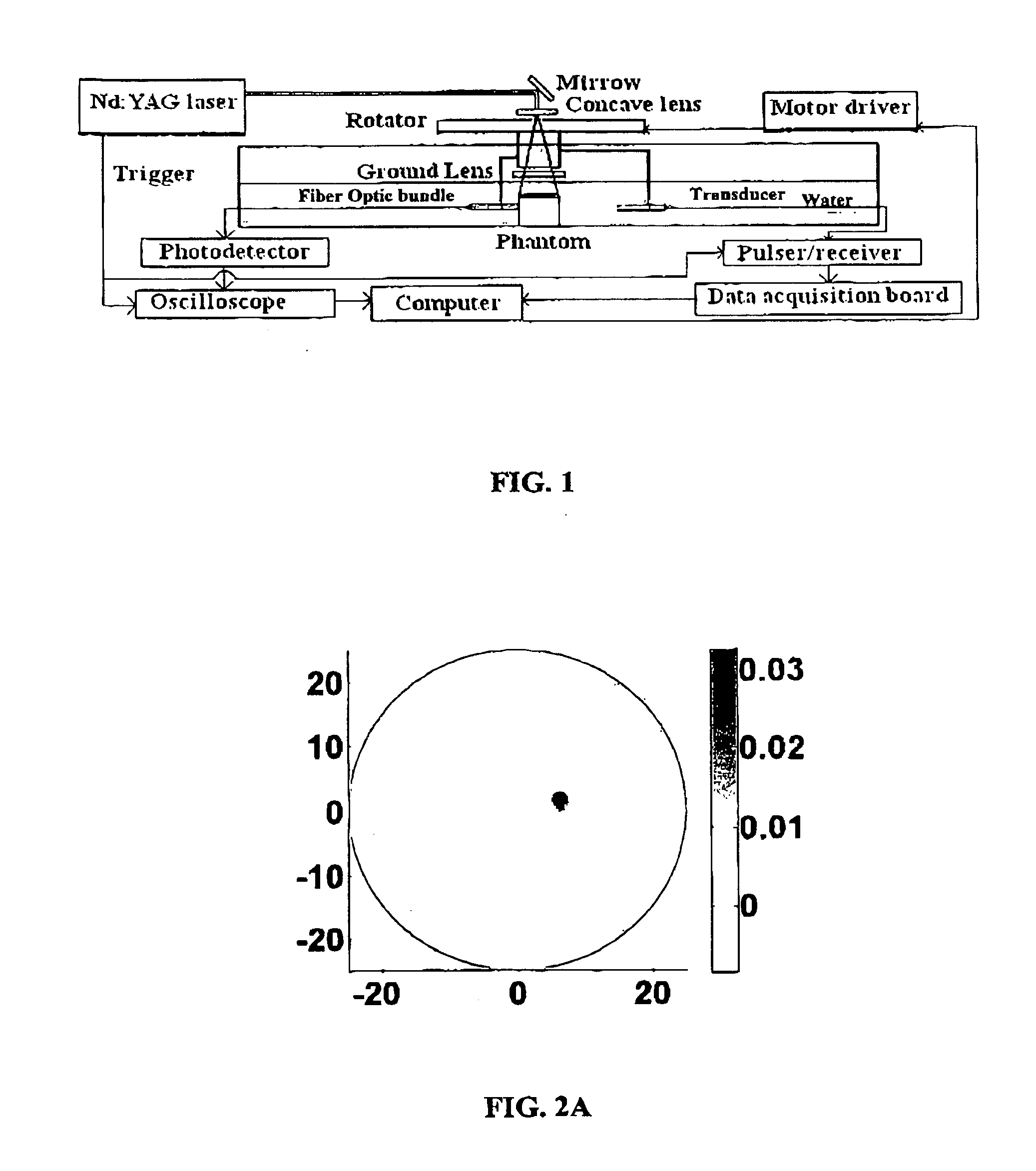
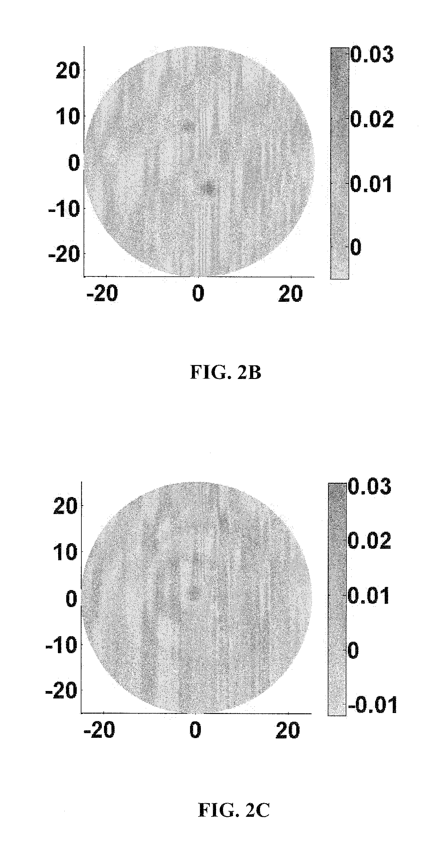
[0023]Embodiments of the disclosure pertain to a method and apparatus for imaging a light absorption coefficient distribution. Embodiments of the subject method can be implemented without knowing the strength of incident light in advance and without requiring careful calibrations in the non-scattering medium. Embodiments of the method can combine conventional photoacoustic tomography (PAT) with diffusing light measurements coupled with an optimization procedure based on the photon diffusion equation. Images of absorbing targets as small as 0.5 mm in diameter embedded in a 50 mm diameter background medium can be quantitatively recovered. Small targets with various optical contrast levels relative to the background can be detected well.

[0024]Embodiments can be utilized to image human, or animal, tissue. Specific embodiments involve imaging of a breast, the brain, a joint, and endoscopic imaging of the GI tract, colon, or esophagus.

[0025]Embodiments of the subject reconstruction method...


PUM


Abstract

Embodiments of the invention pertain to methods for imaging a light absorption coefficient distribution. Embodiments of the subject method can be implemented without knowing the strength of incident light in advance and without requiring careful calibrations in the non-scattering medium. Embodiments of the method can combine conventional photoacoustic tomography (PAT) with diffusing light measurements coupled with an optimization procedure based on the photon diffusion equation. Images of absorbing targets as small as 0.5 mm in diameter embedded in a 50 mm diameter background medium can be quantitatively recovered. Small targets with various optical contrast levels relative to the background can be detected well. Embodiments of the subject reconstruction method can include first obtaining the map of absorbed optical energy density. Embodiments can obtain the map of absorbed optical energy density through a model-based reconstruction algorithm that is based on a finite element solution to the photoacoustic wave equation in frequency domain subject to the radiation or absorbing boundary conditions (BCs). The distribution of optical fluence can then be obtained. Embodiments can obtain the distribution of optical fluence using the photon diffusion equation based optimization procedure. The distribution of optical absorption coefficient can then be recovered from the distribution of optical fluence and the absorbed energy density.

Description

CROSS-REFERENCE TO RELATED APPLICATION[0001]The present application claims the benefit of U.S. Application Ser. No. 60 / 950,304, filed Jul. 17, 2007, which is hereby incorporated by reference herein in its entirety, including any figures, tables, or drawings.[0002]The subject invention was made with government support under a research project supported by a grant from the National Institutes of Health (NIH), Contract No. R01 CA90533.BACKGROUND OF INVENTION[0003]Biomedical photoacoustic tomography (PAT) is a potentially powerful imaging method for visualizing the internal structure of soft tissues with excellent spatial resolution and satisfactory imaging depth. While conventional PAT can image tissues with high spatial resolution, it provides only the distribution of absorbed optical energy density that is the product of both the intrinsic optical absorption coefficient and extrinsic optical fluence distribution, which is a spatially varying function even for a homogeneous medium. Th...

Claims


Application Information

Patent Timeline
Patent Type & Authority Applications(United States)
IPC IPC(8): G06K9/00
CPCA61B5/0073A61B5/4528A61B5/0095
Inventor JIANG, HUABEIYUAN, ZHENWANG, QIANGYIN, LUZHANG, QIZHI
Owner UNIV OF FLORIDA RES FOUNDATION INC
Why Eureka
Try Eureka