Treatment and diagnostic methods for hypersensitive disorders

a hypersensitivity disorder and diagnostic method technology, applied in the field of hypersensitivity disorder treatment and diagnostic methods, can solve the problems of small complexes that form in blood vessel walls, affecting the immune response, so as to reduce the number of days a patient is hospitalized, inhibit or reduce the severity and delay the development of a hypersensitivity disorder.

Active Publication Date: 2010-10-21
PROMEDIOR
View PDF47 Cites 32 Cited by
  • Summary
  • Abstract
  • Description
  • Claims
  • Application Information

AI Technical Summary

Problems solved by technology

These immune responses can be detrimental when they are not appropriately regulated, as in autoimmune diseases, or when excessive, as in allergic diseases.
Production of IFN-γ and TNF-α together with expression of FAS-ligand by Th1 cells leads to epithelial cell activation followed by apoptosis, and compromises barrier function of epithelial cells in the lungs and the skin.
However, small complexes that form tend to deposit in blood vessels walls.
There they can ligate Fc receptors on leukocytes, leading to leukocyte activation and tissue injury.
Unfortunately, many of these drugs still have the disadvantage of producing unwanted side-effects even when administered locally.
As many patients use excessive quantities of these drugs to relieve symptoms quickly, there is an increased risk that the patient will suffer deleterious effects.

Method used


Image

  • Treatment and diagnostic methods for hypersensitive disorders
  • Treatment and diagnostic methods for hypersensitive disorders
  • Treatment and diagnostic methods for hypersensitive disorders

Examples


example 1

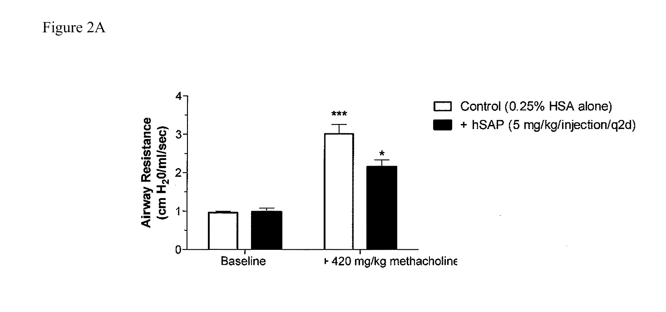
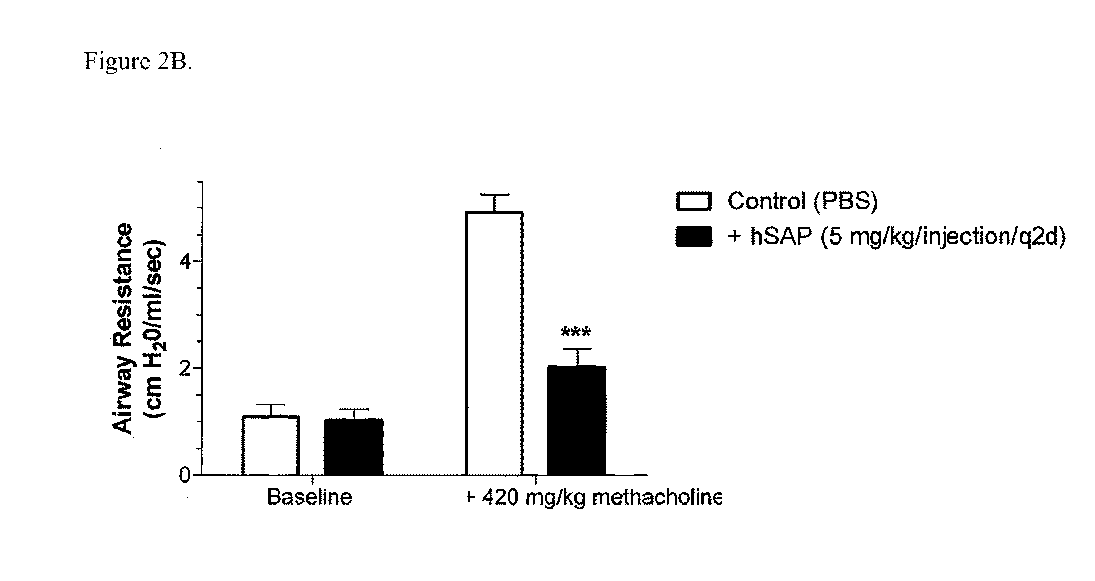
[0196]Chronic allergic airway disease induced by A. fumigatus conidia is characterized by airway hyper-reactivity, lung inflammation, eosinophilia, mucus hypersecretion, goblet cell hyperplasia, and subepithelial fibrosis. C57BL / 6 mice were similarly sensitized to a commercially available preparation of soluble A. fumigatus antigens as previously described (Hogaboam et al. The American Journal of Pathology. 2000; 156: 723-732). Seven days after the third intranasal challenge, each mouse received 5.0×10⁶ A. fumigatus conidia suspended in 30 μl of PBS tween 80 (0.1%, vol / vol) via intratracheal route.

[0197]At day 15- and 30-time points (FIGS. 2A and 2B respectively), groups of five mice treated with SAP or control (PBS) were analyzed for changes in airway hyperresponsiveness (AHR). Bronchial hyperresponsiveness was assessed after an intratracheal A. fumigatus conidia challenge using a Buxco™ plethysmograph (Buxco, Troy, N.Y.). Briefly, sodium pentobarbital (Butler Co., Columbus, Ohio; ...

example 2

[0198]C57BL / 6 mice were similarly sensitized to a commercially available preparation of soluble A. fumigatus antigens as above described. Animals were treated in vivo with hSAP or PBS control for the last two weeks of the model. At day 15- and 30-time points (FIGS. 3A and 3B respectively), groups of five mice treated were analyzed for changes in cytokine production. Spleen cells were isolated from animals at 15 or 30 days after intratracheal conidia challenge, stimulated with aspergillus antigen, and treated in vitro with hSAP. Splenocyte cultures were quantified (pg / mL) for production of IL-4, IL-5, and IL-10.

example 3

[0199]C57BL / 6 mice were similarly sensitized to a commercially available preparation of soluble A. fumigatus antigens as above described. At day 15, the amount of FoxP3 expression was determined in pulmonary draining lymph nodes or splenocyte cultures. Pulmonary lymph nodes were dissected from each mouse and snap frozen in liquid N2 or fixed in 10% formalin for histological analysis. Histological samples from animals treated with PBS (control) or SAP were stained for FoxP3 (FIG. 4A), and the number of FoxP3+ cells were quantified relative to each field examined (FIG. 4B). Purified splenocyte cultures were stimulated with Aspergillus antigen in vitro in the presence or absence of SAP in vitro (0.1-10 m / ml) for 24 hours. Total FoxP3 expression was quantitated using real time RT-PCR (FIG. 4C).


PUM

PropertyMeasurementUnit
concentrationaaaaaaaaaa
affinityaaaaaaaaaa
forced expiratory volume inaaaaaaaaaa

Abstract

The current standard of care for the treatment of allergic airway diseases include short and long acting beta-agonists, and inhaled or systemic corticosteroids, cromolyn and xanthines that all have the potential of detrimental side-effects. The present invention describes a new mechanistic protein-based therapeutic approach for the treatment of allergic airway disease and diseases associated with excessive Th2 pathology. The present invention relates to the surprising discovery that serum amyloid P (SAP) demonstrates a therapeutic affect in the treatment of hypersensitive disorders.

Description

CROSS-REFERENCE TO RELATED APPLICATIONS[0001]This application claims the benefit of U.S. Provisional Application Ser. No. 61 / 209,795 filed Mar. 11, 2009. All the teachings of the above-referenced application is incorporated herein by reference.BACKGROUND OF THE INVENTION[0002]Native CD4 T-cells can differentiate upon activation into either Th1 or Th2 cells, which have significantly different immune functions based on the cytokines they produce. Th1 cells promote type 1 immunity, producing IFN-γ which drives cell-mediated immunity and IgG2A synthesis. Th2 cells enhance type 2 immunity by producing IL-4, IL-5, and IL-13 and promoting antibody-mediated responses and class switching to IgG1 and IgE. These immune responses can be detrimental when they are not appropriately regulated, as in autoimmune diseases, or when excessive, as in allergic diseases.[0003]The Th1 differentiation pathway is normally stimulated in response to infectious microbes that activate macrophages and / or NK cells...

Claims


Application Information

Patent Timeline
Patent Type & Authority Applications(United States)
IPC IPC(8): A61K39/395A61K31/7088A61K38/00A61K31/573A61K31/352A61K31/522A61P37/08
CPCA61K38/1716A61P1/00A61P11/00A61P11/02A61P11/06A61P17/00A61P27/14A61P29/00A61P37/00A61P37/08A61P43/00
Inventor MURRAY, LYNNE ANNE
Owner PROMEDIOR
Why Eureka
Try Eureka