Labeled molecular imaging agents, methods of making and methods of use

a molecular imaging agent and labeling technology, applied in the field of labeling molecular imaging agents, can solve the problems of effective changes in treatment regimens, and achieve the effect of low toxicity/immune respons

Inactive Publication Date: 2010-10-28
GENERAL ELECTRIC CO
View PDF3 Cites 13 Cited by
  • Summary
  • Abstract
  • Description
  • Claims
  • Application Information

AI Technical Summary

Benefits of technology

[0005]The imaging agents and methods of the invention take advantage of the cellular amino acid transporter (cystine / glutamate antiporter, Xc˜), which is activated under conditions of cellular oxidative stress. These labeled molecular imaging agents and methods of the invention provide several benefits, including but not limited to, their use in a wide variety of diagnostic and therapeutic monitoring applications; they are small molecules and comprise labeled variants of a natural compound found in the body, and would be administered at tracer levels with physiological concentrations well below those that generate toxic response, therefore toxicity / immune response is expected to be low; and because the imaging agents act as a transporter substrate, the agents benefit from amplification of the signal because the molecular imaging agent is trapped once inside the cell, unlike other molecular binders in which the imaging agent's signal is limited to the stoichiometric binding of a cell surface epitope. The labeled substrates of the invention for the cystine / glutamate transporter may also be used to introduce compounds in a target for therapeutic purposes.
[0007]Additionally, upregulation of the cystine / glutamate transporter is also associated with chemotherapy resistance in some tumors. Therefore, non-invasive imaging of tumors with basal high cystine uptake could result in identification of tumors likely to be resistant to certain therapies; which could result in efficacious changes in treatment regimens.

Problems solved by technology

Therefore, non-invasive imaging of tumors with basal high cystine uptake could result in identification of tumors likely to be resistant to certain therapies; which could result in efficacious changes in treatment regimens.

Method used


Image

  • Labeled molecular imaging agents, methods of making and methods of use
  • Labeled molecular imaging agents, methods of making and methods of use
  • Labeled molecular imaging agents, methods of making and methods of use

Examples


example 1

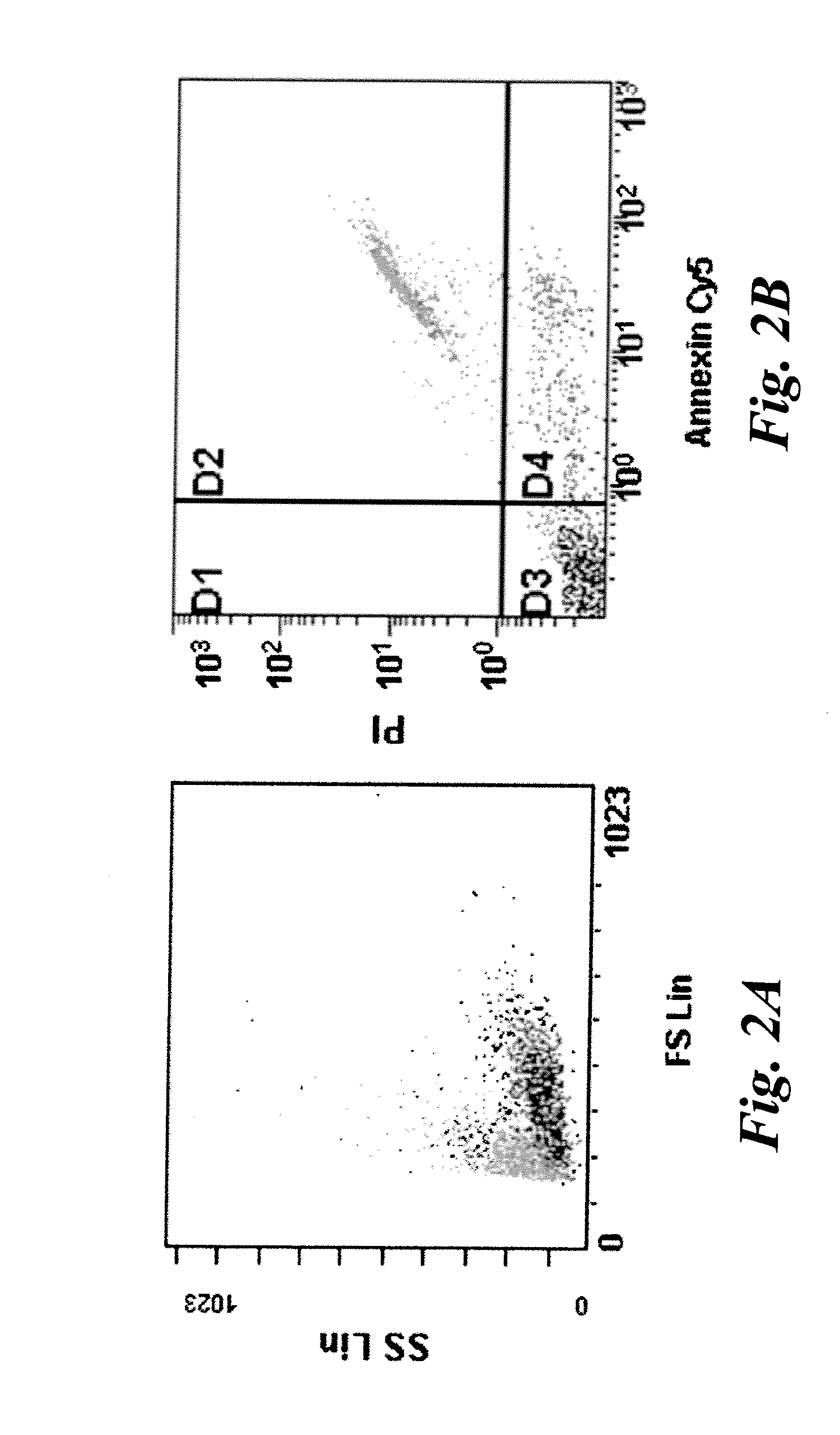
[0039]Human Jurkat cells were cultured with or without 1 uM staurosporine for 16 hours in an in vitro assay for apoptotic cells and stained with propidium iodide(PI) and Cy5-Annexin V (Annexin). Typically ˜30-50% of cells were found to be in some stage of apoptosis or necrosis. Flow cytometry was used to identify population of cells defined as normal (PI and Annexin negative), early apoptotic (PI negative, Annexin positive), and late apoptosis and necrosis (PI and Annexin positive). The results are shown in FIGS. 2A and 2B. FIG. 2A is a forward scatter / side scatter plot showing some late apoptotic cells (light gray) with a different granularity than non-apoptotic cells (darker gray). In FIG. 2B, Annexin V and PI negative cells are shown in quadrant D3 and represent non-apoptotic cells. Cells in quadrant D4 indicate early apoptotic cells that are positive for Annexin V staining, but negative for PI staining. Cells in quadrant D2 indicate late apoptotic and necrotic cells that are pos...

example 2

[0040]Jurkat cells were incubated with (FIG. 3B) and without (FIG. 3A) 1 μM staurosprine (STN) for 16-18 hours, stained with Annexin V-Cy5 and propidium iodide, and incubated with DBC for 30 minutes. DBC is a commercially available product from Invitrogen (catalog #B20340), which is sold for the purpose of reversible thiol labeling of nucleotides, proteins and cells via a disulfide exchange reaction at acidic conditions. DBC has the structure shown below.

[0041]In FIG. 3A, untreated cells show some low intensity fluorescence corresponding to uptake or nonspecific binding of the DBC molecule. In FIG. 3B, staurosporine induced cells have a subpopulation of cells with high intensity DBC staining and this correlated primarily to the late apoptotic / necrotic cells (+Annexin V, +PI). A population of cells with intermediate DBC staining also appeared, which correlates well with the early apoptotic cells (+Annexin V, −PI). To more specifically address the role of the the xc- transporter in th...

example 3

[0042]The apoptotic Jurkat cell uptake of DBC shows not only that the xc- transporter is activated in cells undergoing oxidative stress (and in this case apoptosis), but also that the xc- transporter is promiscuous enough to allow this substrate (cystine) with amine appended green fluorophores (bodipyFL) into the cell. A labeled cystine derivative was synthesized with a red fluorophore (bodipy650) to further determine the promiscuity of the transporter. In a side-by-side comparison to the bodipyFL labeled cystine, DBC-650 did not appear to label apoptotic cells any more than normal cells, as shown in FIGS. 4A and 4B, indicating that some labels are not sufficient for maintaining xc- transporter substrate status. In this example, the bodipy-650 fluorophore and associated linker are larger than that of DBC (FIG. 4A), and therefore size represents one limitation of what groups are appended to the amines while retaining the ability to pass through the transporter.


PUM

PropertyMeasurementUnit
timeaaaaaaaaaa
timeaaaaaaaaaa
timeaaaaaaaaaa

Abstract

Imaging agents that comprise labeled substrates of the cystine / glutamate antiporter of cells, whereby the methods of use comprise introducing the labeled agents into cells via the cystine / glutamate antiporter, which are then reduced to a labeled cysteine, and subsequently detected in the cell.

Description

BACKGROUND[0001]The invention relates generally to labeled molecular imaging agents and more particularly to imaging agents that are taken up by the cells via the cystine / glutamate transporter.[0002]The concept of molecular imaging promises specific contrast enhancement of molecular signatures of pathology and requires targetable biomarkers that are specifically regulated in certain pathological indications. While such a specific molecular contrast agent could have great utility for imaging and diagnosing disease; validation of a truly specific biomarker has proven to be very difficult. Even if an agent to such a specific biomarker is created, the market for such an agent will be limited to the prevalence of this indication. Therefore there is great interest in developing molecular contrast agents that can be utilized to image a variety of pathological indications. Most imaging agents target specific tissue or cell types, or specific therapies, or they degrade rapidly over time. One...

Claims


Application Information

Patent Timeline
Patent Type & Authority Applications(United States)
IPC IPC(8): A61K51/04C07C321/00C07F5/02
CPCA61K49/0019A61K49/0052A61K51/0406C07F5/022C07B2200/05C07C323/58C07C323/59C07B59/001
Inventor WEBSTER, JACK MATHEWSYUD, FAISAL AHMEDLEE, BRIAN DUH-LANSCHAFFER, PAULZHANG, RONGHUNTINGTON, JENNIFER LYNNEAMARASINGHE, KANDE KANKANAMALAGE DAYARATHNA
Owner GENERAL ELECTRIC CO
Why Eureka
Try Eureka