Detection of Changes in Cell Populations and Mixed Cell Populations

a cell population and cell technology, applied in the field of mixed cell population analysis, can solve the problems of damage to cells, time-consuming and complicated preparation of biological samples for analysis, and limited field of cell analysis, in particular, stem cell analysis, primary cell analysis,

Inactive Publication Date: 2010-11-18
X BODY
View PDF101 Cites 35 Cited by
  • Summary
  • Abstract
  • Description
  • Claims
  • Application Information

AI Technical Summary

Benefits of technology

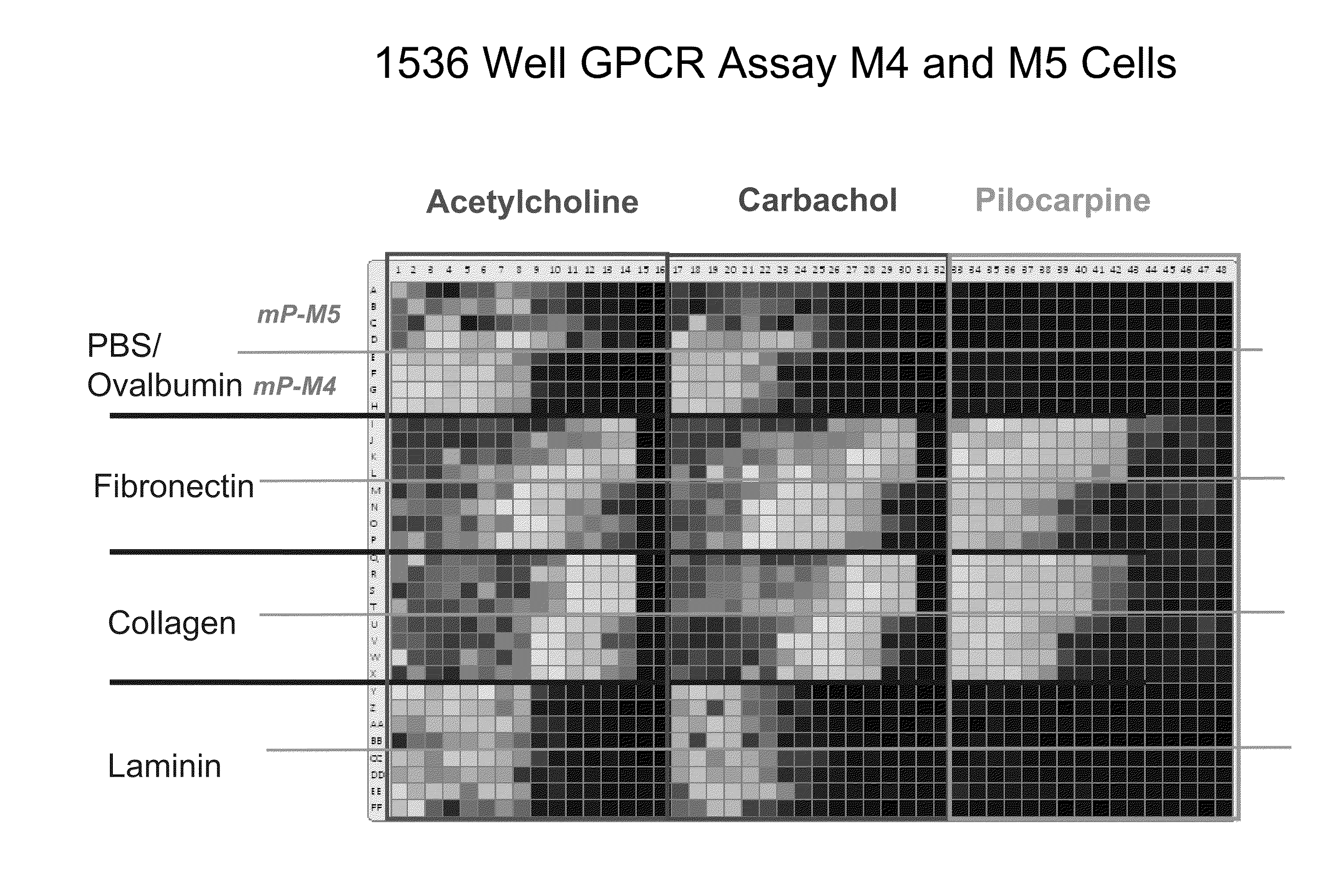
[0146]One functional advantage of the invention is that by screening multiple cells against the same test reagent in a single well, the assay has a built-in test of test reagent specificity. If the same test reagent is found to inhibit the activation of multiple cell lines expressing different receptors, then the test reagent is likely promiscuous or cytotoxic. Currently, this can be done by screening one test reagent in multiple wells containing different cells. The instant invention allows the user to screen one test reagent against mixed cell populations (such as cardiomyocytes and hepatocytes) in a single well. The per well cost of screening is effectively divided by the number of targets being screened simultaneously.

Problems solved by technology

Cell analysis, in particular, stem cell analysis, primary cell analysis, and mixed cell population analysis, is currently limited in the field due to the lack of tools available to accurately measure real time biological processes, such as adhesion, cell migration and chemotaxis, invasion into basement membranes or tissues, differentiation, differentiation mediated by cellular adhesion, differentiation mediated by tertiary environments (3-D cell culture), and differentiation mediated by co-culture with different cell types, in particular when cell numbers are scarce.
Additionally, preparation of biological samples for analysis can be time consuming and complicated.
However, these methods are expensive, require detectable labels, can damage the cells leaving them unusable for further analysis, and require relatively large sample volumes.
Furthermore, the devices are difficult to sterilize, mechanically complicated, and can only be operated and maintained by trained personnel.

Method used


Image

  • Detection of Changes in Cell Populations and Mixed Cell Populations
  • Detection of Changes in Cell Populations and Mixed Cell Populations
  • Detection of Changes in Cell Populations and Mixed Cell Populations

Examples


Embodiment Construction

[0050]As used herein, the singular forms “a,”“an”, and “the” include plural referents unless the context clearly dictates otherwise.

Biosensors

[0051]Biosensors of the invention can be colorimetric resonant reflectance biosensors. See e.g., Cunningham et al., “Colorimetric resonant reflection as a direct biochemical assay technique,” Sensors and Actuators B, Volume 81, p. 316-328, Jan. 5, 2002; U.S. Pat. Publ. No. 2004 / 0091397; U.S. Pat. No. 7,094,595; U.S. Pat. No. 7,264,973. Colorimetric resonant biosensors are not surface plasmon resonant (SPR) biosensors. SPR biosensors have a thin metal layer, such as silver, gold, copper, aluminum, sodium, and indium. The metal must have conduction band electrons capable of resonating with light at a suitable wavelength. A SPR biosensor surface exposed to light must be pure metal. Oxides, sulfides and other films interfere with SPR. Colorimetric resonant biosensors do not have a metal layer, rather they have a dielectric coating of high refracti...


PUM

PropertyMeasurementUnit
wavelengthsaaaaaaaaaa
grating periodaaaaaaaaaa
grating depthaaaaaaaaaa

Abstract

The invention provides methods of label-free detection of changes in cell populations and mixed cell populations.

Description

PRIORITY[0001]This application claims the benefit of the following provisional applications: U.S. Ser. No. 61 / 178,787, filed May 15, 2009, U.S. Ser. No. 61 / 257,345, filed Nov. 2, 2009, U.S. Ser. No. 61 / 296,099, filed Jan. 19, 2010, U.S. Ser. No. 61 / 315,144, filed Mar. 18, 2010, and U.S. Ser. No. 61 / 323,070, filed Apr. 12, 2010, all of which are incorporated by reference in their entirety.BACKGROUND OF THE INVENTION[0002]Cell analysis, in particular, stem cell analysis, primary cell analysis, and mixed cell population analysis, is currently limited in the field due to the lack of tools available to accurately measure real time biological processes, such as adhesion, cell migration and chemotaxis, invasion into basement membranes or tissues, differentiation, differentiation mediated by cellular adhesion, differentiation mediated by tertiary environments (3-D cell culture), and differentiation mediated by co-culture with different cell types, in particular when cell numbers are scarce....

Claims


Application Information

Patent Timeline
Patent Type & Authority Applications(United States)
IPC IPC(8): C12Q1/68G01N33/567C12Q1/02
CPCG01N33/5044G01N33/5064G01N21/7743G01N33/54373G01N33/5073C12Q1/02G01N33/53G01N33/543G01N33/68
Inventor SHAMAH, STEVENLAING, LANCE G.YUZHAKOV, ALEXANDERWAGNER, RICKABODEELY, MARLAROCKNEY, BENNETTSCHULZ, STEPHEN C.PADALIA, ZINKALGETMAN, MICHAELSANDBERG, ERIC
Owner X BODY
Why Eureka
Try Eureka