Pharmaceutical compositions for cell therapy of pigmentation disorders
A technology of pigmentation and composition, applied in the direction of drug combination, cell culture support/coating, animal cells, etc., can solve the problems of low recoloration and side effects
- Summary
- Abstract
- Description
- Claims
- Application Information
AI Technical Summary
Problems solved by technology
Method used
Image
Examples
Embodiment 1
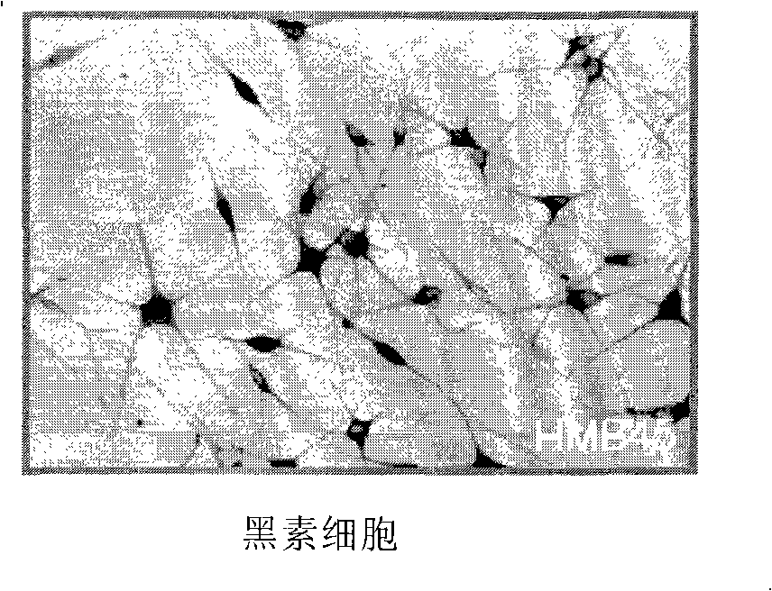
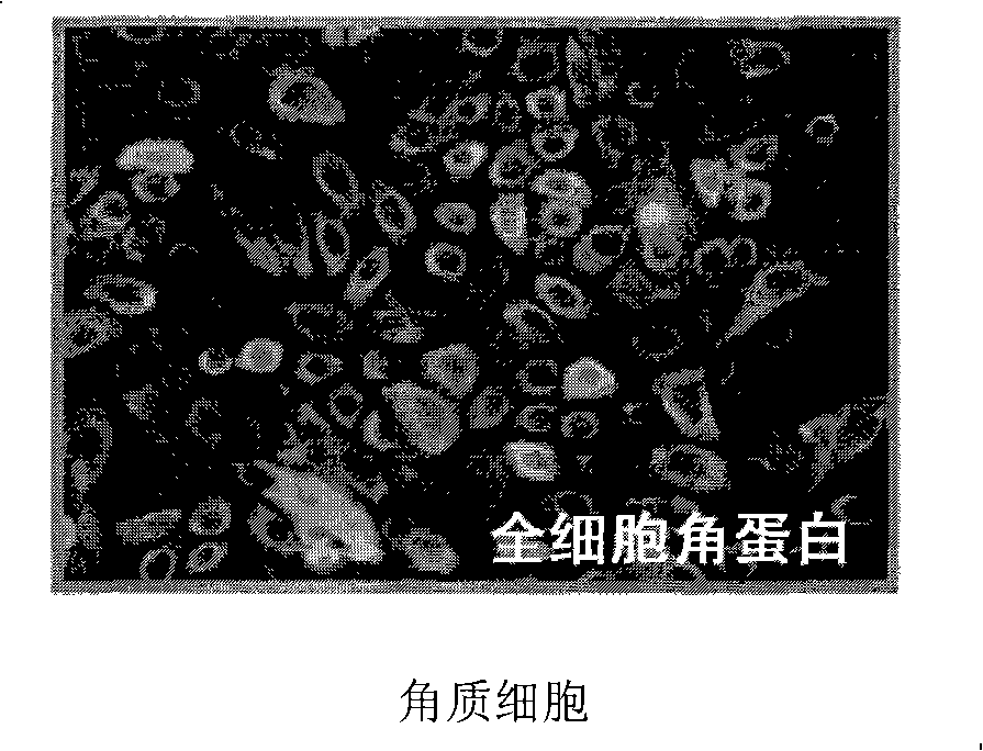
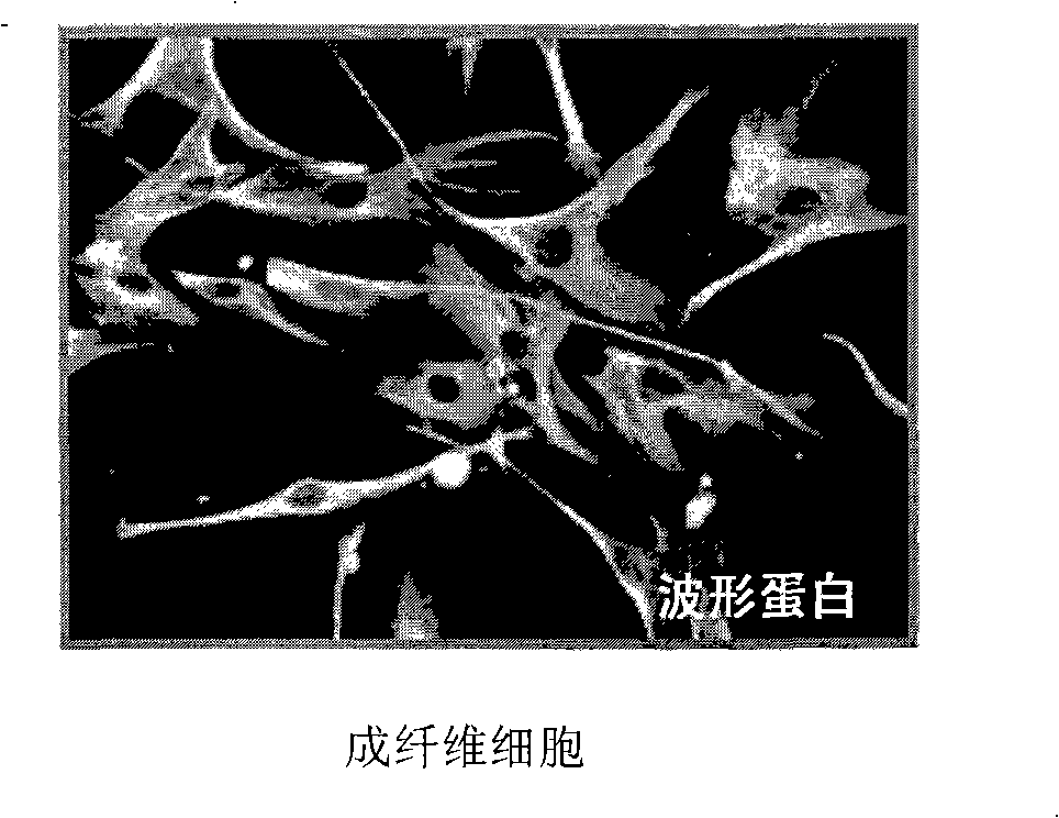
[0050] Example 1: Isolation and culture of melanocytes, keratinocytes and fibroblasts
[0051] Melanocytes, keratinocytes, and fibroblasts isolated from normal human skin tissue and cultured.
Embodiment 1-1
[0052] Example 1-1: Isolation and culture of melanocytes
[0053] The skin tissue was washed 8 times with phosphate buffered saline (WelGENE) containing 50 μg / ml antibiotic (gentamycin, Gibco) to remove coagulated blood and contaminants. The fat layer in the dermis of the skin tissue was removed, and the tissue was cut into 5 mm size, and the cut tissue was incubated with 1 mg / ml dispase (Roche) at 4° C. for 16 hours. Then, the epidermis was separated from the skin tissue, washed with phosphate buffer solution and incubated with trypsin-EDTA solution (0.05% trypsin-0.53 mM EDTA·4Na, Gibco) for 30 minutes at 37°C. Several aspirations of the trypsin-EDTA solution containing the epidermis were used to separate the cells (melanocytes and keratinocytes) from the epidermis.
[0054] MGM-3 (Cambrex) was added as a melanocyte medium to the trypsin-EDTA solution containing isolated melanocytes and keratinocytes and the resultant was transferred to a 50 ml test tube, followed by centr...
Embodiment 1-2
[0055] Embodiment 1-2: Isolation and culture of keratinocytes
[0056] The skin tissue was washed 8 times with phosphate buffered saline (WelGENE) containing 50 μg / ml antibiotic (gentamycin, Gibco) to remove coagulated blood and contaminants. The fat layer in the dermis of the skin tissue was removed, and the tissue was cut into a size of 5 mm, and the cut tissue was physically stimulated with 10 ml of trypsin solution (0.125% trypsin: versene=1: 1, Gibco ) at room temperature for 45 minutes to separate keratinocytes. To the trypsin solution containing the isolated cells, 0.1 mg / ml of trypsin inhibitor (Gibco) was added and transferred to a 50 ml tube, followed by centrifugation at 300 xg. After removing the supernatant, KGM (Cambrex) as a keratinocyte medium was added to homogenize the isolated cells and the homogenization solution (containing 3 × 10 5 cells) were aliquoted into 100mm Petri dishes. at 37°C in CO 2 Cell cultures were performed in an incubator with medium...
PUM
 Abstract
Description
Claims
Application Information
 - R&D
- Intellectual Property
- Life Sciences
- Materials
- Tech Scout
- Unparalleled Data Quality
- Higher Quality Content
- 60% Fewer Hallucinations
Browse by: Latest US Patents, China's latest patents, Technical Efficacy Thesaurus, Application Domain, Technology Topic, Popular Technical Reports.
© 2025 PatSnap. All rights reserved.Legal|Privacy policy|Modern Slavery Act Transparency Statement|Sitemap|About US| Contact US: help@patsnap.com


