In-vitro construction method for simulating blood brain barrier through human brain microangiogenesis

A blood-brain barrier and construction method technology, applied in artificial cell constructs, biochemical equipment and methods, microorganisms, etc., can solve the problem that 2D models are difficult to obtain in vivo cells, and achieve the goal of reducing sample consumption and reducing test costs. Effect

Active Publication Date: 2018-11-16
WUHAN CHOPPER BIOLOGY
View PDF6 Cites 9 Cited by
  • Summary
  • Abstract
  • Description
  • Claims
  • Application Information

AI Technical Summary

Problems solved by technology

At present, although animal models have been widely used in the research of various diseases, some scholars have realized in recent years that there are still huge differences between animal models and humans. Even animals that are very similar to humans cannot be directly used to predict human diseases.
Recently, the field of cell biology has begun to recognize the dissimilarity between the environment in which cells grow on these flat surfaces and the complex environment in which 3D cells grow in vivo, so it is difficult for 2D models to faithfully capture the physiological behavior of cells in vivo

Method used


Image

  • In-vitro construction method for simulating blood brain barrier through human brain microangiogenesis
  • In-vitro construction method for simulating blood brain barrier through human brain microangiogenesis
  • In-vitro construction method for simulating blood brain barrier through human brain microangiogenesis

Examples


Embodiment 1

[0073] Example 1 Preparation and cultivation of 3D cell culture chip

[0074] (1) Preparation of cell solution: Primary GFP-HBVEC cells (purchased from Science cell, USA) and primary mCherry-HA cells (purchased from Science cell, USA) were respectively used in endothelial cell growth medium EGM-2 (purchased from Lonza) and astrocyte culture medium AM (purchased from Science cell, USA) were resuscitated and cultured. When the cell confluence reached 80-90%, GFP-HBVEC cell suspension and mCherry-HA cell suspension were collected respectively Each 1mL, corresponding to obtain endothelial cell suspension and astrocyte suspension, for use; wherein, the resuscitation culture conditions are: culture temperature 37°C, culture humidity 95%, CO 2 The concentration is 5%; the cell concentration in the GFP-HBVEC cell suspension and mCherry-HA cell suspension are both 1×10 6 cell / mL.

[0075] (2) Preparation of fibrinogen mother liquor: After preheating DPBS (purchased from Gibco, USA) to 37°C,...

Embodiment 2

[0080] Example 2 Morphological characterization of 3D cell culture model

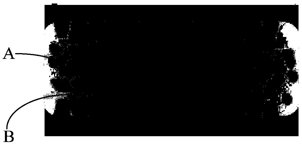
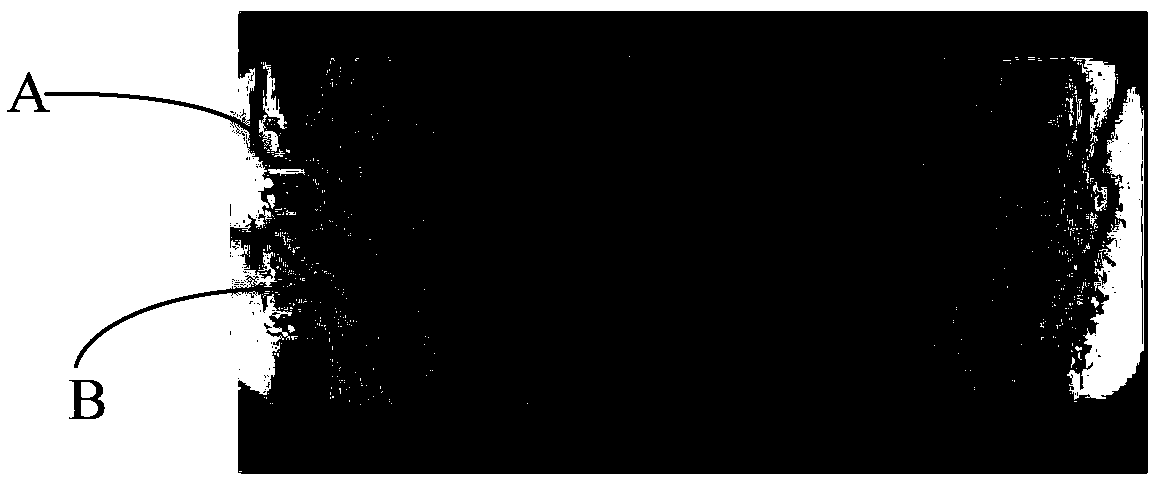
[0081] During the continuous culture period in step (6) of the above-mentioned Example 1, the 3D cell culture chip was scanned by a laser confocal microscope, and 3D fluorescence images were collected for cell morphology identification (GFP green fluorescence was excited at 488nm, and 509nm Emission light imaging; mCherry red fluorescence is excited at 587nm and imaging with 610nm emission light), the 3D fluorescence image acquisition results are as follows Figure 4 to Figure 8 As shown, Figure 4 to Figure 6 These are the 3D fluorescence images collected when the cells are grown on the microfluidic chip for 1, 2, and 3 days. Figure 7 to Figure 8 Is the 3D fluorescence image collected when the cells are grown in the microfluidic chip for 4 days, Figure 7 This is the result of cell growth in the microfluidic chip for 4 days showing the formation of vascular tubule-like structures, Figure 8 This is the re...

Embodiment 3

[0086] Example 3 Functional fluidity verification of 3D cell culture model

[0087] After step (6) of the above-mentioned embodiment 1, particles are loaded into the 3D cell culture chip formed with the brain microvascular network structure, and the trajectory of the loaded particles in the microvascular network is observed by tomography, and the particles are observed The microvascular network moves and migrates through the blood vessels as the fluid flows. The monitoring results of part of its trajectory are as follows Picture 9 Shown ( Picture 9 The granular material shown at C in the middle is partially loaded with particles and evenly distributed in the formed vascular network), indicating that the brain microvascular network formed by endothelial cells and astrocytes exhibits good structural integrity and Vascular permeability.

[0088] In summary, the present invention uses the mixed cells of primary human brain microvascular endothelial cells and primary human brain astro...


PUM


Abstract

The invention discloses an in-vitro construction method for simulating a blood brain barrier through human brain microangiogenesis. The method comprises the following steps: preparing human brain microvascular endothelial cell suspension and human astrocyte suspension, and preparing fibrinogen mother liquor and thrombin mother liquor; mixing the endothelial cell suspension, the astrocyte suspension, a DMEM (Dulbecco's Modified Eagle Medium) medium, the fibrinogen mother liquor and the thrombin mother liquor, and preparing a mixed cell gel solution; injecting the mixed cell gel solution into amicro-fluidic chip, performing thermostatic incubation to gelation, adding an endothelial growth medium into the micro-fluidic chip, and constructing a 3D cell culture chip; performing continuous culture on the 3D cell culture chip, and enabling the endothelial cells and astrocyte to grow into a brain microvascular network structure, namely correspondingly producing the simulated blood brain barrier. According to the technical scheme provided by the invention, the in-vitro model of the blood brain barrier is successfully constructed, and the characteristics of the blood brain barrier are clearly and accurately reflected.

Description

Technical field [0001] The present invention relates to the technical field of tissue engineering, in particular to an in vitro construction method of human brain microangiogenesis to simulate the blood-brain barrier. Background technique [0002] The blood-brain barrier refers to the barrier between blood plasma and brain cells formed by brain capillary walls and glial cells, and the barrier between plasma and cerebrospinal fluid formed by the choroid plexus. These barriers can prevent harmful substances from entering the brain tissue from the blood. It plays an important biological role in maintaining the normal physiological state of the central nervous system. Therefore, studying the formation of the blood-brain barrier is of great significance to the study of related diseases. [0003] At present, two-dimensional (2D) blood brain barrier (BBB) ​​models are mostly composed of single cultured brain microvascular endothelial cells, or developed and formed by glial cells or astro...

Claims


Application Information

Patent Timeline
Patent Type & Authority Applications(China)
IPC IPC(8): C12N5/071
CPCC12N5/0697C12N2502/086C12N2502/28C12N2513/00C12N2533/56
Inventor 吴扬宫智勇李岩
Owner WUHAN CHOPPER BIOLOGY
Why Eureka
Try Eureka