
Adaptive radiotherapy treatment using ultrasound

a radiotherapy treatment and ultrasound technology, applied in the field of monitoring therapy treatments, can solve the problems of deteriorating the actual delivered dose to the target organ, risks of exposing surrounding sensitive structures to unwanted radiation, and inability to achieve the effect of avoiding potential radiation damage to the ultrasound prob

Active Publication Date: 2011-01-13
ELEKTA AB
View PDF103 Cites 22 Cited by
  • Summary
  • Abstract
  • Description
  • Claims
  • Application Information

AI Technical Summary

Benefits of technology

[0008]Embodiments of the invention permit simultaneous use of radiation therapy devices (e.g., a LINAC) and ultrasound imaging probes. To avoid the constraints described above, the radiation and imaging entry points are maintained in mutually exclusive spaces and, in some cases, travel along mutually exclusive paths to the target tissue. In this way, the ultrasound probe can acquire a temporally distinct set of two-dimensional and / or three-dimensional images while the radiation beam is active and administering radiotherapy, in addition to just prior to and after a fraction. This approach to detecting and compensating for intra- and interfractional motion before, during, and after radiotherapy fractions allows for the real-time adaptation of a radiation treatment plan without risking mechanical or electromagnetic interference between the radiotherapy device and the diagnostic imaging device, and avoids potential radiation damage to the ultrasound probe. As a result, it is possible to plan and / or correct for interfractional motion, and particularly intrafractional motion while the probe remains stationary, allowing technicians to remain outside the radiation delivery room.
[0009]More specifically, corrections may be implemented by detecting the location and movement of target tissue and surrounding sensitive structures at many points in time before, during and after treatment with little or no direct involvement by a human. As a result, physicians, technicians and radiobiologists can safely and quickly review the effectiveness of radiotherapy or other treatments to determine if treatment modifications are warranted, as well as to document and understand the relationship between dosages and in-vivo damage to diseased cells and surrounding healthy tissue. Deviations from the original plan can be compensated, for example, by moving the treatment couch, stopping the beam if the organ positions are out of tolerance, or modifying the beam delivery parameters such as beam apertures and angles in real time or at various discrete time points throughout the treatment. Compensations can be made during the fraction itself or can be carried out by modifying the treatment plan for subsequent fractions.
[0011]The diagnostic imaging device may be a two-dimensional ultrasound imaging device, in which case the images reflect changes within the two-dimensional plane being acquired. In some cases, the diagnostic imaging device is a three-dimensional ultrasound imaging device, which can capture fully-formed three-dimensional images, or, in some cases, multiple two-dimensional slices from varying angles or positions. These slices may then be reconstructed into three-dimensional images. In either case, the ultrasound imaging device may include one or more tracking devices (typically reflectors or emitters attached to the handle or head of the device) which allow for real-time positional tracking of the device. By tracking the device over time, the multiple temporally-displaced, three-dimensional ultrasound images may be used to create a “four-dimensional” ultrasound image.
[0013]In another aspect of the invention, an apparatus for monitoring the delivery of radiation treatment being delivered to a patient includes an imaging device (e.g., a two or three-dimensional ultrasound imaging device) and a brace for holding the device. The brace includes a housing for accepting the imaging device and may be secured to a patient support platform (e.g., a table) such that the brace and the diagnostic imaging device remain outside of a radiation treatment beam as radiation treatment is delivered to the patient. As a result, the diagnostic imaging device obtains images during, but without interfering with, the delivery of radiation to the patient.

Problems solved by technology

Having the target tissue move or change shape relative to the treatment plan can cause a deterioration of the actual delivered dose to the target organs and risks exposing surrounding sensitive structures to unwanted radiation.
However, due to various constraints these techniques are not technically feasible.
One constraint arises from the geometry of treatment and imaging apparatus; an ultrasound probe, for example, generally must maintain contact with the patient while the radiation is being delivered, but in most cases, the probe sits in the path of commonly used beam angles.
This will affect the radiation dose distribution inside the patient, which in turn cannot be compensated for standard radiotherapy beams.
Moreover, radiation beams are typically directed at the patient from multiple intersecting directions, and the probe must not be in the path of the radiation beam, which would cause attenuation of the radiation, and potentially increase the skin dose to the patient.
Furthermore, a user cannot remain in the radiation room to operate the ultrasound device while the beam is on due to radiation safety concerns.

Method used


Image

  • Adaptive radiotherapy treatment using ultrasound
  • Adaptive radiotherapy treatment using ultrasound
  • Adaptive radiotherapy treatment using ultrasound

Examples


Embodiment Construction

[0025]Throughout the following descriptions and examples, the invention is described in the context of monitoring changes to patient anatomy during treatment delivery, and / or monitoring and measuring the effects of radiotherapy as administered to a lesion or tumor. However, it is to be understood that the present invention may be applied to monitoring various physical and / or biological attributes of virtually any mass within or on a patient in response to and / or during any form of treatment. For example, the therapy can include one or more of radiation, cryotherapy, or any other treatment method.

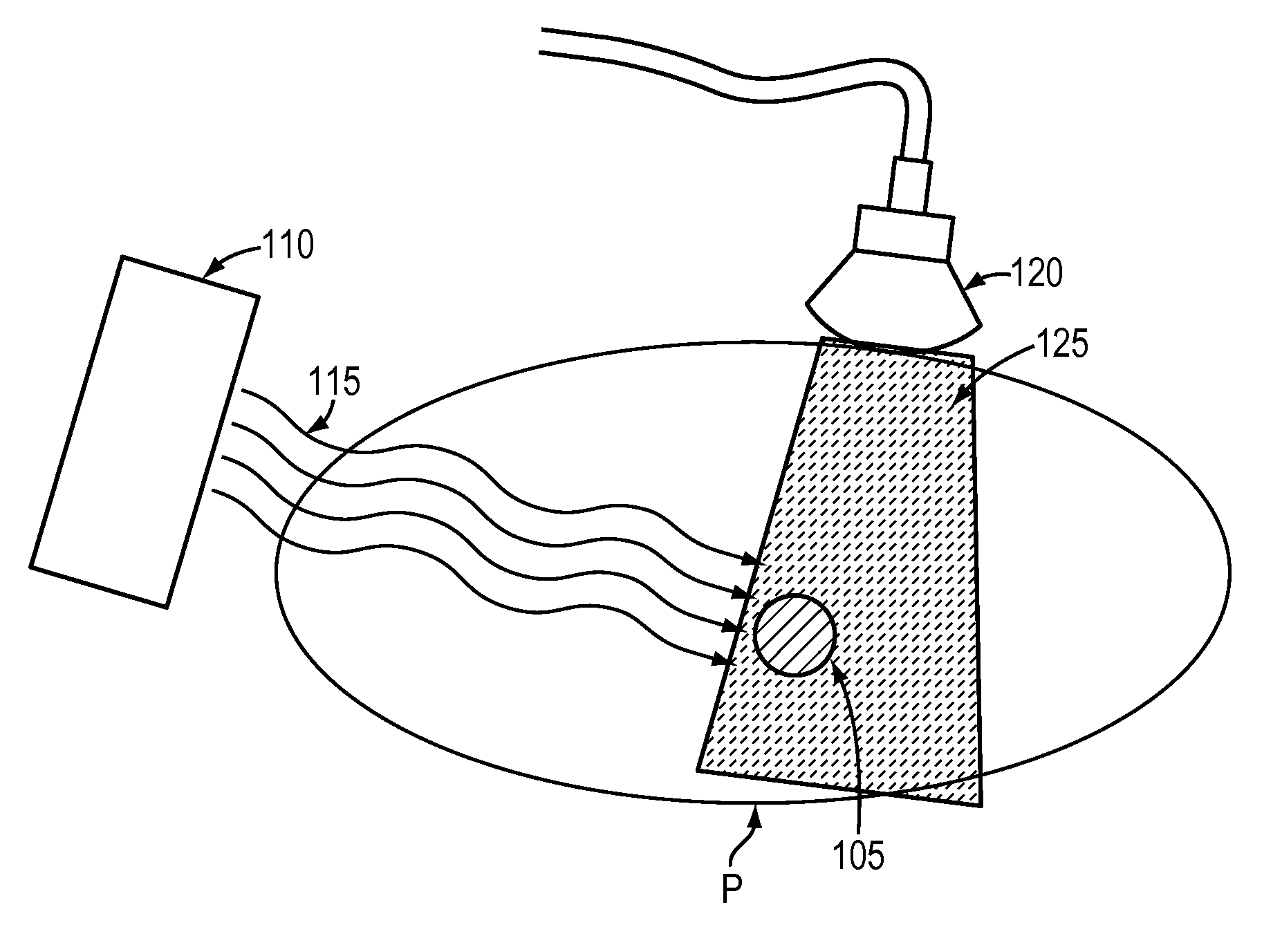
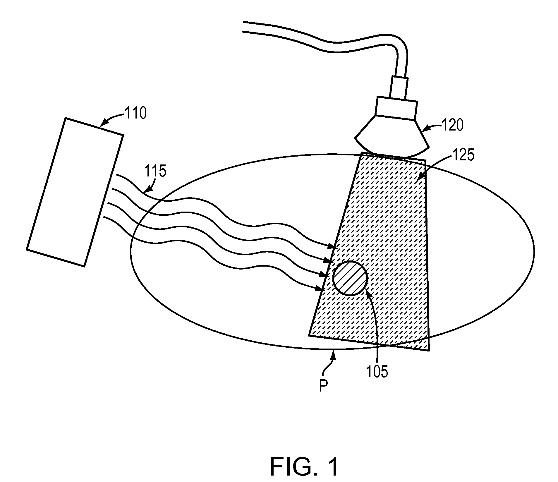
[0026]Referring to FIG. 1, one or more ultrasound scans are acquired during the delivery of radiotherapy treatment, which is typically given in many fractions over an extended period of time. For example, an ultrasound scan is taken of a target organ or lesion 105 within the illustrated region R of the patient. Radiotherapy treatment is administered to the lesion 105 using, for example, an e...


PUM


Abstract

Radiation treatment is delivered to a patient by positioning the patient such that a radiation beam is delivered to a lesion within the patient along a beam-delivery path while securing a diagnostic imaging device about the patient such that the diagnostic imaging device does not intersect the beam-delivery path. Radiation therapy is simultaneously delivered along the beam-delivery path while diagnostic images are obtained using the imaging device.

Description

CROSS-REFERENCE TO RELATED APPLICATIONS[0001]This application claims priority to provisional patent application Ser. No. 61 / 224,582, entitled “Adaptive Radiotherapy Treatment Using Ultrasound” filed on Jul. 10, 2009, the entire contents of which are incorporated herein by reference.TECHNICAL FIELD[0002]This invention relates to methods and systems for monitoring therapy treatments, and more specifically to observing the effects of radiation and anatomical changes during radiotherapy treatment fractions.BACKGROUND INFORMATION[0003]While a patient undergoes radiotherapy treatment, the location, arrangement or shape of anatomical structures or organs can vary relative to the treatment coordinate system being used to delivery the therapy. This is especially true with respect to a planning stage, when a treatment plan is initially devised, and when they are set up for each treatment fraction (commonly referred to as interfractional motion). Furthermore, the anatomy can change during the ...

Claims


Application Information

Patent Timeline
IPC IPC(8): A61B5/05A61B8/00
CPCA61B6/4225A61B6/4441A61B8/08A61B8/42A61B8/4472A61B8/483A61N5/1067A61N2005/1058A61B8/085A61B8/4227A61B8/4245A61N5/1049
Inventor LACHAINE, MARTINTREMBLAY, SEBASTIENFORTIER, PHILIPPEKOPTENKO, SERGEIFALCO, TONY
Owner ELEKTA AB
Learn More