Electrosurgical systems and methods for removing and modifying tissue

a tissue removal and electrosurgical technology, applied in the field of electrosurgical systems, can solve the problems of undesirable collateral tissue damage in the region, rapid tissue heating, and the like of conventional electrosurgical devices and procedures

Inactive Publication Date: 2011-02-03
ARTHROCARE
View PDF99 Cites 19 Cited by
  • Summary
  • Abstract
  • Description
  • Claims
  • Application Information

AI Technical Summary

Benefits of technology

[0015]In one aspect, tissue is cut or otherwise modified by molecular dissociation or disintegration processes. (In contrast, in conventional electrosurgery tissue is cut by rapidly heating the tissue until cellular fluids explode, producing a cutting effect along the pathway of localized heating.) The present invention volumetrically removes the tissue along the cutting pathway in a cool ablation process that minimizes thermal damage to surrounding tissue. In these embodiments, the high frequency voltage applied to the active electrode(s) is sufficient to vaporize the electrically conductive fluid (e.g., gel or saline) between the active electrode(s) and the tissue. Within the vaporized fluid, a plasma is formed and charged particles (e.g., electrons) cause the molecular breakdown or disintegration of the tissue, perhaps to a depth of several cell layers. This molecular dissociation is accompanied by the volumetric removal of the tissue, e.g., along the incision of the tissue. This process can be precisely controlled to effect the volumetric removal of tissue as thin as 10 microns to 150 microns with minimal heating of, or damage to, surrounding or underlying tissue structures. A more complete description of this phenomenon is described in commonly assigned U.S. Pat. No. 5,683,366, the complete disclosure of which is incorporated herein by reference.
[0017]In another exemplary embodiment, the present invention provides a method for harvesting the IMA from a patient. The electrosurgical probe is positioned adjacent the IMA and high frequency electrical energy is applied between one or more active electrode(s) and one or more return electrode(s). The probe is then moved so that the active electrode(s) volumetrically removes connective tissue adjacent to the IMA so that the IMA is free from connective tissue along a portion of its length. In an exemplary embodiment, the probe is positioned adjacent to the IMA, and advanced along the length of the IMA while high frequency electrical energy is applied between the active electrode(s) and a return electrode to remove or cut the connective tissue or other structures surrounding the IMA. The residual heat from the electrical energy also provides simultaneous hemostasis of severed blood vessels, which increases visualization and improves recovery time for the patient. In addition, the ability to simultaneously cut through tissue on either side of the IMA decreases the length of the procedure, which further improves patient recovery time. After a suitable length of the IMA has been dissected, it may be transected, and anastomosed to a diseased coronary artery using known methods. In some embodiments, an electrically conductive fluid (liquid, gas, or gel) is placed at the target site adjacent to the IMA so as to provide a current flow path between the return electrode and the active electrode.
[0019]The apparatus can further include a fluid delivery element for delivering electrically conductive fluid to the active electrode(s) and the target site. The fluid delivery element may be located on the probe, e.g., a fluid lumen or tube, or it may be part of a separate instrument. Alternatively, an electrically conductive gel or spray, such as a saline electrolyte or other conductive gel, may be applied the target site. In this embodiment, the apparatus may not have a fluid delivery element. In both embodiments, the electrically conductive fluid preferably provides a current flow path between the active electrode(s) and one or more return electrode(s). In an exemplary embodiment, the return electrode is located on the probe and spaced a sufficient distance from the active electrode(s) to substantially avoid or minimize current shorting therebetween and to shield the return electrode from tissue at the target site.
[0022]In another aspect of the invention, there is provided an electrosurgical probe having a blade-like active electrode affixed to an electrically insulating electrode support on the distal end of a shaft. In a specific configuration, the active electrode is in the form of a plasma blade comprising a substantially flat metal blade having at least one active edge and first and second blade sides. In one embodiment, the active electrode comprises a hook. The hook may include a curved portion. One or more portions of the hook may have a serrated edge. The return electrode is typically located on the shaft distal end proximal to the electrode support. In use, the active electrode and the return electrode are coupled to opposite poles of a high frequency power supply. The active edge may have a variety of shapes, and is adapted for generating high current densities thereon, and for precisely severing or ablating tissue or an organ in a highly controlled manner via molecular dissociation of tissue components. The first and second blade sides are adapted for engaging with tissue, such as tissue severed by the active edge, and for coagulating tissue engaged therewith.
[0024]According to one aspect of the invention, there is provided a method for modifying a tissue using an electrosurgical probe having an active electrode in the form of a single blade which includes at least one active edge and first and second blade sides. The method involves positioning the probe such that the active electrode makes contact with, or is in close proximity to, a target tissue; and applying a high frequency voltage between the active and return electrodes sufficient to precisely sever or remove target tissue via molecular dissociation of tissue components adjacent to the active edge. The probe may be manipulated during the application of the high frequency voltage such that the active electrode is moved with respect to the target tissue. According to one aspect of the invention, the configuration of the active electrode (e.g., a hook shaped electrode) is adapted for severing tissue as the probe distal end is drawn or pulled towards the operator of the probe. In this manner, the extent to which the tissue is severed can be precisely controlled. Thereafter, the severed tissue may be coagulated upon engagement of the tissue against the first and second blade sides of the active electrode.
[0025]In another aspect of the invention, there is provided a method of harvesting a tissue or organ using an electrosurgical probe having an active electrode in the form of a single blade, wherein the single blade electrode includes an active edge and first and second blade sides. In situations where the tissue to be harvested is concealed by an overlying tissue, the tissue to be harvested must first be accessed by incising or removing the overlying tissue. Removal of the overlying tissue may be performed in various ways, including: 1) mechanically, e.g. using a scalpel, rongeur, surgical saw or drill, etc. or a combination thereof; 2) via conventional electrosurgery, e.g., a Bovie; or 3) using an electrosurgical probe of the instant invention adapted for severing tissue in a cool ablation process. Once the tissue or organ to be harvested is accessible, the tissue or organ to be harvested may be dissected by juxtaposing the active edge of the active electrode against the surrounding connective tissue, and applying a high frequency voltage between the active and return electrodes sufficient to cause molecular dissociation of connective tissue components. In this way, the connective tissue adjacent to the active electrode is ablated at a temperature in the range of 40° C. to 70° C., with no, or minimal, thermal damage to the tissue to be harvested.

Problems solved by technology

Conventional electrosurgical devices and procedures, however, suffer from a number of disadvantages.
At the point of contact of the electric arcs with the tissue, rapid tissue heating occurs due to high current density between the electrode and the tissue.
Thus, the tissue is parted along the pathway of evaporated cellular fluid, inducing undesirable collateral tissue damage in regions surrounding the target tissue.
This creates the potential danger that the electric current will flow through undefined paths in the patient's body, thereby increasing the risk of unwanted electrical stimulation to portions of the patient's body.
This current, however, may inadvertently flow along body paths having less impedance than the defined electrical path, which will substantially increase the current flowing through these paths, possibly causing damage to or destroying surrounding tissue.
One drawback with this configuration, however, is that the return electrode may cause tissue desiccation or destruction at its contact point with the patient's tissue.
The close proximity of these electrodes generates the danger that the current will short across the electrodes, possibly impairing the electrical control system and / or damaging or destroying surrounding tissue.
In addition, conventional electrosurgical methods are generally ineffective for ablating certain types of tissue, and in certain types of environments within the body.
For example, loose or elastic connective tissue, such as the synovial tissue in joints, is extremely difficult (if not impossible) to remove with conventional electrosurgical instruments because the flexible tissue tends to move away from the instrument when it is brought against this tissue.
Since conventional techniques rely mainly on conducting current through the tissue, they are not effective when the instrument cannot be brought adjacent to, or in contact with, the elastic tissue for a sufficient period of time to energize the electrode and conduct current through the tissue.
Vascular complications produced by atherosclerosis, such as stenosis, aneurysm, rupture, or occlusion, increase the likelihood of angina, stroke, and heart attacks.
However, such methods suffer from numerous drawbacks.
For example, the longitudinal incision in the sternum often results in bone bleeding, which is difficult to stop.
The bone bleeding can produce a high degree of trauma, a larger risk of complications, an extended hospital stay, and a painful recovery period for the patient.
A number of disadvantages inherent in conventional electrosurgical devices have been set forth hereinabove.

Method used


Image

  • Electrosurgical systems and methods for removing and modifying tissue
  • Electrosurgical systems and methods for removing and modifying tissue
  • Electrosurgical systems and methods for removing and modifying tissue

Examples


Embodiment Construction

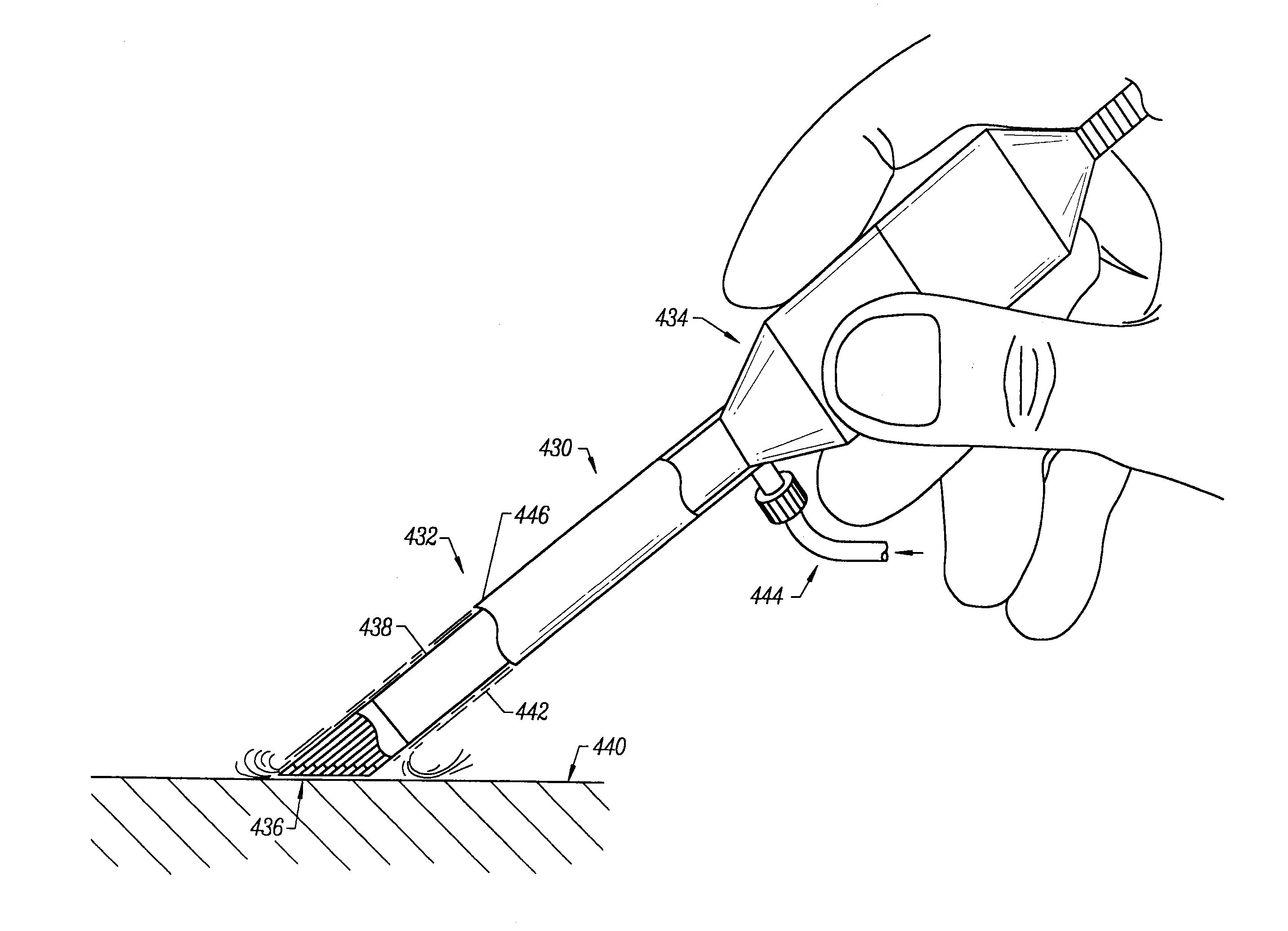
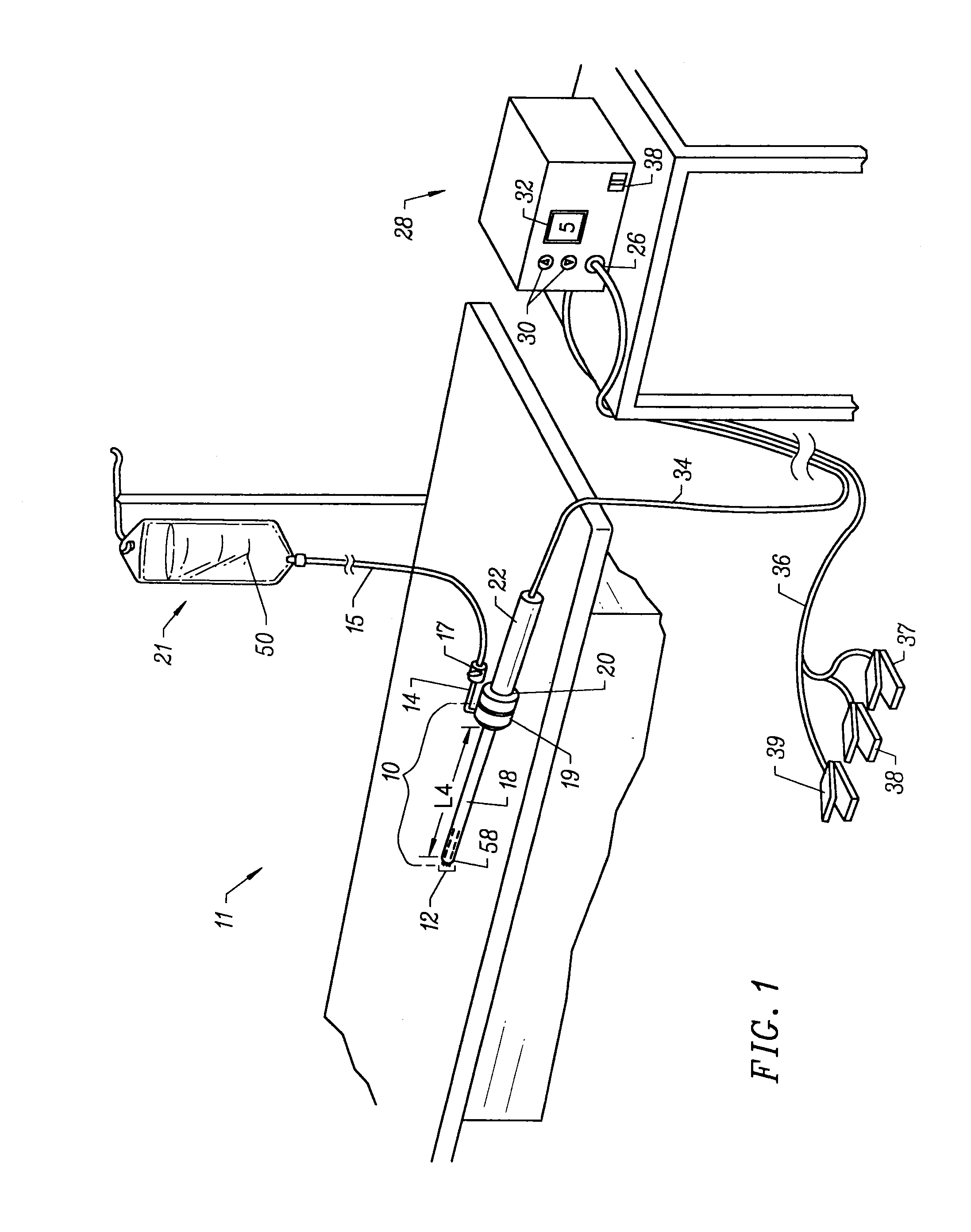
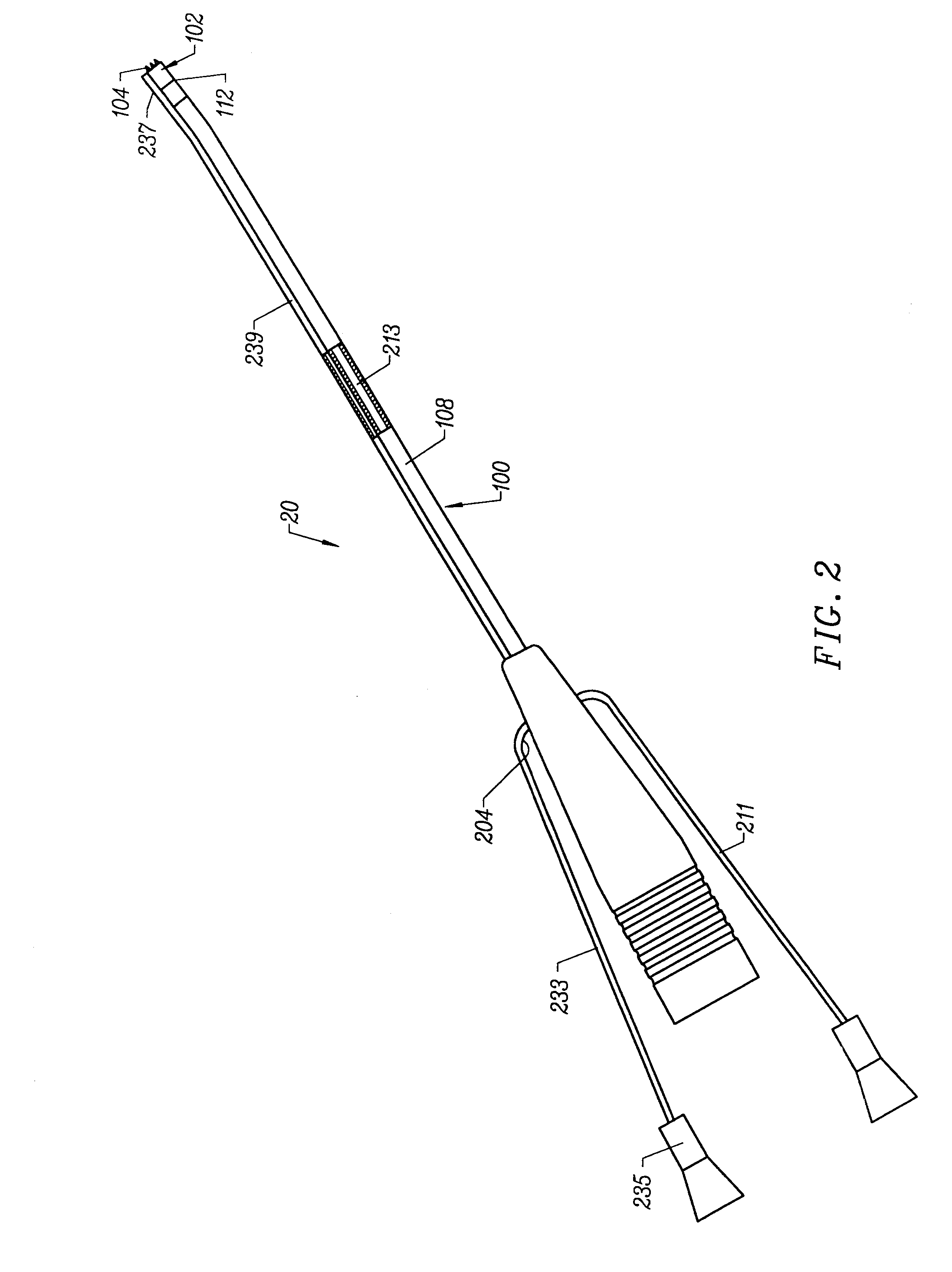
[0071]The present invention provides systems and methods for selectively applying electrical energy to a target location within or on a patient's body, particularly for cutting, ablating, and / or coagulating a tissue using a blade-like electrode. The instant invention also provides apparatus and methods for making incisions to access a tissue or organ within a patient's body, to dissect or harvest the tissue or organ from the patient, and to transect or otherwise modify the tissue or organ. In one aspect, the invention provides apparatus and methods for dissecting and harvesting blood vessels from a patient.

[0072]The present invention is useful in procedures where the target tissue or organ is, or can be, flooded or submerged with an electrically conductive fluid, such as isotonic saline. In addition, tissues which may be treated by the system and method of the present invention further include, but are not limited to, tissues of the heart, chest, knee, shoulder, ankle, hip, elbow, h...


PUM

PropertyMeasurementUnit
distanceaaaaaaaaaa
temperatureaaaaaaaaaa
energy levelaaaaaaaaaa

Abstract

The present invention provides systems, apparatus and methods for selectively applying electrical energy to body tissue in order to ablate, contract, coagulate, or otherwise modify a tissue or organ of a patients. An electrosurgical apparatus includes an electrode support bearing an active electrode in the form of a plasma blade or hook having an active edge and first and second blade sides. The active edge is adapted for severing a target tissue via localized molecular dissociation of tissue components. The first and second blade sides are adapted for engaging against, and coagulating, the severed tissue. A method of the present invention comprises positioning an electrosurgical probe adjacent to the target tissue so that a blade- or hook-like active electrode is brought into at least close proximity to the target tissue in the presence of an electrically conductive fluid. A high frequency voltage is applied between the active electrode and a return electrode to effect cool ablation or other modification of the target tissue. During application of the high frequency voltage, the electrosurgical apparatus may be translated, reciprocated, or otherwise manipulated such that the active edge is moved with respect to the tissue. The present invention volumetrically ablates or otherwise modifies the target tissue with minimal or no damage to surrounding, non-target tissue.

Description

CROSS-REFERENCES TO RELATED APPLICATIONS[0001]This application is a continuation of U.S. patent application Ser. No. 10 / 339,470, filed Jan. 9, 2003, which is a divisional of U.S. patent application Ser. No. 09 / 780,745, filed Feb. 9, 2001, now U.S. Pat. No. 6,770,071, which claims the benefit of U.S. Provisional Patent Application No. 60 / 182,751 filed Feb. 16, 2000. application Ser. No. 09 / 780,745 is also a continuation-in-part of U.S. patent application Ser. No. 09 / 162,117, filed Sep. 28, 1998, now U.S. Pat. No. 6,117,109, which is a continuation in part of U.S. patent application Ser. No. 08 / 977,845, filed Nov. 25, 1997, now U.S. Pat. No. 6,210,402, which is a continuation-in-part of U.S. patent application Ser. No. 08 / 562,332, filed Nov. 22, 1995, now U.S. Pat. No. 6,024,733, all of which are herein incorporated by reference for all purposes.[0002]The present invention is also related to commonly assigned U.S. Provisional Patent Application No. 60 / 062,996, filed Oct. 23, 1997, and...

Claims


Application Information

Patent Timeline
Patent Type & Authority Applications(United States)
IPC IPC(8): A61B18/14A61B17/00A61B18/00A61F2/02A61M3/02
CPCA61B18/042A61M3/0283A61B18/1402A61B18/148A61B18/1482A61B18/1485A61B18/149A61B18/1492A61B2017/00026A61B2017/00084A61B2017/00101A61B2017/00247A61B2018/00029A61B2018/00083A61B2018/00119A61B2018/00148A61B2018/0016A61B2018/00178A61B2018/00327A61B2018/00392A61B2018/00404A61B2018/0047A61B2018/00476A61B2018/00505A61B2018/00577A61B2018/00583A61B2018/00601A61B2018/00625A61B2018/00678A61B2018/00702A61B2018/00726A61B2018/00791A61B2018/00797A61B2018/00815A61B2018/00821A61B2018/00827A61B2018/00875A61B2018/1213A61B2018/124A61B2018/1253A61B2018/126A61B2018/1273A61B2018/1407A61B2018/1412A61B2018/1422A61B2018/1467A61B2018/1472A61B2018/162A61B2018/165A61B2218/002A61B2218/003A61B2218/007A61F2/2493A61B18/1206
Inventor WOLOSZKO, JEANTSUJI, CRAIGTAPLIYAL, HIRA V.EGGERS, PHILIP E.
Owner ARTHROCARE
Why Eureka
Try Eureka