Reduced area imaging device incorporated within wireless endoscopic devices

a wireless endoscope and imaging device technology, applied in the field of solid-state image sensors incorporated within wireless endoscopes, can solve the problem of low power supply (5 volts), and achieve the effect of facilitating precise and accurate control of the distal end and not enlarge the capsule profil

Inactive Publication Date: 2011-02-10
MICRO IMAGING SOLUTIONS
View PDF39 Cites 166 Cited by
  • Summary
  • Abstract
  • Description
  • Claims
  • Application Information

AI Technical Summary

Benefits of technology

[0027]In another aspect of the invention, the imaging device is housed within an endoscopic instrument in which the endoscope is steerable by incorporating a steering connector assembly located near the imaging device. The connector assembly is constructed so that it does not enlarge the profile of the capsule that houses the imaging device, yet the connector assembly facilitates precise and accurate control of the distal end of the endoscope with a four-way deflection capability. More specifically, the image sensor has a frontal profile defined by a length and width dimension, and the connector assembly does not extend beyond this frontal profile or a slightly larger frontal profile defined by the length and width dimension of the capsule.

Problems solved by technology

Additionally, this hybrid is able to run on a low power source (5 volts) which is normally not possible on standard CCD imagers which require 10 to 30 volt power supplies.

Method used


Image

  • Reduced area imaging device incorporated  within wireless endoscopic devices
  • Reduced area imaging device incorporated  within wireless endoscopic devices
  • Reduced area imaging device incorporated  within wireless endoscopic devices

Examples


first embodiment

[0062]In all of the arrangements of the imaging device discussed above with respect to the endoscope, each of the elements or components of the imaging device electrically communicate with one another through a wired connection.

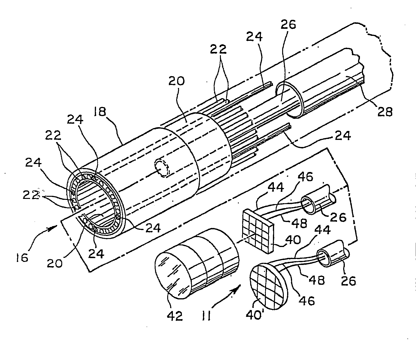
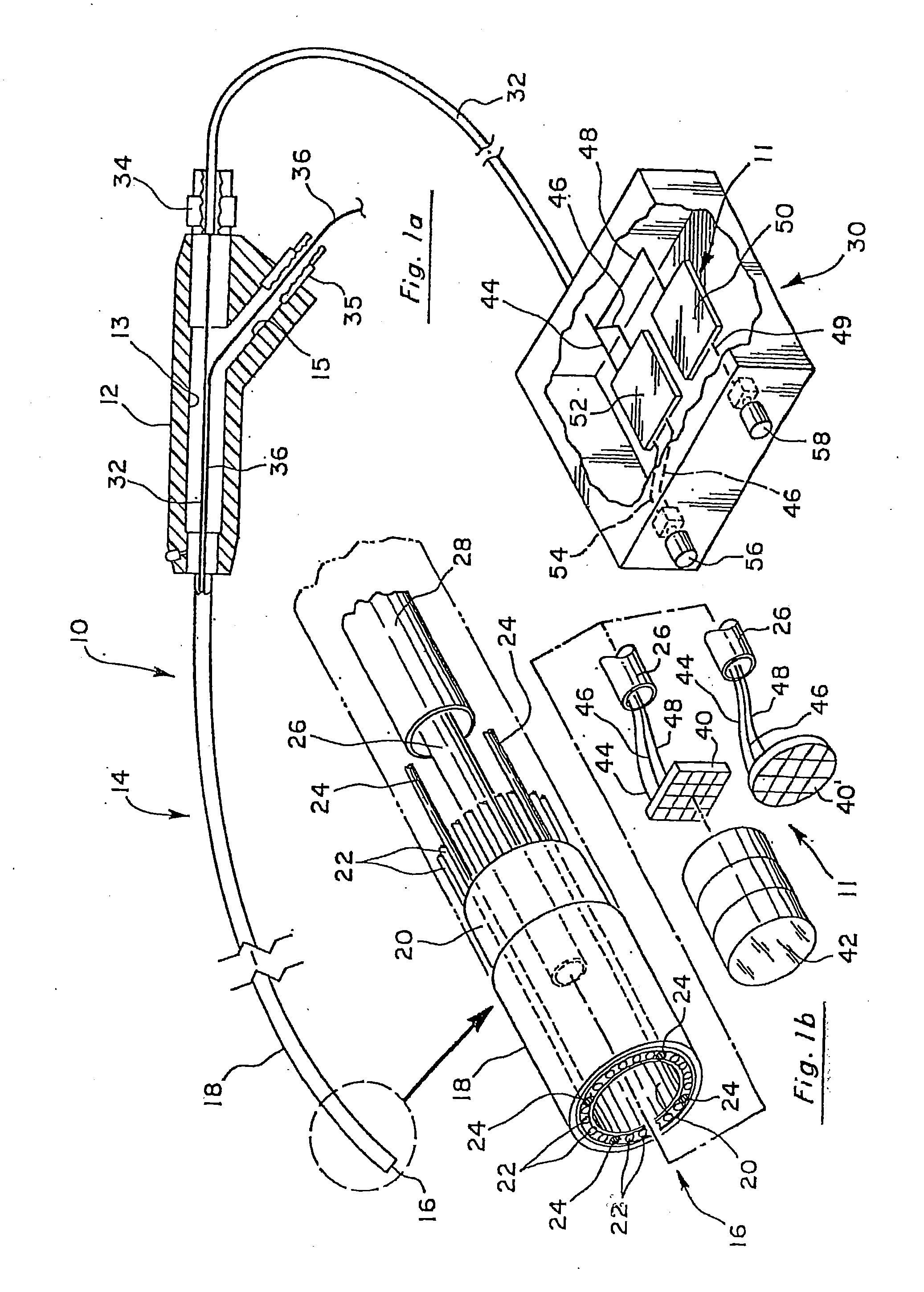
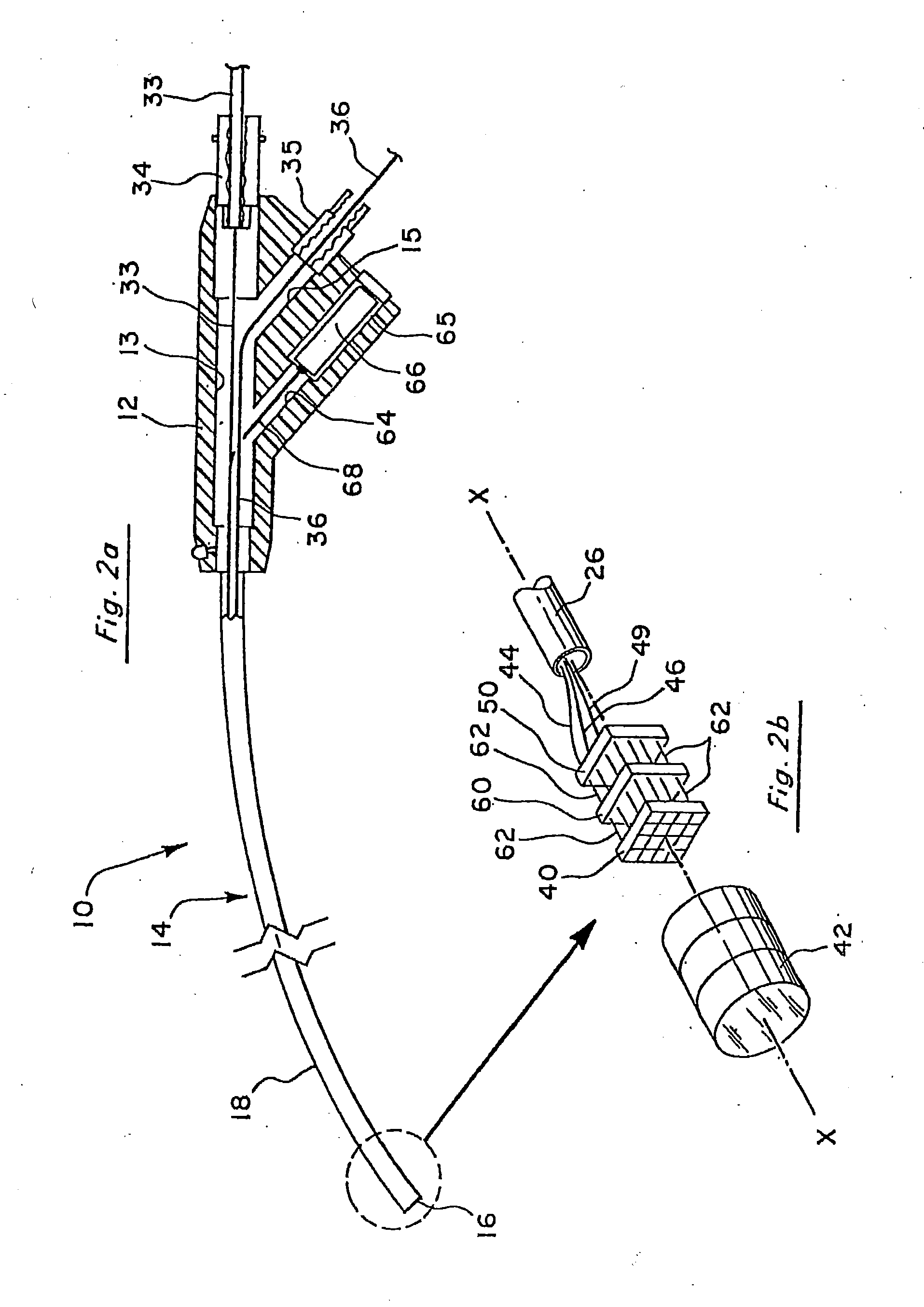
[0063]FIG. 4 is a schematic diagram illustrating one way in which the imaging device 11 may be constructed. As illustrated, the image sensor 40 may include the timing and control circuits on the same planar structure. Power is supplied to image sensor 40 by power supply board 52. The connection between image sensor 40 and board 52 may simply be a cable having two conductors therein, one for ground and another for transmitting the desired voltage. These are illustrated as conductors 44 and 46. The output from image sensor 40 in the form of the pre-video signal is input to video processor board 50 by means of the conductor 48. In the configuration of FIG. 4, conductor 48 may simply be a 50 ohm conductor. Power and ground also are supplied to video processing bo...

second embodiment

[0086]Transceiver radio module 178 receives the post-video signals via antennae 180, decodes the signals, and then electrically transmits them to the monitor 196 for viewing by the user. The endoscope in this second embodiment is powered by a battery 176 which is housed adjacent the antennae 174. Electrical leads (not shown) extend from the battery 176 to power the image sensor and the transceiver radio element 170. As discussed further below, antennae 174 and battery 176 may be secured within their own casing or housing 172 which then connects to the handle 12 of the endoscope. Transceiver radio module 178 may simply be powered by the same electrical power source (not shown) which powers the display monitor 196, such as conventional 110 volt, 3 phase power. In order to recharge the battery 176 of the endoscope, the transceiver radio module may be a combination unit which also has a battery charge circuit 182 for recharging battery 176. Charge circuit 182 would also be powered by a ...


PUM


Abstract

A reduced area imaging device is provided for use in medical or dental instruments such as an endoscope. The imaging device is provided in various configurations, and connections between the imaging device elements and a video display may be achieved by wired or wireless connections. A connector assembly located near the imaging device interconnects the imaging device to an image/power cable extending through the endoscope. The connector provides strain relief and stabilization for electrically interconnecting the imager to the cable. The connector also serves as the structure for anchoring the distal ends of steering wires extending through the body of the endoscopic device. The connector includes a strain relief member mounted over a body of the connector. The connector allows a steering wire capability without enlarging the profile of the distal tip of the endoscopic device.

Description

[0001]This application is a continuation-in-part of U.S. Ser. No. 11 / 245,960, filed on Oct. 6, 2005, and entitled “Reduced Area Imaging Device Incorporated Within Wireless Endoscopic Devices”, which is a continuation of U.S. patent application Ser. No. 09 / 929,531, filed on Aug. 13, 2001, and entitled “Reduced Area Imaging Device Incorporated Within Wireless Endoscopic Devices”, which is a continuation-in-part of U.S. Ser. No. 09 / 496,312 filed on Feb. 1, 2000, and entitled “Reduced Area Imaging Devices”, which is a continuation of U.S. Ser. No. 09 / 175,685 filed Oct. 20, 1998 entitled “Reduced Area Imaging Devices”, now U.S. Pat. No. 6,043,839, which is a continuation-in-part of U.S. Ser. No. 08 / 944,322, filed Oct. 6, 1997, and entitled “Reduced Area Imaging Devices Incorporated Within Surgical Instruments”, now U.S. Pat. No. 5,929,901. This application is also a continuation-in-part of U.S. Ser. No. 09 / 368,246 filed on Aug. 3, 1999, and entitled “Reduced Area Imaging Devices Incorpor...

Claims


Application Information

Patent Timeline
Patent Type & Authority Applications(United States)
IPC IPC(8): A61B1/04
CPCA61B1/00016A61B1/051A61B1/042A61B1/05A61B1/055A61B1/0607H04N5/23203H04N5/374H04N2005/2255A61B1/00064A61B1/00009A61B1/00045A61B1/045H04N5/3745A61B1/008A61B1/002A61B1/00101A61B1/00124A61B1/00128A61B1/000094A61B1/000095H04N23/555H04N23/55H04N23/66H04N25/76H04N23/51H04N23/54H04N25/77H04N25/745A61B1/00032
Inventor ADAIR, JEFFREY L.WIGGINS, KEVIN EWING
Owner MICRO IMAGING SOLUTIONS
Why Eureka
Try Eureka