Serum-based, diagnostic, biological assay to predict pregnancy disorders

Active Publication Date: 2011-03-10
WOMEN & INFANTS HOSPITAL OF RHODE ISLAND
View PDF1 Cites 7 Cited by
  • Summary
  • Abstract
  • Description
  • Claims
  • Application Information

AI Technical Summary

Benefits of technology

[0039]The first trimester human trophoblast cell line HTR8 (representing normal invasive extravillous trophoblasts), the third trimester human trophoblast cell line TCl-1, and the first trimester human villous trophoblast cell line 3A were used in the following studies. Human HTR-8 cells and TCl-1 cells were a gift from Dr. Charles Graham (Queens University, Canada) and human 3A cells r were a gift from Dr. Gil Mor, Yale University, USA.
[0040]Cytotrophoblasts from human placental tissue were isolated according to published methods. Briefly, placental tissues were digested with decreasing concentrations of trypsin-DNase 1 (trypsin, 1 mg/ml; and DNase, 1.5 mg/ml) at least four times at 37° C. for 20 min each. The cells from the first digestion were excluded. The cell mass collected in the following steps was treated with a lysis buffer (0.15 M NH4Cl, 1 mM KHCO3, and 0.1 mM EDTA (pH 7.3) for 5 min at room temper

Problems solved by technology

Surprisingly, serum from pregnant female humans that go on to acquire complications

Method used


Image

  • Serum-based, diagnostic, biological assay to predict pregnancy disorders
  • Serum-based, diagnostic, biological assay to predict pregnancy disorders
  • Serum-based, diagnostic, biological assay to predict pregnancy disorders

Examples


example 1

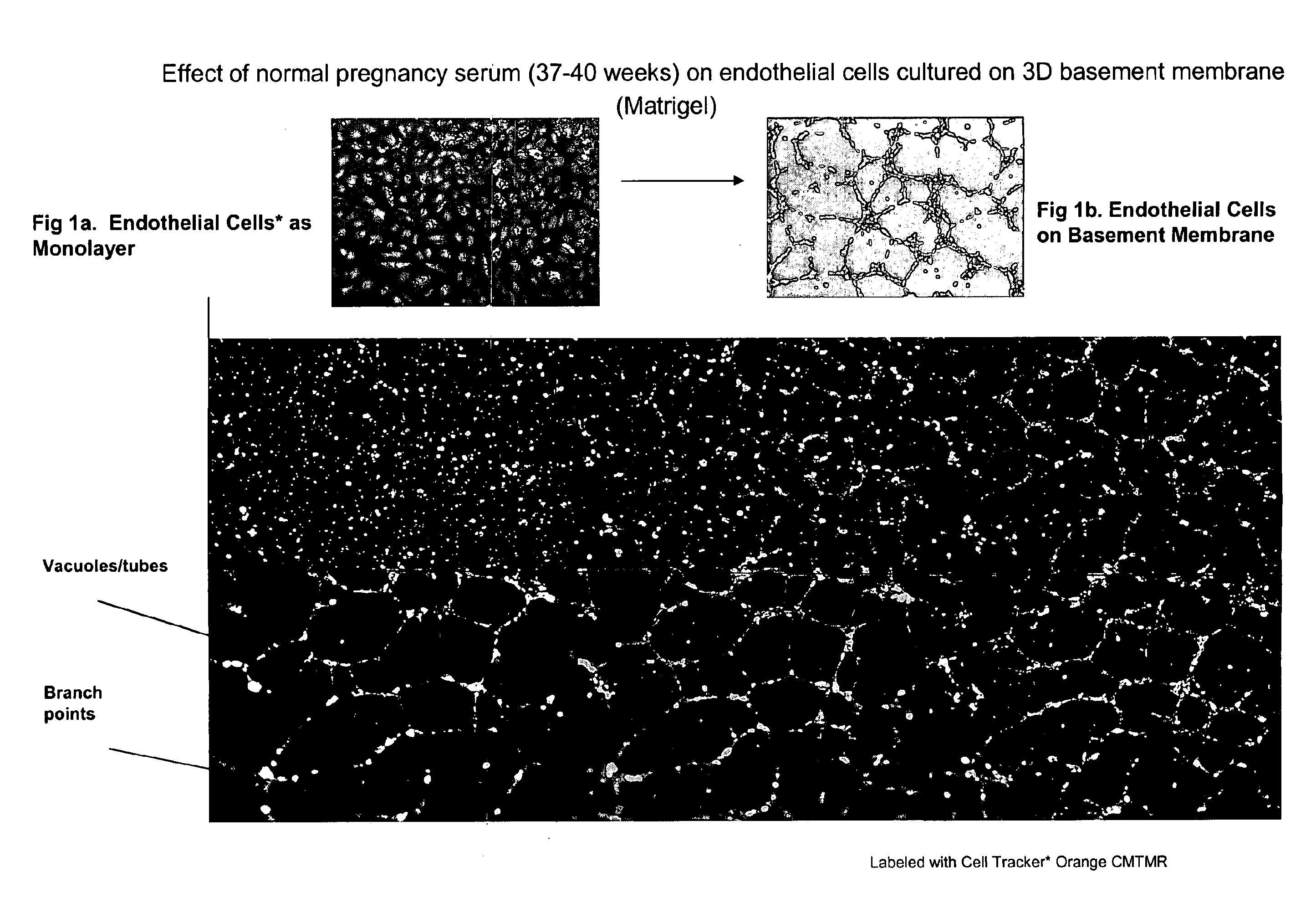
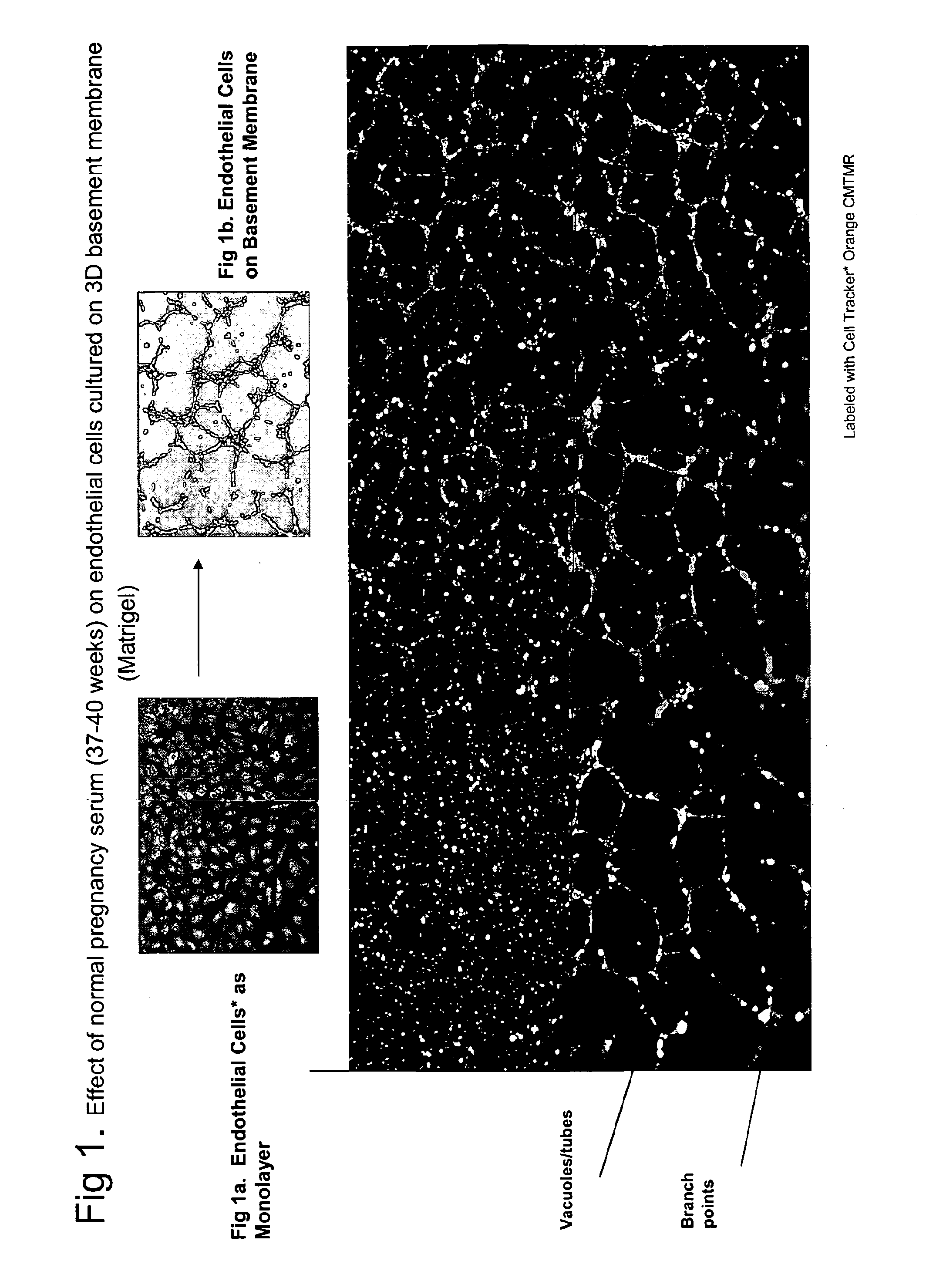
Effect of Normal Pregnancy Serum (NPS, 37-40 weeks) on Endothelial Cells

[0038]Human umbilical cord endothelial cells (HUVEC) and human uterine endothelial cells (HUtEC) were obtained from Cambrex (East Rutherford, N.J., USA). Growth factor-reduced Matrigel representing basement membrane (BD Biosciences, San Diego, Calif., USA) was thawed overnight at 4° C. and mixed to homogeneity. The 48-well culture plates (Costar) were coated with 0.1 ml of Matrigel and allowed to gelatinize at 37° C. for 30 min. 2.5×10⁴ endothelial cells (HUtEC or HUVEC) labeled with cell tracker orange CMTMR (Molecular Probes, Eugene, Oreg.) were plated (1:1) in the presence of RPMI media containing 10% human pregnancy serum on the matrigel-coated plates. The architectural and morphological changes that took place were monitored and recorded 12-14 hrs after incubation under standard culture conditions using florescence microscopy (4× magnifications, Nikon Eclipse TS 100 coupled with CCD camera; photographs take...

example 2

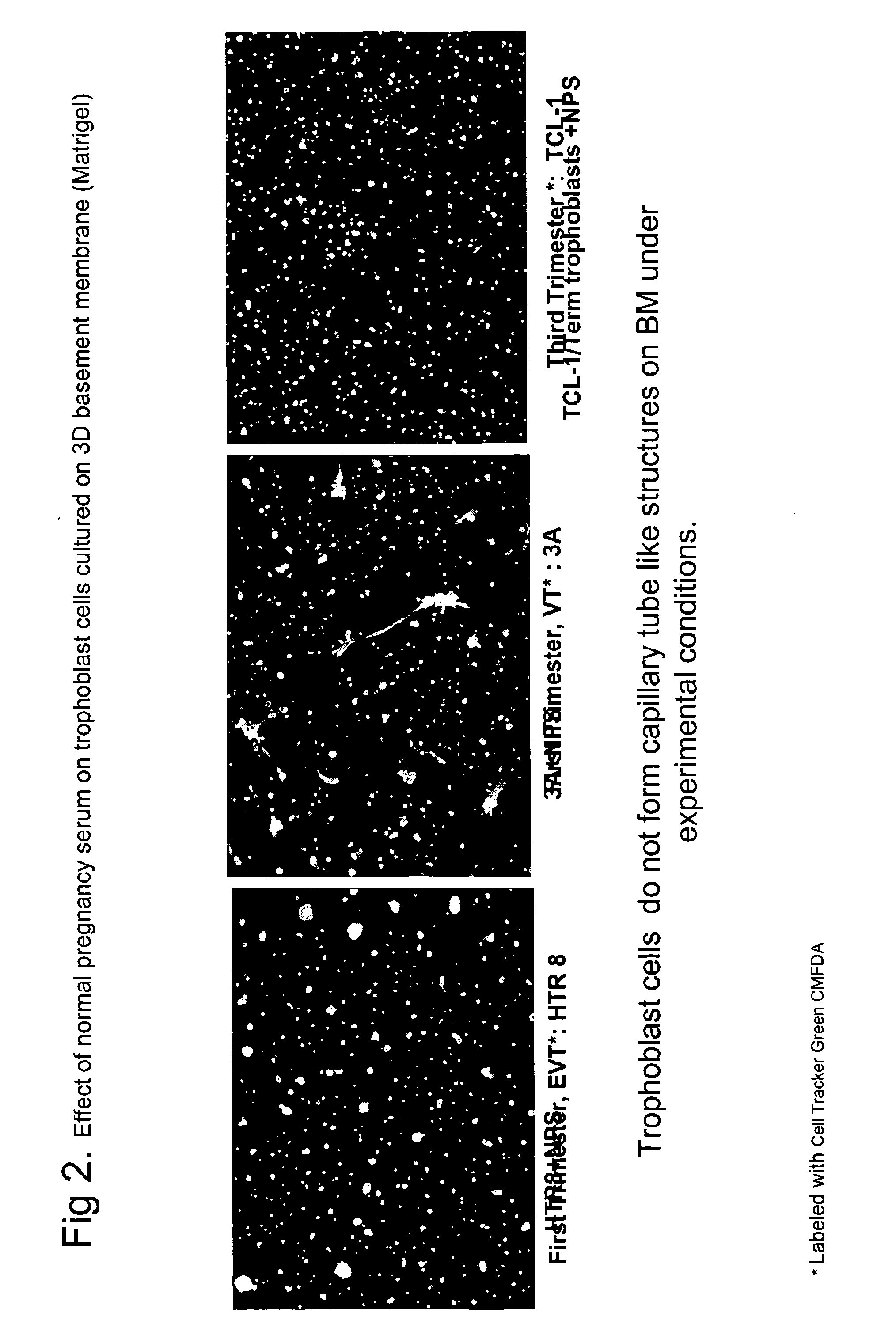
Effect of Normal Pregnancy Serum on Trophoblast Cells

[0039]The first trimester human trophoblast cell line HTR8 (representing normal invasive extravillous trophoblasts), the third trimester human trophoblast cell line TCl-1, and the first trimester human villous trophoblast cell line 3A were used in the following studies. Human HTR-8 cells and TCl-1 cells were a gift from Dr. Charles Graham (Queens University, Canada) and human 3A cells r were a gift from Dr. Gil Mor, Yale University, USA.

[0040]Cytotrophoblasts from human placental tissue were isolated according to published methods. Briefly, placental tissues were digested with decreasing concentrations of trypsin-DNase 1 (trypsin, 1 mg / ml; and DNase, 1.5 mg / ml) at least four times at 37° C. for 20 min each. The cells from the first digestion were excluded. The cell mass collected in the following steps was treated with a lysis buffer (0.15 M NH4Cl, 1 mM KHCO3, and 0.1 mM EDTA (pH 7.3) for 5 min at room temperature with constant sh...

example 3

NPS Supports Trimester-Specific Differential Interaction with Endothelial Cells

[0043]2.5×10⁴ endothelial cells labeled red and trophoblasts from human subjects, each from different trimesters labeled green, were co-cultured on matrigel-coated plates and stimulated with Normal Pregnancy Serum (NPS) (sample L31, see Table 1) as described in Examples 1 and 2.

[0044]Gestational age specific architectural patterns were observed. As seen in FIG. 3, left panel (which is labeled “EC+HTR8+L31”), surprisingly only the first trimester HTR8 trophoblasts spontaneously interact and align with endothelial cell architecture. S3A cells, representing villous trophoblasts from first trimester, attract endothelial cells and form massive branch points that allow reduced vacuolization only around its vicinity. (See FIG. 3, middle panel, labeled “EC+3A+L31.”) In contrast, term trophoblasts and third trimester TCL-1 trophoblasts tend to naturally impede the capillary formation by endothelial cells. Smaller ...


PUM

PropertyMeasurementUnit
Temperatureaaaaaaaaaa
Timeaaaaaaaaaa
Volumeaaaaaaaaaa

Abstract

The invention provides serum-based, diagnostic, biological assays for predicting disorders of pregnancy resulting from poor trophoblast and/or placental ischemia, including preeclampsia. Serum samples from such subjects exhibit an ability to disrupt the architecture involving fetal trophoblasts and maternal endothelial cells in a three-dimensional, dual cell co-culture system provided herein, in contrast to normal pregnancy serum samples. Based on these distinctions, the assays are employed to predict pregnancy outcomes as early as first trimester.

Description

BACKGROUND[0001]Disorders of pregnancy pose a major public health problem because they involve approximately 10% of human pregnancies. Preeclampsia (PE) is one such disorder which presents itself with maternal symptoms of global endothelial disease, including glomeruloendotheliosis, liver and cerebral vascularitis. Diagnostic symptoms are high blood pressure (>140), proteinuria (>0.3 gm / ml) and general edema. (ACOG Committee on Obstetric Practice, 2002) PE is a pregnancy-specific systemic disorder characterized by a cascade of events and symptoms, including impaired trophoblast invasion, decreased placental perfusion, placental ischemia, oxidative stress and imbalance in angiogenic and prothrombotic factors which can lead to apoptosis of trophoblasts(20-23). Studies have also shown that in preeclampsia, there are elevated levels of circulating or placental TNFα, IL-6, IL-8. IFNγ, leptin, perturbed renin angiotensin system, complement split products, antibodies to phospholipids...

Claims


Application Information

Patent Timeline
IPC IPC(8): C12Q1/02A61K38/12
CPCG01N2800/368G01N33/5091G01N33/5032
Inventor SHARMA, SURENDRAKALKUNTE, SATYAN
Owner WOMEN & INFANTS HOSPITAL OF RHODE ISLAND
Why Eureka
Try Eureka