Device and methods for detecting analytes in saliva

a technology of analytes and saliva, which is applied in the field of devices and methods for detecting analytes in saliva, can solve the problems of many unsatisfactory tests, interfere with many detection methods, and contain components of saliva, and achieves the effects of less chance of errors, high specificity and sensitivity, and ease of manipulation

Active Publication Date: 2011-03-17
KONINKLIJKE PHILIPS ELECTRONICS NV +1
View PDF15 Cites 9 Cited by
  • Summary
  • Abstract
  • Description
  • Claims
  • Application Information

AI Technical Summary

Benefits of technology

[0009]The present invention provides devices and methods whereby pre-treatment of the sample and detection are performed subsequently in the same setting, offering ease of manipulation as well as high specificity and sensitivity. These methods and devices are of particular use in the testing of analytes in saliva. Moreover, the methods and devices allow use outside a laboratory environment.
[0010]The integration of a pre-treatment region and a detection region in one device offers several advantages. There is less need for user intervention with the sample. As a consequence, there is less chance of errors introduced by sample manipulation. Also, less sample material is required. Indeed, there is no loss of sample as is the case when transferring sample material from one device to another. Moreover, this ensures minimal contact of the user with the sample material, minimizing risk of contamination of the sample.
[0011]The methods and devices of the present invention envisage a pretreatment of the sample prior to detection, whereby components interfering with detection are removed. This allows the use of magnetizable particles in the device without affecting sensitivity or specificity. Indeed, the interfering fraction which typically induces aggregation of magnetizable particles is removed prior to contacting with the magnetizable particles. This results in a reduction of the background signal or a specific binding reduced. Moreover the methods and devices of the present invention allow for detection based on magnetic actuation. This is of particular importance as magnetic actuation can be used to further speed up the diagnostic tests and simplify fluidics in e.g. a microfluidic device. Accordingly the methods and devices described in the present invention provide rapid, sensitive, and robust tools for determining analytes in saliva.

Problems solved by technology

Many of these tests have however been demonstrated to be unsatisfactory, primarily with respect to the sensitivity of the assays.
More importantly however, saliva contains components which interfere with many detection methods.

Method used


Image

  • Device and methods for detecting analytes in saliva
  • Device and methods for detecting analytes in saliva
  • Device and methods for detecting analytes in saliva

Examples


example 1

Removal of Interfering Mucin by Repeating the Pre-Treatment Step

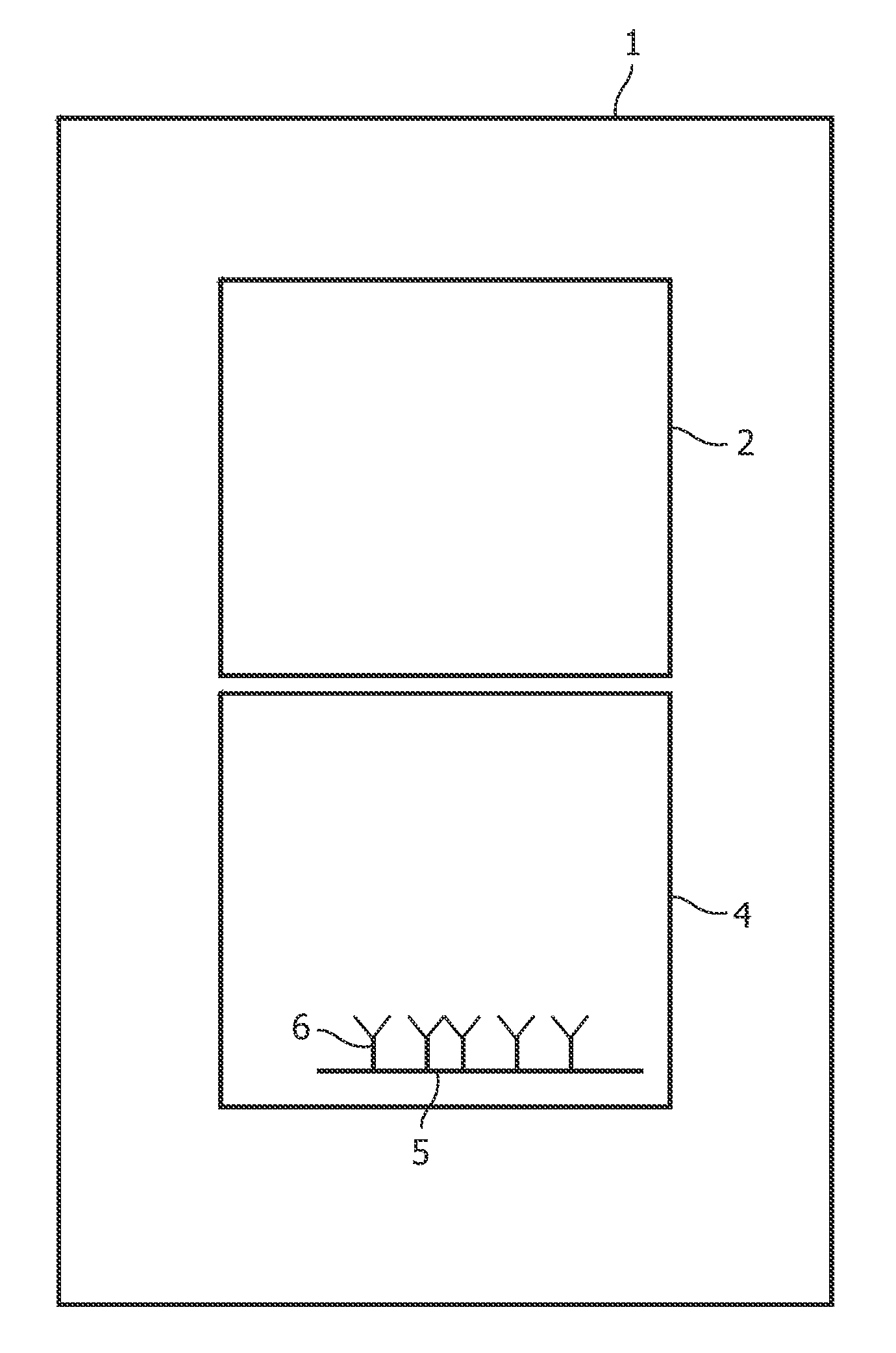
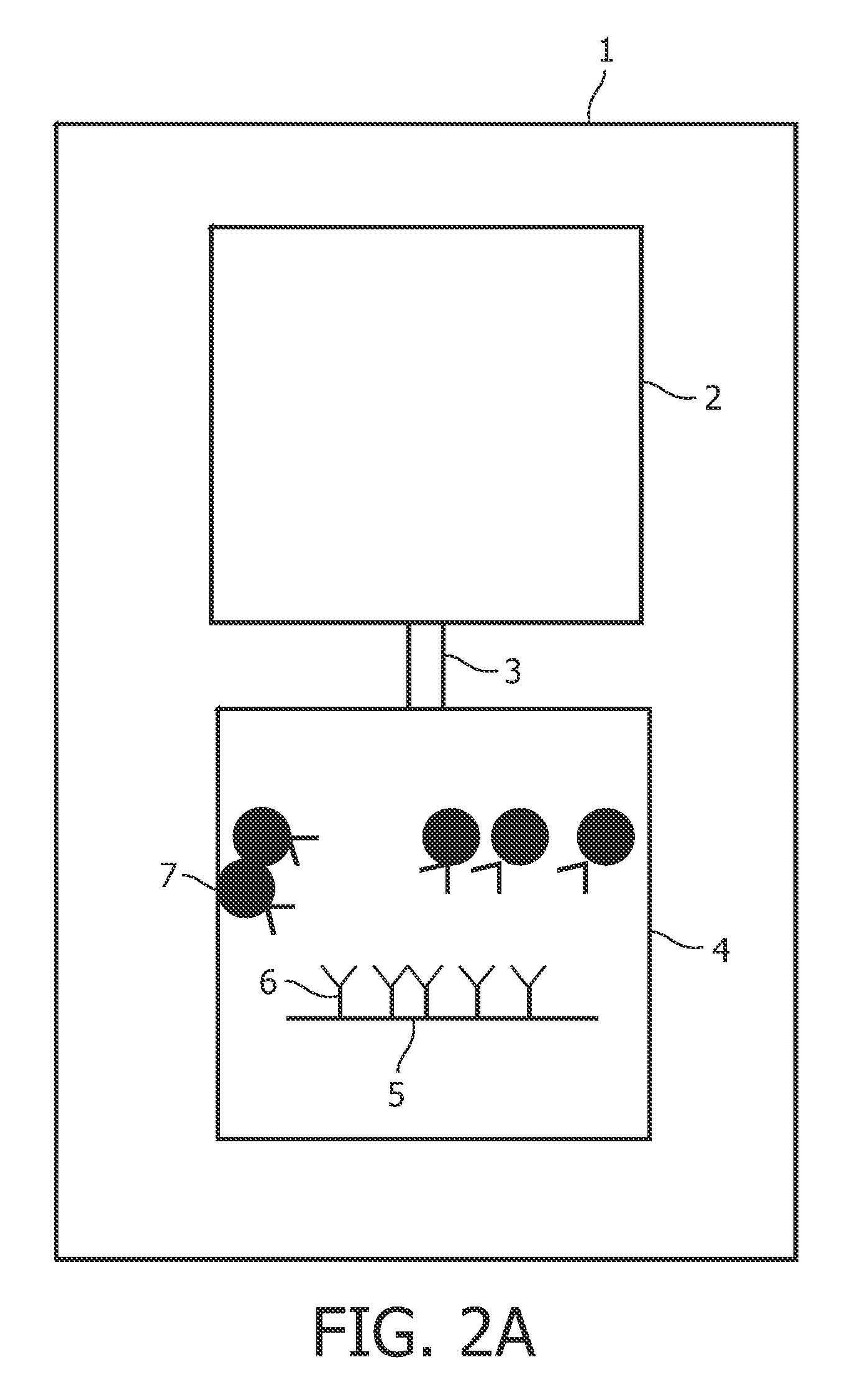
[0138]Super paramagnetic beads are coated with a molecule capable of specifically binding an interfering compound of saliva, in this case an anti-mucin antibody. In a first step a saliva sample is contacted with a first pre-treatment region comprising the paramagnetic bead coated with the anti-mucin antibody. By application of a magnetic field the beads are removed from the fluid. The sample is then moved to a second pre-treatment region. The effect of the contacting of the sample with the paramagnetic beads (e.g. the clustering or aggregation status of the superparamagnetic beads caused by the mucins can be monitored during the contacting with the pre-treatment region. This is done by studying the sample, or an aliquot thereof, under a microscope, or for example by using a Nanosizer (Malvern instruments). When aggregation or clustering is observed, the pre-treatment step is repeated (either by applying the sample to a ...

example 2

Rapid Detection of Amphetamines using a Lateral Flow Assay

[0143]A saliva sample is added to a pre-treatment region comprising a sample pad (a porous structure) with anti-mucin antibodies immobilized onto the surface of the sample pad material. The mucins present in the saliva will bind to the immobilized anti-mucin antibodies. The incubation time can be influenced by the size of the sample pad and / or the pore size of the sample pad. The number of immobilized antibodies and the incubation time with the sample pad is adjusted such that the interfering matrix components are sufficiently removed and therefore the a-specific binding or aggregation / clustering of the mobile solid phase is no longer an issue.

[0144]After moving through the pre-treatment zone, the sample transits to a zone where it is contacted with colored particles which are labeled with amphetamine or an amphetamine analogue. The sample then further moves to the detection region, comprising a detection region with anti-amp...

example 3

Detection of Opiate in Assays Making use of HAP Filtration

[0145]Superparamagnetic particles (Ademtech 500 nm COOH coated particles) were coated with monoclonal anti-opiate antibodies. The detection region is prepared by providing a surface coated with BSA-opiate. The top and bottom part of the biosensor was assembled by using tape, and the sensors were kept under lab conditions at room temperature. Four different pre-treatment conditions were used: no pre-treatment, Sponge Bob filter material (Filtrona, density 0.29 g / cm3), and contacting with 50 mg / ml HAP followed by a filter. Next, the particles were redispersed at 0.2 wt % in the pretreated buffer or saliva.

[0146]Samples comprising either assay buffer or 70% saliva (saliva donor 1-3) in assay buffer were subjected to the different pre-treatment regions and subsequently mixed with the magnetizable particles.

[0147]The detection of opiates in samples obtained from the different pre-treatment conditions was performed using a competit...


PUM

PropertyMeasurementUnit
diameteraaaaaaaaaa
pore sizeaaaaaaaaaa
pore sizeaaaaaaaaaa

Abstract

The invention provides a device for detecting drugs of abuse or other compounds in saliva. The invention thus provides a device for detecting the presence of one or more analytes in a saliva sample, comprising: (a) One or more pre-treatment regions for specifically or non-specifically removing at least a part of the fraction of the saliva sample interfering with detection of the one or more analytes; and (b) A detection region comprising a biosensor surface, the surface comprising: molecules capable of specifically binding the one or more analytes; or the one or more analytes and/or analyte analogues.

Description

FIELD OF THE INVENTION[0001]The present invention relates to devices and methods for detecting and / or quantifying molecules in a saliva sample. The methods and devices combine pre-treatment and detection.BACKGROUND OF THE INVENTION[0002]In a diagnostic test one or more specific target molecules or analytes have to be measured. Often this is done by allowing the target molecule to interaction in a (bio-chemical) reaction, which directly or indirectly results in a detectable signal.SUMMARY OF THE INVENTION[0003]A number of devices and methods are currently commercially available to perform road-side testing of drugs. Most of these are based on immunological detection of the target, which is performed on the sample directly in WO 02 / 082040, a “one-device” system is described for determining different analytes, in particular drugs, in body fluids. The bodily fluid is collected via a collection end of the device with an absorbent pad, a series of pressure heads inside the cap of the coll...

Claims


Application Information

Patent Timeline
Patent Type & Authority Applications(United States)
IPC IPC(8): G01N33/566G01N30/00
CPCG01N27/745G01N33/54326B01L3/502753G01N33/946G01N33/9486G01N33/54393
Inventor NIEUWENHUIS, JEROEN HANSJEHANLI, AHMED MOHAMMED TAKIDE THEIJE, FEMKE KARINAPELSSERS, EDUARD GERARD MARIE
Owner KONINKLIJKE PHILIPS ELECTRONICS NV
Why Eureka
Try Eureka