Method and apparatus for high-sensitivity Single-Photon Emission Computed Tomography

a computed tomography and high-sensitivity technology, applied in tomography, instruments, nuclear engineering, etc., can solve the problems of low image reconstruction quality, low image data quality, and use of collimators with very narrow apertures, so as to facilitate the reconstruction of image quality, accurate clinical diagnosis of patients, and low ionizing radiation

Active Publication Date: 2011-03-31
SUBBARAO MURALIDHARA
View PDF11 Cites 38 Cited by
  • Summary
  • Abstract
  • Description
  • Claims
  • Application Information

AI Technical Summary

Benefits of technology

[0043]It is an object of the present invention to provide a high-sensitivity SPECT method and apparatus that facilitates higher quality of reconstructed image in terms of the signal to noise ratio. Therefore the present invention provides a much more accurate clinical diagnosis of patients due to the more accurate reconstruction of 3D images of emission sources.
[0044]It is another object of the present invention to provide a safer SPECT imaging apparatus and method that uses a much lower dosage of radiophamaceuticals so that patients are exposed to lower ionizing radiation.
[0045]Another object of the present invention is to provide a cheaper a SPECT imaging method that uses lower dosage of radiopharmaceuticals and the cost of the radiopharmaceuticals per patient is recuced.
[0046]It is another object of the present invention to provide a high-sensitivity PET method and apparatus that facilitates more accurate, lower dosage, and cheaper PET imaging of patients. The requirement of timing circuitry for detecting coincident photons in opposite directions is avoided.
[0047]An advantage of the present invention is a high-sensitivity apparatus for use in medical imaging, particularly SPECT imaging, wherein a large aperture collimator is used to admit more gamma rays to strike the photon detector, thus increasing sensitivity by a factor of at least 5 and likely much more than 10.
[0048]Another advantage of the present invention is a nuclear medical imaging device that uses much less dosage of radiopharmaceuticals on patients thus reducing harm to the patients. Reduced dosage will be adequate for SPECT imaging due to the increased sensitivity of the SPECT apparatus.

Problems solved by technology

A fundamental drawback of prior art is the use of collimators with very narrow apertures (see FIG. 4).
In summary, SPECT apparatus in prior art use collimators with small apertures (FIG. 4) resulting in very low quality image data and low quality of image reconstruction.
It also requires using a high dosage of radiopharmaceuticals resulting in harm to the patient as well as increased costs due to the need for larger amounts of radiopharmaceuticals being used per patient per SPECT scan.

Method used


Image

  • Method and apparatus for high-sensitivity Single-Photon Emission Computed Tomography
  • Method and apparatus for high-sensitivity Single-Photon Emission Computed Tomography
  • Method and apparatus for high-sensitivity Single-Photon Emission Computed Tomography

Examples


Embodiment Construction

7.1 Apparatus of the Invention

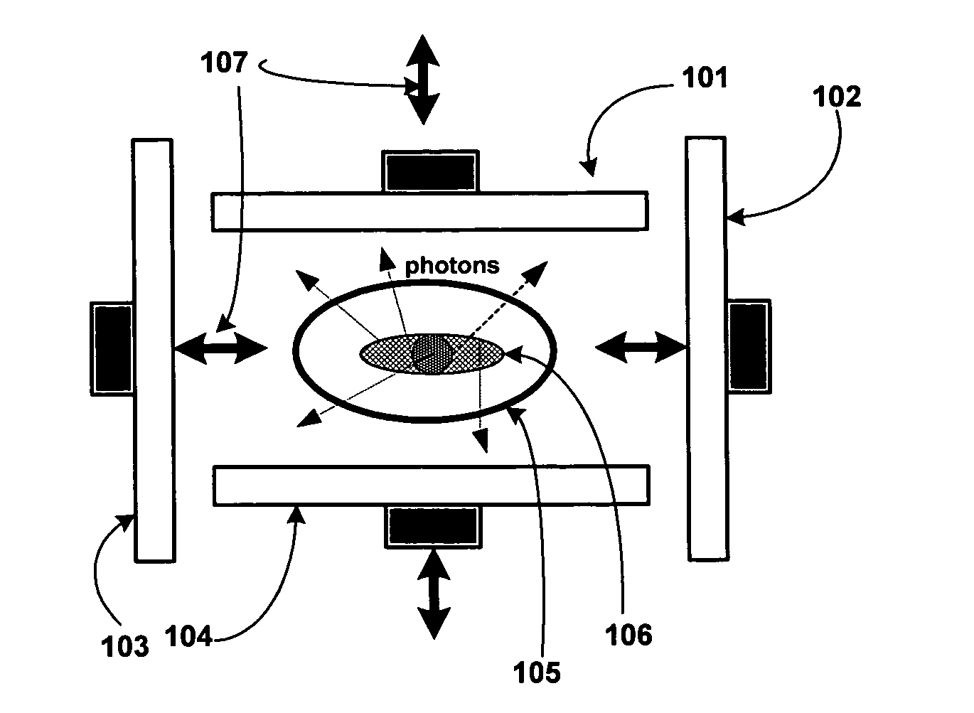
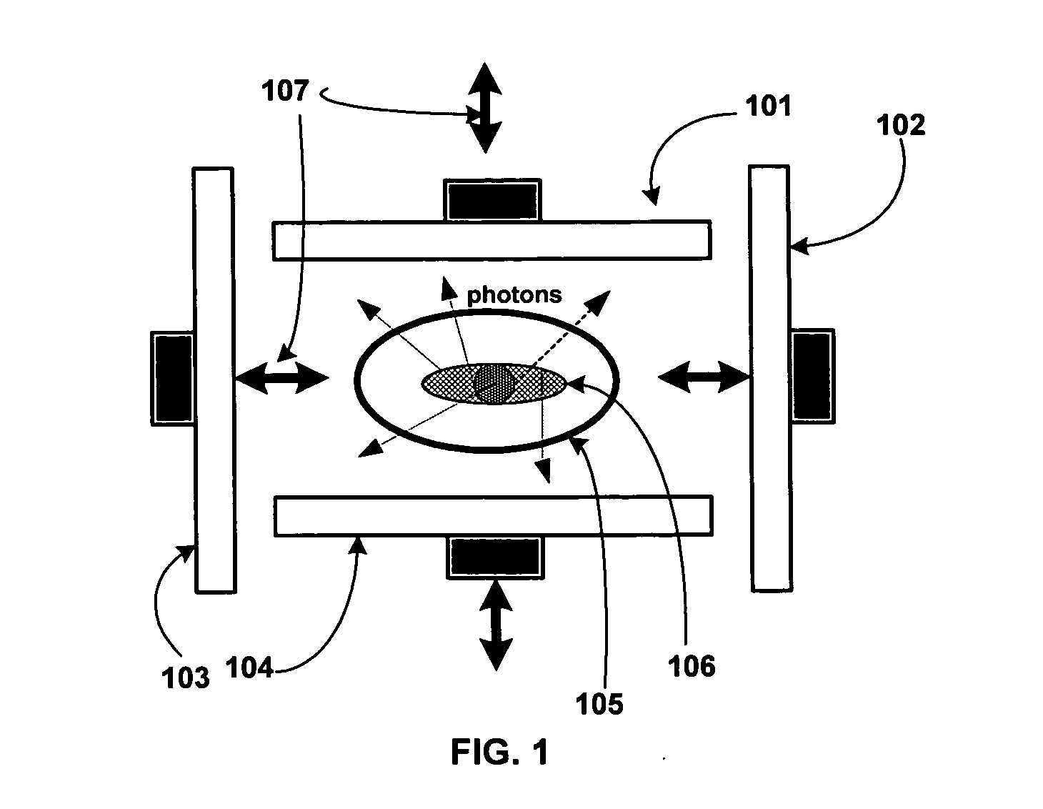
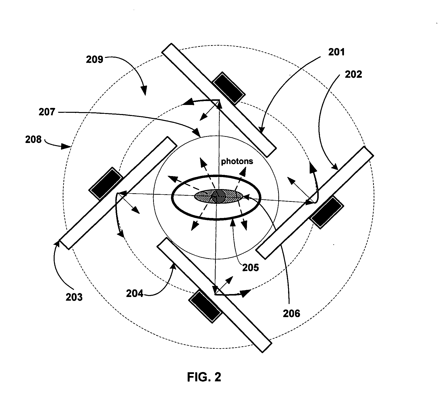
[0102]A high-sensitivity Single-Photon Emission Computed

[0103]Tomography (SPECT) apparatus includes a two-dimensional (2D) gamma detector array. This is typically a planar photon sensitive array of elements that are collimated. A cross section of such an array is shown in FIG. 5 with parallel-hole collimators, but collimators with other geometries such as converging / diverging holes, fan-beam, and cone-beam arrangements can also be used. In addition, unlike a conventional SPECT machine, each photon detector element or pixel in the 2D gamma detector array is provided with a very large collimator aperture corresponding to a field of view of 5 degrees to 180 degrees. See FIG. 4 and FIG. 5. This increases the sensitivity of the novel SPECT apparatus by a factor of at least 5 and often by over 20 in comparison with a conventional SPECT machine. The photon sensitive detector elements can be sensitive to gamma photons with different energies including photons e...


PUM


Abstract

A method and apparatus are disclosed for high-sensitivity Single-Photon Emission Computed Tomography (SPECT), and Positron Emission Tomography (PET). The apparatus includes a two-dimensional (2D) gamma detector array that, unlike a conventional SPECT machine, moves to different positions in a three-dimensional (3D) volume space near an emission source and records a data vector g which is a measure of gamma emission field. In particular, the 3D volume space in which emission data g is measured extends substantially along a radial direction r pointing away from the emission source, and unlike a conventional SPECT machine, each photon detector element in the 2D gamma detector array is provided with a very large collimator aperture. Data g is related to the 3D spatial density distribution f of the emission source, noise vector n, and a system matrix H of the SPECT/PET apparatus through the linear system of equations g=Hf+n. This equation is solved for f by a method that reduces the effect of noise.

Description

[0001]This patent application is a continuation of the following U.S. Provisional Patent Application filed by this inventor:[0002]M. Subbarao, “Method and Apparatus for High-Sensitivity Single-Photon Emission Computed Tomography (SPECT)”, U.S. Provisional Patent Application No. 61 / 276,570, filed on Sep. 14, 2009.This patent application provides details of the description of the above invention.1. FIELD OF THE INVENTION[0003]The present invention relates to a method and apparatus for Single Photon Emission Computed Tomography (SPECT) in Nuclear Medicine. This method and apparatus are also applicable to Positron Emission Tomography (PET). The method and apparatus are specifically related to exploiting the information in the spatial intensity distribution of the three-dimensional (3D) photon emission field in a 3D volume space around an emission source. This information can reveal the 3D spatial density distribution of the emission source. In particular, the 3D volume space is designed...

Claims


Application Information

Patent Timeline
Patent Type & Authority Applications(United States)
IPC IPC(8): G01T1/20G01T1/164A61B6/03
CPCA61B6/037G01T1/1648G01T1/1611
Inventor SUBBARAO, MURALIDHARA
Owner SUBBARAO MURALIDHARA
Why Eureka
Try Eureka