Eureka AIR delivers breakthrough ideas for toughest innovation challenges, trusted by R&D personnel around the world.

Method for chromogenic detection of two or more target molecules in a single sample

a chromogenic detection and target molecule technology, applied in biochemistry apparatus and processes, instruments, material analysis, etc., can solve the problems of difficult tracing to either instruments or reagents, and the background was not always reproducibl

Active Publication Date: 2011-06-09
VENTANA MEDICAL SYST INC
View PDF2 Cites 20 Cited by
  • Summary
  • Abstract
  • Description
  • Claims
  • Application Information

AI Technical Summary

Benefits of technology

[0012]For example, the second, hapten-labeled specific binding moiety may be a hapten-labeled nucleic acid probe, such as a hapten-labeled DNA probe (e.g., a DNP-labeled DNA probe). In some examples, the concentration of the DNP nucleic acid-labeled probe is sufficient to prevent or reduce background staining due to the DNP-labeled nucleic acid probe binding non-specifically to a chromogen product associated with a first target molecule. In certain examples, the concentration of DNP-labeled probe is greater than 1 and typically at least 5 μg / ml. For example, the concentration of the DNP nucleic acid-labeled probe ranges from 10 μg / ml to 15 μg / ml.
[0018]In a particular embodiment, a method for chromogenic detection of two or more target molecules in a single tissue sample comprises: contacting the tissue sample with a first specific binding moiety that specifically binds a first target molecule; detecting the first target molecule in the tissue sample by depositing an insoluble, electron-rich aromatic chromogen product; contacting the tissue sample with a second, hapten-labeled specific binding moiety that specifically binds a second target molecule, where a hapten of the second, hapten-labeled specific binding moiety comprises an electron-deficient aromatic compound; treating the tissue sample with a solution comprising a soluble, electron-rich aromatic compound prior to or concomitantly with contacting the second, hapten-labeled specific binding moiety with the tissue sample; and detecting the second target molecule by depositing a second, insoluble chromogen product that is distinguishable from the insoluble, electron-rich aromatic compound deposited to detect the first target molecule, where treating the tissue sample with the solution containing the soluble, electron-rich aromatic compound reduces background due to non-specific binding of the hapten-labeled specific binding moiety to the insoluble electron rich compound deposited near the first target molecule.

Problems solved by technology

The silver background was not always reproducible and it varied from instrument to instrument and from run to run making it difficult to trace to either instrument or reagent related causes.

Method used


Image

  • Method for chromogenic detection of two or more target molecules in a single sample
  • Method for chromogenic detection of two or more target molecules in a single sample
  • Method for chromogenic detection of two or more target molecules in a single sample

Examples


working examples

XI. WORKING EXAMPLES

[0179]The following examples are provided to illustrate certain specific features of working embodiments. The scope of the present invention is not limited to those features exemplified by the following examples.

example 1

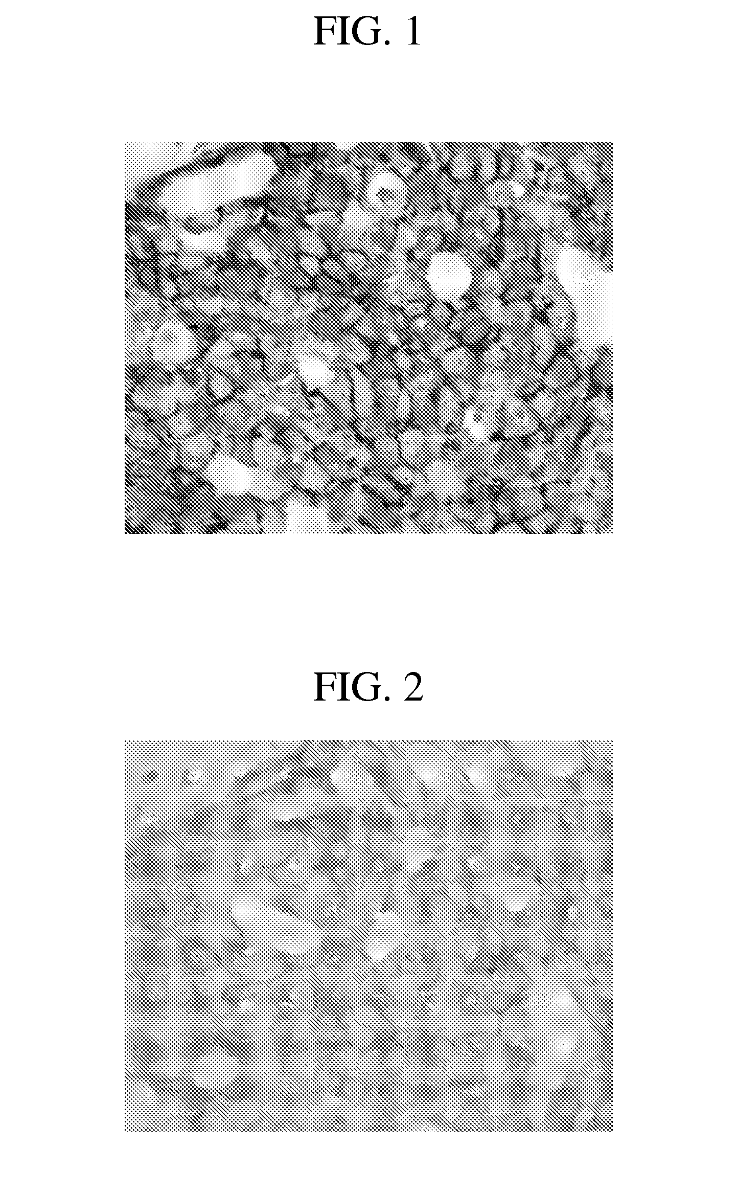
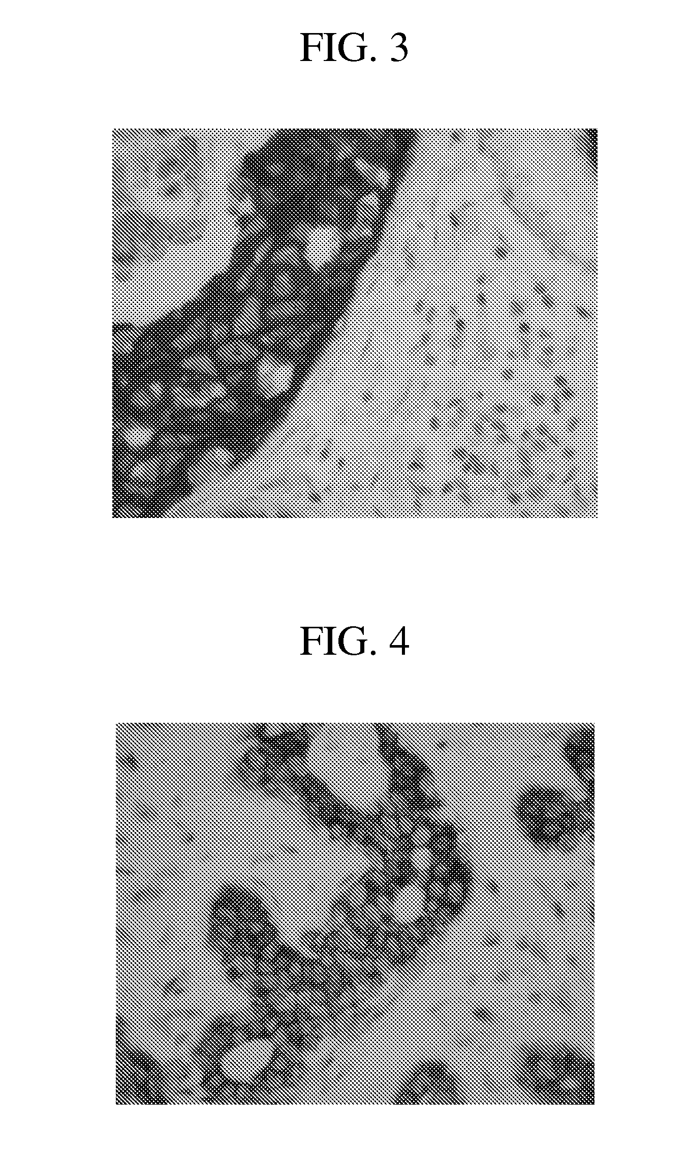
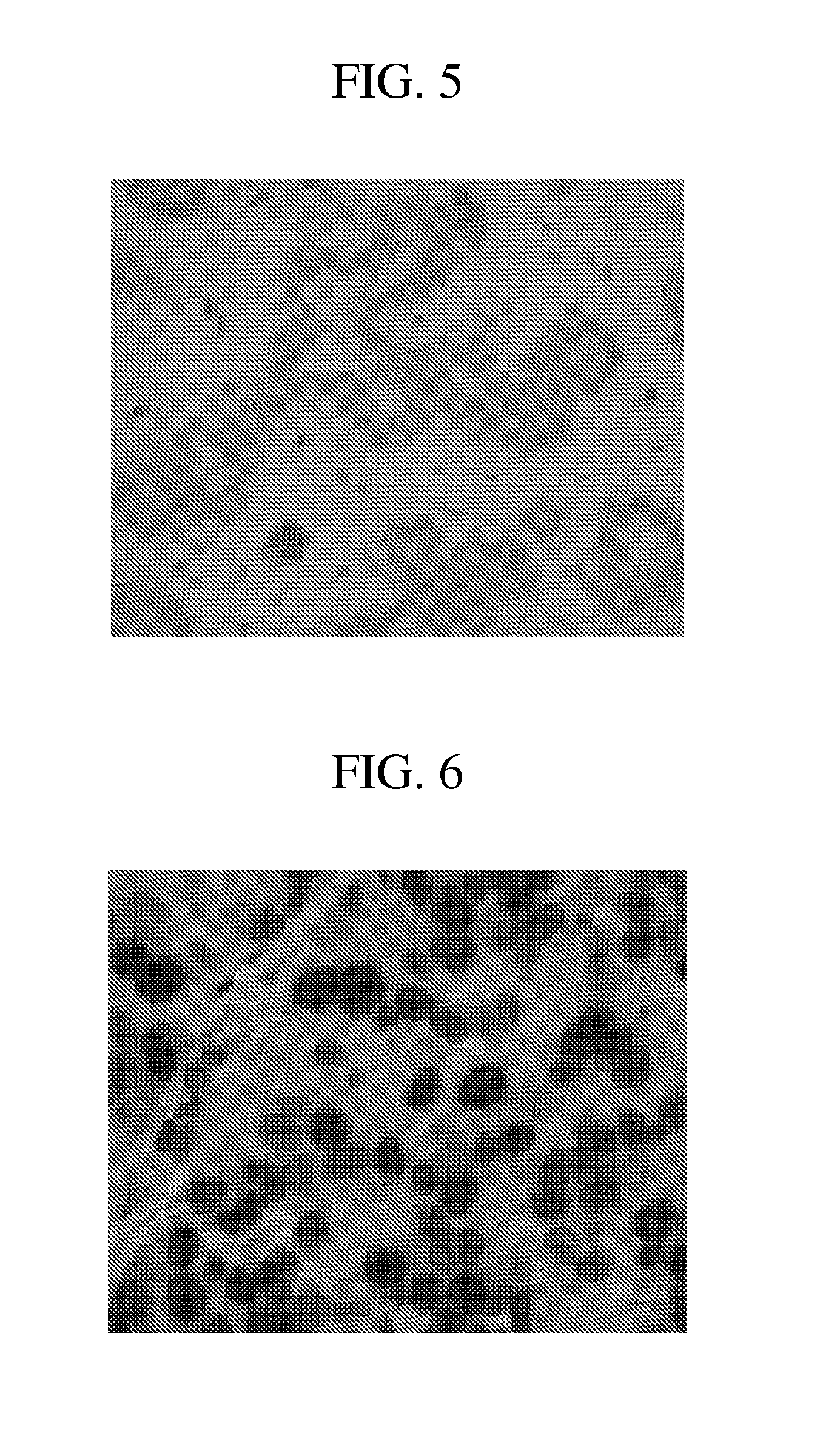
[0180]This example provides a staining assay that allows nucleic acids and protein to be detected in a single sample.

A. Material and Methods

[0181]Reagents utilized to perform the dual nucleic acid / protein hybridization and detection assays included the ULTRAVIEW SISH Detection Kit (Ventana Medical Systems, Inc., p / n 780-001), the INFORM HER2 DNA Probe (Ventana Medical Systems, Inc., p / n 780-4332), the Rabbit Anti-DNP Antibody (Ventana Medical Systems, Inc., p / n 780-4335), the Rabbit Anti-HER2 (4B5) Antibody (Ventana Medical Systems, Inc., p / n 800-2996), and the ULTRAVIEW Universal Alkaline Phosphatase Red Detection Kit (Ventana Medical Systems, Inc., p / n 760-501). Standard bulk solutions were used on the BENCHMARK XT instrument. The NexES software programs were modified as needed to establish the order of addition of reagents, temperature and incubation times.

[0182]Tissues: Dual hybridization and detection studies were performed on breast carcinoma tissues and xenograft material (HE...

example 2

Optimal Cell Conditioning

[0191]This example provides conditions for optimal cell conditioning for dual gene protein staining procedures.

A. Materials and Methods

[0192]Cell Conditioning: Optimal cell conditioning for each assay was determined by comparing different types of cell conditioning. Cell conditioning options included CC1 (Tris / Boric acid / EDTA, pH 8.6), CC2 (citric acid, pH 6.0), and a Reaction Buffer. The extent of cell conditioning was adjusted by selecting different times for cell conditioning, i.e., mild, standard, or extended. Tissue was also digested for ISH staining with either ISH protease 3 for approximately 4 minutes, protease 3 for approximately 8 minutes, or ISH protease 2 for approximately 4 minutes.

B. Results

[0193]Experiments were performed to determine the optimal cell conditioning conditions for anti-HER2 4B5 antibody staining according to the methods previously described. Optimal anti-HER2 4B5 antibody target detection was observed when the CC1 standard was s...


PUM

PropertyMeasurementUnit
Concentrationaaaaaaaaaa

Abstract

The present invention provides a method and kit for detection of two or more target molecules in a single tissue sample, such as for gene and protein dual detection in a single tissue sample. Methods comprise treating a tissue sample with a first binding moiety that specifically binds a first target molecule. Methods further comprise treating the tissue sample with a solution containing a soluble electron-rich aromatic compound prior to or concomitantly with contacting the tissue sample with a hapten-labeled binding moiety and detecting a second target molecule. In one example, the first target molecule is a protein and the second is a nucleic acid sequence, the first target molecule being detected by immunohistochemistry and the second by in situ hybridization. The disclosed method reduces background due to non-specific binding of the hapten-labeled specific binding moiety to an insoluble electron rich compound deposited near the first target molecule.

Description

CROSS REFERENCE TO RELATED APPLICATION[0001]This application claims the benefit of U.S. Provisional Application Ser. No. 61 / 189,752, filed on Aug. 22, 2008, which is incorporated herein by reference in its entirety.FIELD[0002]This disclosure relates to immunohistochemistry (IHC) and in situ hybridization (ISH), and specifically to embodiments of a method for chromogenic detection of two or more target molecules in a single sample.BACKGROUND[0003]Immunohistochemistry (IHC) employs specific binding agents, such as antibodies, to detect an antigen of interest that may be present in a tissue sample. IHC is widely used in clinical and diagnostic applications, such as to diagnose particular disease states or conditions. For example, particular cancer types can be diagnosed based on the presence of a particular marker molecule in a sample obtained from a subject. IHC is also widely used in basic research to understand biomarker distribution and localization in different tissues.[0004]Biolo...

Claims


Application Information

Patent Timeline
IPC IPC(8): C12Q1/68G01N33/53
CPCG01N33/581G01N33/53C12Q1/6841G01N33/583
Inventor GNIEWEK, RICHARDFARRELL, MICHAELNITTA, HIROAKILEHRKAMP, MEGANKOSMEDER, JEROMEKELLY, BRIAN DANIELGAIRE, FABIENPADILLA, MARYGROGAN, THOMASBIENIARZ, CHRISTOPHER
Owner VENTANA MEDICAL SYST INC
Why Eureka
Eureka Blog
Learn More