
Methods for Preparing Human Skin Substitutes from Human Pluripotent Stem Cells

a human skin substitute and pluripotent stem cell technology, applied in the field of human skin substitutes can solve the problems of inability to obtain human keratinocytes derived from reduced microbial defences, and cosmetic concerns, and achieve the effect of reducing the risk of desiccation, and reducing the number of human pluripotent stem cells

Inactive Publication Date: 2011-07-07
INST NAT DE LA SANTE & DE LA RECHERCHE MEDICALE (INSERM)
View PDF0 Cites 8 Cited by
  • Summary
  • Abstract
  • Description
  • Claims
  • Application Information

AI Technical Summary

Benefits of technology

[0069]In a particular embodiment, the substantially pure homogenous population of human keratinocytes derived from human pluripotent stem cells is previously seeded on a cell culture matrix populated with human dermis fibroblasts before providing an organotypic culture of it as above described. This particular embodiment allows obtaining a human skin substitute which comprises dermis and epidermis. Such a method may be performed through the protocol as described by Del Rio M. et al. (2002) or Larcher F. et al. (2007). For example, the substantially pure homogenous population of human keratinocytes derived from human pluripotent stem cells of the invention may be seeded on a fibrin matrix populated with live dermis fibroblasts. Organotypic cultures are then grown submerged up to keratinocyte confluence, and finally maintained at the air-liquid interface for 7 days to enhance stratification and differentiation of the epithelium.

Problems solved by technology

Loss of epidermal function leads to loss of thermal regulation, reduced microbial defences, risks of desiccation, inhibited wound repair, and cosmetic concerns.
However, up to now, the methods of the prior art have failed to obtain human keratinocytes derived from human pluripotent stem cells that would demonstrate an ability to form a pluristratified epidermis (in vitro or following xenografting in animals), when treated according to techniques that were shown instrumental when using adult basal keratinocytes from donors (see, e.g., Green, 2008).

Method used


Image

  • Methods for Preparing Human Skin Substitutes from Human Pluripotent Stem Cells
  • Methods for Preparing Human Skin Substitutes from Human Pluripotent Stem Cells
  • Methods for Preparing Human Skin Substitutes from Human Pluripotent Stem Cells

Examples


example 1

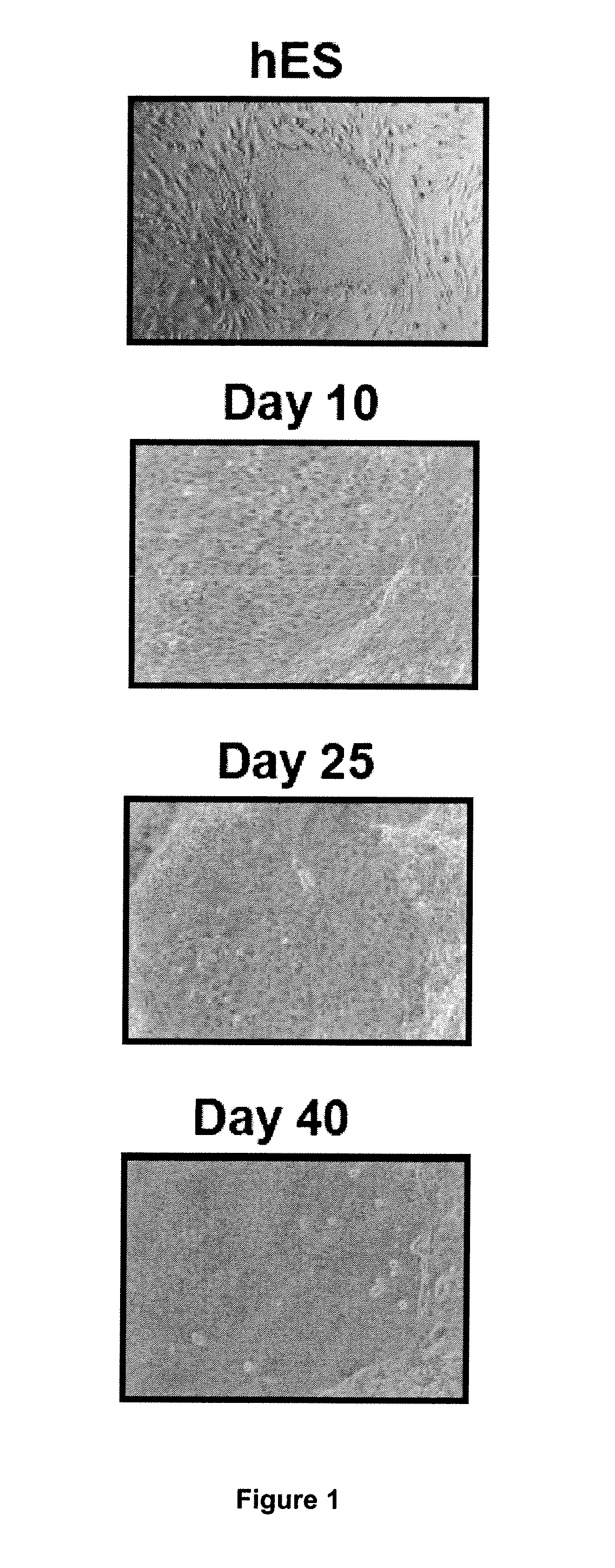
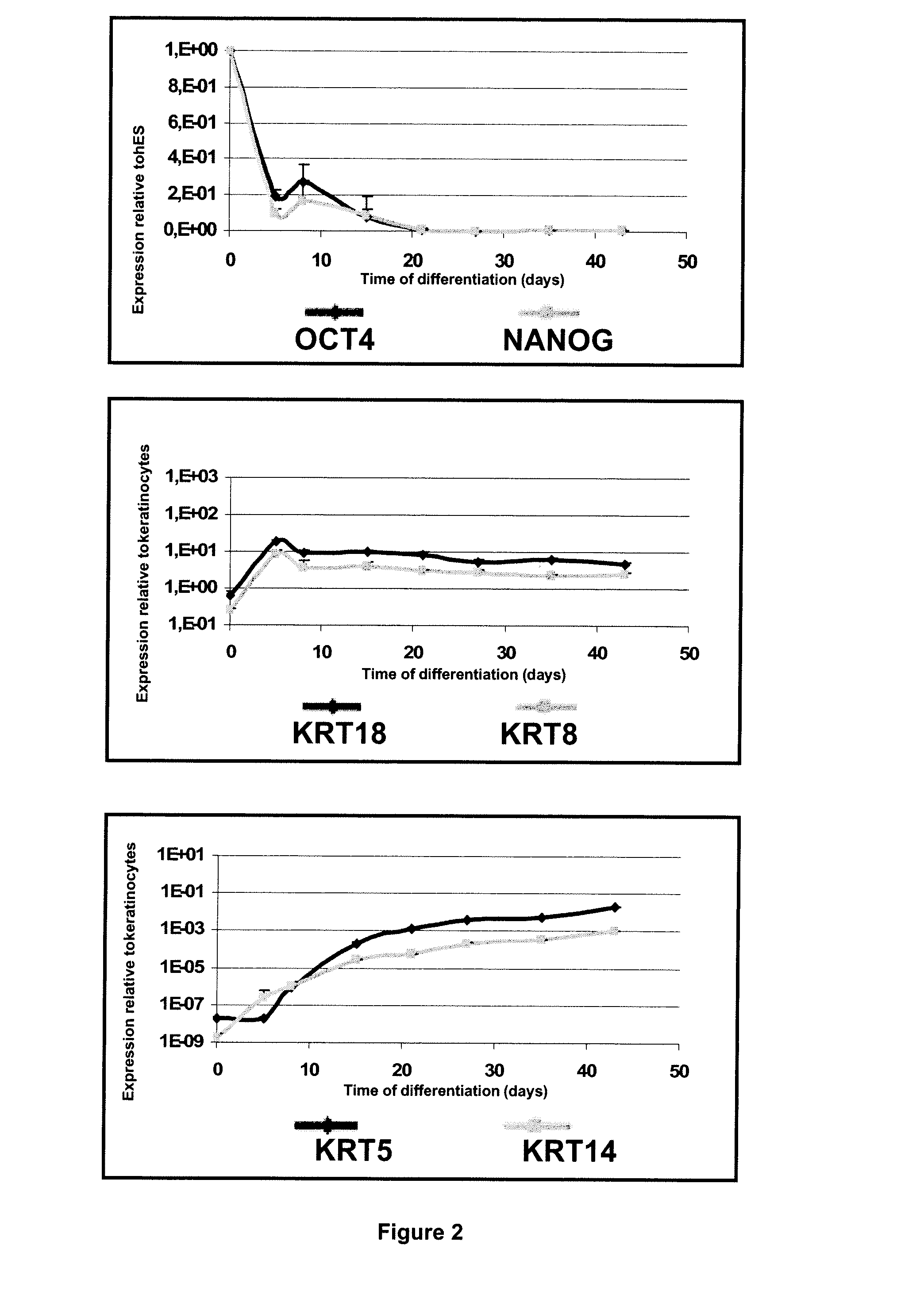
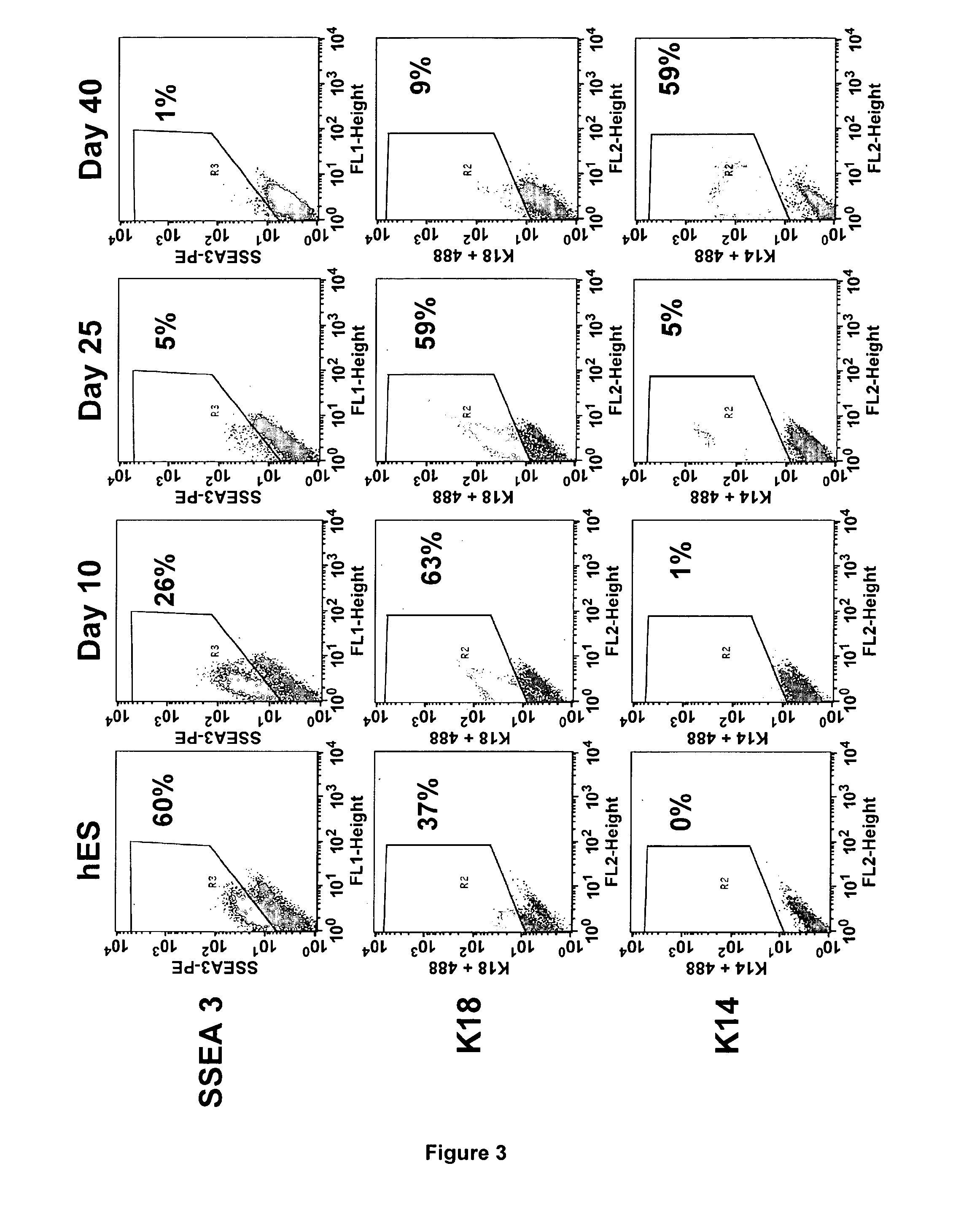
Method for Preparing a Population of Keratinocytes and a human skin substitute from hES

[0107]Material & Methods

[0108]Maintenance Culture of hES Cells.

[0109]The hESC(SA-01 and H9) were grown on a feeder layer of mouse fibroblast cells, STO (inactivated with 10 mg / ml mitomycin C and seeded at 30000 / cm2) in DMEM / F12 (Sigma) supplemented with 20% (vol / vol) Knockout Serum Replacement (KSR, Invitrogen), 1 mM glutamine, 0.1 mM nonessential amino acids (Invitrogen), 4 ng / ml recombinant human bFGF (PeProTech) and 0.1 mM 2-mercaptoethanol at 37° C. under 5% CO2. For passaging, hESC colonies were cut and passages were done every 5 days.

[0110]Derivation of hES Cells in Keratinocytes.

[0111]For derivation, clumps were seeded onto mitomycin C-treated 3T3 fibroblasts in FAD medium composed of ⅔ DMEM, ⅓ HAM:F12 and 10% of fetal calf serum (FCII, Hyclone) supplemented with 5 μg / ml insulin, 0.5 μg / ml hydrocortisone, 10−10 M cholera toxin, 1.37 ng / ml T3, 24 μg / ml adenine and 10 ng / ml recombinant human ...

example 2

Method for Preparing a Population of Keratinocytes and a human skin substitute from iPS

[0141]The same protocol of differentiation as described in Example 1 was performed with human induced pluripotent stem cells (iPS). Briefly, iPS were seeded onto mitomycin C-treated 3T3 fibroblasts in FAD medium composed of ⅔ DMEM, ⅓ HAM:F12 and 10% of fetal calf serum (FCII, Hyclone) supplemented with 5 μg / ml insulin, 0.5 μg / ml hydrocortisone, 10−10M cholera toxin, 1.37 ng / ml triodothyronin, 24 μg / ml adenine and 10 ng / ml recombinant human EGF. The induction of ectodermal differentiation was done when 0.5 nM of human recombinant BMP-4 (R&D Systems Europe, UK) and 0.3 mM ascorbic acid (Sigma) were added. Cells were grown in the same medium until clones of epithelial cells were isolated. Cells were then seeded in the same feeder layer in FAD medium devoid of BMP4 and ascorbic acid. As a control, primary human keratinocytes (HK) were cultured on mitomycin C treated 3T3 fibroblasts in FAD medium.

[0142...


PUM


Abstract

The present invention relates to an ex vivo method for obtaining a population of human keratinocytes derived from human pluripotent stem cells comprising a step of co-culturing human pluripotent stem cells with cells that support ectodermal differentiation in presence of an agent that stimulates epidermal induction and a agent that stimulates terminal differentiation of keratinocytes. A further object of the invention relates to a method for preparing a human skin substitute comprising a step of providing an organotypic culture of the substantially pure homogenous population of human keratinocytes derived from human pluripotent stem cells obtained according to the method of the invention.

Description

FIELD OF THE INVENTION[0001]The present invention relates to ex vivo methods for obtaining populations of human keratinocytes derived from human pluripotent stem cells and methods for preparing human skin substitutes.BACKGROUND OF THE INVENTION[0002]The skin consists of self-renewing layers organized into functional units of differentiating cells with their origin in a single basal stratum of proliferating keratinocytes. The dead and dying cells that comprise the stratum corneum are continually shed during desquamation and replaced by cells derived from epidermal stem cells found in the stratum germinativum. Loss of epidermal function leads to loss of thermal regulation, reduced microbial defences, risks of desiccation, inhibited wound repair, and cosmetic concerns. In the absence of sufficient autologous donor for skin grafts, coverage of wounds with cultured human keratinocytes represents a promising option for treatment.[0003]Furthermore, in vitro and in vivo models for human ski...

Claims


Application Information

Patent Timeline
IPC IPC(8): A61K35/36C12Q1/02C12N5/071A61P17/02
CPCC12N5/0629C12N2500/40C12N2501/01C12N2501/11C12N2502/13C12N2501/33C12N2501/395C12N2506/02C12N2506/45C12N2501/155A61P17/02A61F2/10A61K35/36A61L27/38C12N5/0606
Inventor GUENOU, HIND
Owner INST NAT DE LA SANTE & DE LA RECHERCHE MEDICALE (INSERM)
Learn More