Method for preparing contact lens-shaped amniotic dressing

a technology of amniotic dressing and contact lens, which is applied in the field of preparing amniotic dressing with contact lens, can solve the problems of insatisfactory conditions, inconvenient patient treatment, and method using amniotic membran

Inactive Publication Date: 2011-08-04
SK BIOLAND CO LTD
View PDF3 Cites 70 Cited by
  • Summary
  • Abstract
  • Description
  • Claims
  • Application Information

AI Technical Summary

Benefits of technology

The contact lens-shaped amniotic dressing prepared by the inventive method of the present invention is capable of solving the problems associated with suturing an amniotic membrane, e.g., highly delicate surgical techniques of suturing, long surgery time, stitch abscess,

Problems solved by technology

However, the conventional methods, except for the method using an amniotic membrane, do not satisfy said conditions.
In order to apply an amniotic membrane to the treatment of ocular surface diseases, it is necessary to perform 10-0 nylon suturing requiring highly delicate surgical techniques and a long surgery time, which often causes side effects, such as stitch abscess, granuloma formation, tissue necrosis, an

Method used


Image

  • Method for preparing contact lens-shaped amniotic dressing
  • Method for preparing contact lens-shaped amniotic dressing
  • Method for preparing contact lens-shaped amniotic dressing

Examples


example 1

Preparation of an Amniotic Membrane

(1-1) Preparation of Human Amniotic Membrane

Human amniotic membrane was treated by the method disclosed by an article of Kim and Tseng (Kim J C and Tseng S C, Cornea, 14:473-84 (1995)). An amniotic membrane was isolated from the placenta of a pregnant who had taken Caesarean section. The pregnants had been prescreened for hepatitis B and C viruses, HIV and syphilis, and only those placentas of which the maternal bloods reveal negative serological results were used. The amniotic membrane thus obtained was washed sequentially several times with sterile saline solution and sodium hypochlorite, and washed repeatedly with purified water. The resulting membrane was stored under a cold condition (2˜8° C.) until used.

(1-2) Preparation of Bovine Amniotic Membrane

Bovine amniotic membrane was treated by the following procedure. The bovine placenta was obtained upon delivery, and the amniotic membrane was separated therefrom and washed several times with salin...

example 2

Preparation of a Mold and a Ring

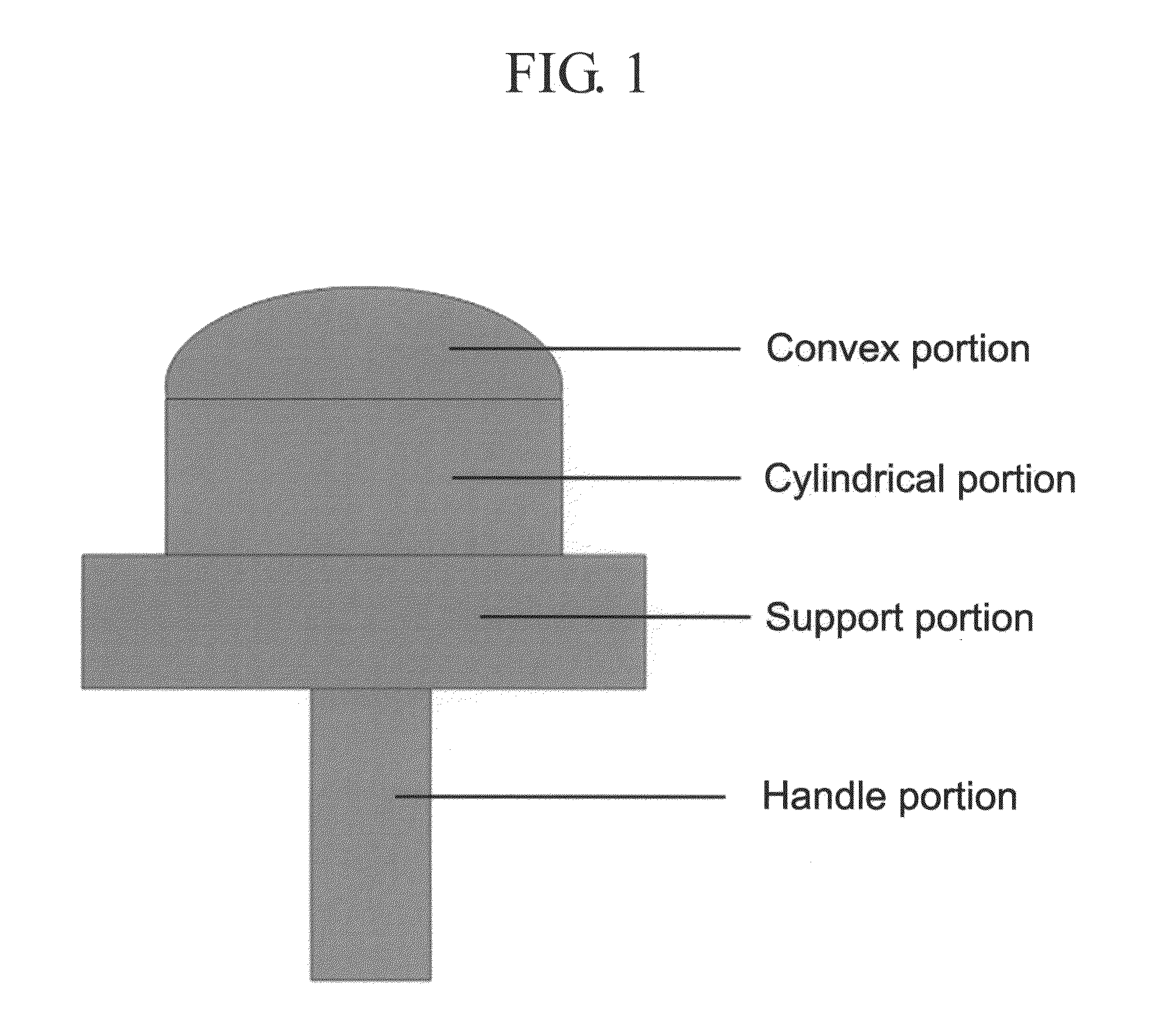
A mold for forming a contact lens-shaped amniotic membrane and a ring (or “O-ring”) for contacting an amniotic membrane with the mold were manufactured as shown in FIGS. 1 and 2. The mold was composed of a cylindrical portion having a diameter of 14 mm, a convex portion having a base curve of 8.4 to 8.8 mm, a support portion, and a handle portion. The support portion comprises a support which supports the ring used for contacting and fastening the amniotic membrane, and the handle portion has a cylindrical shape connected to the middle of the bottom surface of the support portion. The mold was made of an acetal resin. The O-ring of FIG. 2 was manufactured to fit the respective mold having a specific base curve. The O-ring was made of Teflon (14.1 mm internal diameter) or a perfluoroelastomer (12 mm internal diameter).

example 3

Arrangement of the Amniotic Membrane

In order to make the amniotic membrane obtained in (1-1) to (1-3) have a contact lens-shape, the amniotic membrane was brought in contact with the contact lens-shaped mold obtained in Example 2, and then fixed on the mold with the O-ring.

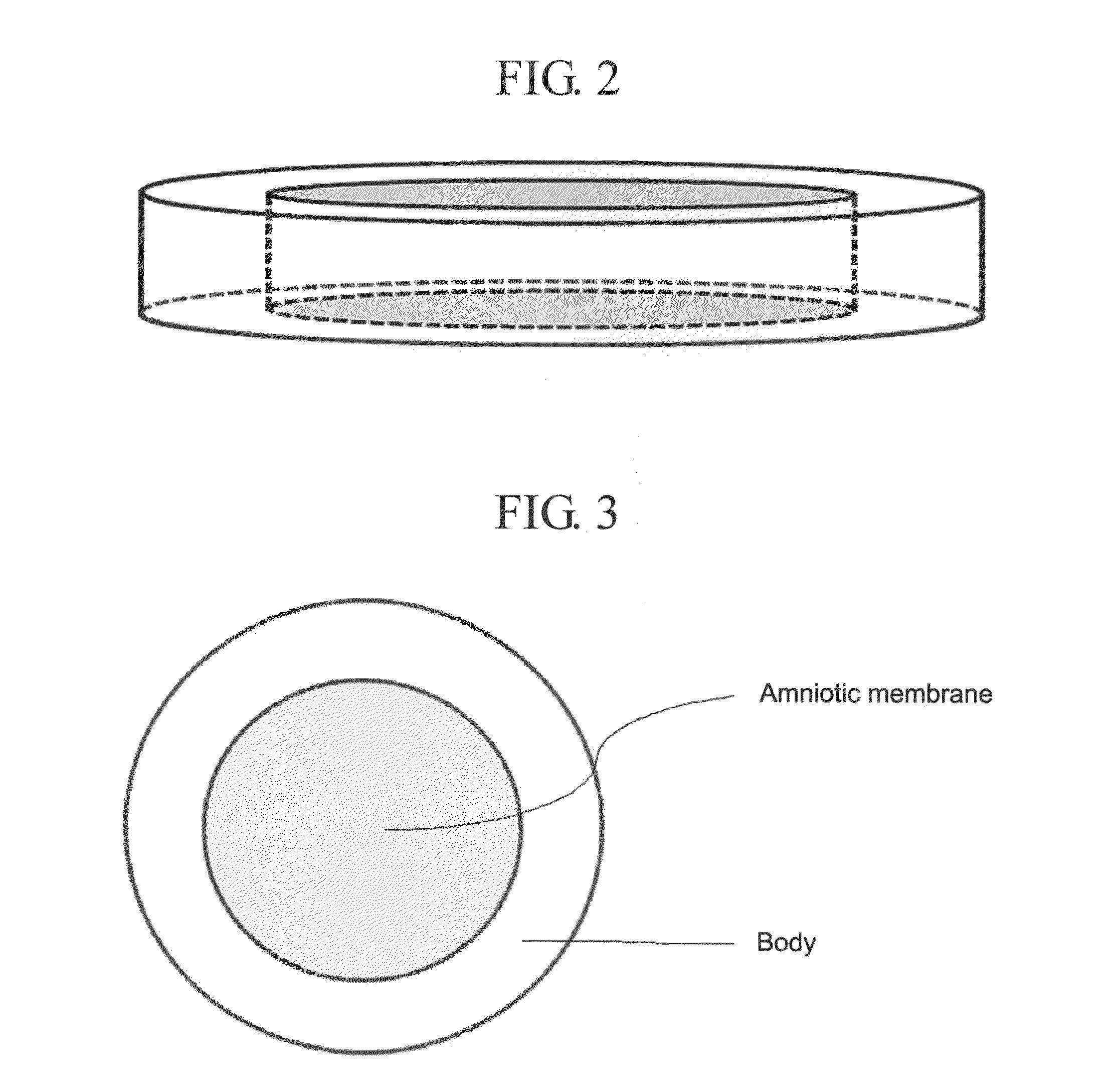
Specifically, the amniotic membrane was cut to the size of the mold, and then was brought in contact with the convex portion of the mold (FIG. 1) with its epithelial layer facing downwards. In case of overlapping the amniotic membrane, the last amniotic membrane was overlapped so that its epithelial layer faced outside. The O-ring (FIG. 2) was placed thereafter on the amniotic membrane or overlapping amniotic membranes to completely cover the mold, followed by natural drying, freeze drying, or vacuum drying in a clean bench. A top perspective view of the dried amniotic membrane (including the mold) is shown in FIG. 3.


PUM

PropertyMeasurementUnit
Diameteraaaaaaaaaa
Diameteraaaaaaaaaa
Diameteraaaaaaaaaa

Abstract

The present invention relates to a method for preparing a contact lens-shaped amniotic dressing and a contact lens-shaped amniotic dressing prepared therefrom for treating ocular surface diseases, which does not require the use of sutures or an adhesion material. The inventive contact lens-shaped amniotic dressing is capable of solving the problems associated with suturing an amniotic membrane, e.g., highly delicate surgical techniques of suturing, long surgery time, stitch abscess, granuloma formation, tissue necrosis, and discomfort of patients; and the problems associated with the use of a support, e.g., the elimination of the support by eye blinking, breaking of the support, and discomfort.

Description

FIELD OF THE INVENTIONThe present invention relates to a method for preparing a contact lens-shaped amniotic dressing for treating ocular surface diseases, which does not require the use of sutures or an adhesion material.BACKGROUND OF THE INVENTIONAn amniotic membrane, a membrane having the thickness of 0.02 to 0.5 mm that is positioned in the inner surface of the placenta, has a stacked structure of an epithelium, a basement membrane, a compact layer, a fibroblast layer, and a sponge layer. An amniotic membrane is avascular and does not express histocompatible antigens, and hence immunological rejection after its transplantation does not occur (Akle C A et al., Lancet, 2:1003-5 (1981); Dua H S et al., Br. J. Ophthalmol., 83:748-52 (1999)). Since the basement membrane of the amniotic membrane is composed of type IV collagen, laminin, and α6 / β4 integrin, and an interstitium thereof is composed of type I collagen, type III collagen, hyaluronic acid, fibronectin, and α5 / β1 integrin, a...

Claims


Application Information

Patent Timeline
IPC IPC(8): A61K35/48A61P27/02B29C63/02
CPCB29C63/02A61K35/48B29D11/00038A61L27/3604A61F9/0017A61L2/081
Inventor YANG, EUN KYUNGJUNG, SAM HYUNKIM, SUNG POJOO, JI HOONLEE, JONG WONHONG, JONG MYOUNGKIM, JAE CHANCHUN, YEOUN SOOKLEE, SUNG CHUNBAIK, KWANG III
Owner SK BIOLAND CO LTD
Why Eureka
Try Eureka