Device and method for detection of analyte from a sample

a technology for analyte detection and sample, applied in the field of devices and methods for detection of analyte particles, can solve the problems of not being able to efficiently aid, not having a simple and robust method for circulating epcs, and being highly time-consuming, so as to reduce the non-specific adhesion of analyte particles, improve the sensitivity of impedance detection, and improve the detection efficiency.

Inactive Publication Date: 2011-08-11
SINGAPORE HEALTH SERVICES PTE +1
View PDF11 Cites 21 Cited by
  • Summary
  • Abstract
  • Description
  • Claims
  • Application Information

AI Technical Summary

Benefits of technology

[0018]The device and methods of using the device of the present invention therefore may provide a fast, efficient and label-free approach to detecting and quantifying a particular type of target analyte particle in a sample. As well, since the sample may be deposited directly into the chamber rather than using a flow-through system, small sample volumes may be used along with sequential batch loading, avoiding large dead volumes within the device and the resulting loss of target analyte particles through non-specific adhesion or sedimentation of analyte particles.
[0019]The methods of the present invention use DEP to trap analyte particles contained within the sample, including negative DEP, which may allow for the use of a buffer solution for sample preparation that can also serve as a conductive medium, for example phosphate buffered saline (PBS) solution for trapping and detecting cells, which can be used for the cell trapping step (DEP) and the cell detecting step (impedance measurement).
[0020]The use of negative DEP results in trapping of target analyte particles at

Problems solved by technology

However, there is currently no simple and robust method to detect circulating EPCs and stent selection is therefore often based on the individual physician's experience rather than knowledge of the actual EPC count.
However, this technique usually requires off-site analysis and therefore is not an efficient aid when deciding on the type of stent to deploy in the patient.
Using FACS to determine EPC count involves two major time-

Method used


Image

  • Device and method for detection of analyte from a sample
  • Device and method for detection of analyte from a sample
  • Device and method for detection of analyte from a sample

Examples


example 1

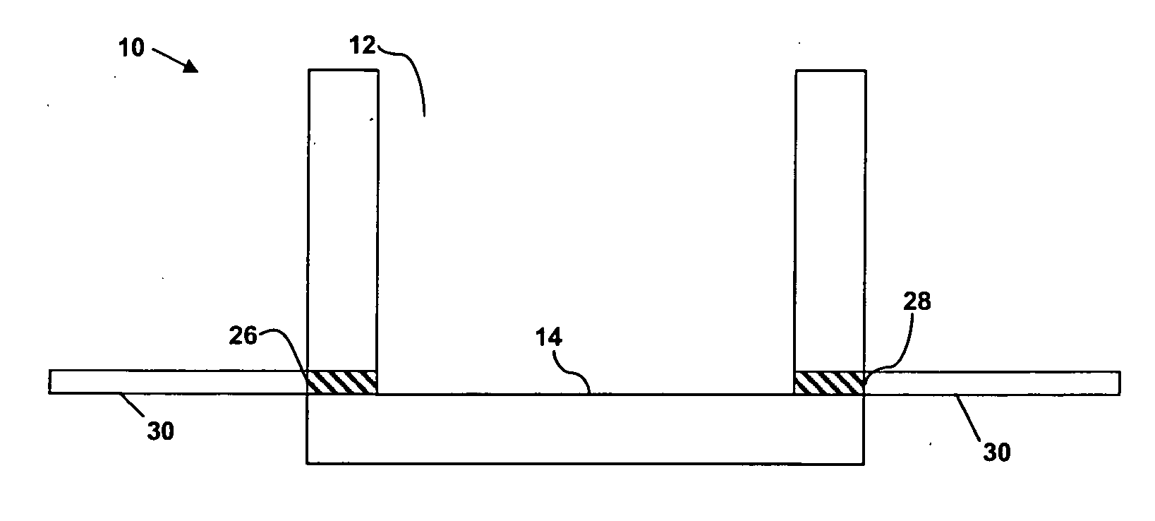
[0111]FIG. 7 shows florescent images of the antibody specifically immobilized on the electrode areas of the microchip, which is surrounded by a silicon oxide biocompatible layer that has been coated with a cell-repellent material to prevent non-specific adhesion of anti-CD34 antibodies prior to immobilization and of cells during cell trapping step.

[0112]The surface coating process and polyethylene glycol (PEG) passivation were adapted from work done by M. Zhang's group (Mandana, V., Wickes, B. T., Castner, D. G., Zhang, M. (2004) Biomaterials 25(16), 3315-3324; Lan, S., Veiseh, M., Zhang, M. (2005) Biosensors and Bioelectronics 20(9), 1697-1708). A mixture of COOH-terminated alkanethiols (a 20 mM solution of 1 / 10 v / v mercapto-undecanoic acid (MUA) / mercapto-propionic acid (MPA) in ethanol) were used to create a self-assembled monolayer (SAM) on the gold electrodes that can be further modified to bind to NH2-amino acids of proteins through activation with N-(3′-dimethylaminopropyl)-N′...

example 2

[0118]FIG. 13 demonstrates the results of testing the lower detection limit of the device using a sample containing a total of 15,000 cells with Jurkat cells as non-target cells and CD34+ cells as target cells. As can be seen, at least a lower limit of 150 CD34+ cells in a total of a mixed sample containing 15,000 cells can be detected in a single batch loading.

[0119]FIGS. 14 and 15 demonstrate the improved detection using multiple batch loading of cells. For each batch, a mixture of Jurkat and CD34+ cells (for a total of 15,000 cells containing 750 CD34+ cells) were loaded into the chamber. The results indicate that the CD34+ cells are retained on the inner electrode from batch to batch loading, cell trapping and washing procedures, resulting in overall increase in % impedance change with increasing number of batches loaded. Thus, if a single batch contains a target cell concentration that falls below the lower detection limit, multiple batch loading may be used to allow for loadin...

example 3

[0121]The described method is performed on a device having three separate chambers each with a separate electrode array. Chamber 1 has antibody A immobilised on the electrode surfaces, chamber 2 has antibody B immobilised on the electrode surfaces, and chamber 3 has antibodies A and B immobilised on the electrode surfaces, as shown in FIG. 18.

[0122]Thus, in chamber 1, cells expressing antigen A (specifically bound by antibody A) and antigen A and B together will be detected and quantified; in chamber 2, cells expressing antigen B (specifically bound by antibody B) and antigen A and B together will be detected and quantified; and in chamber 3, cells expressing antigen A, antigen B or antigen A and B together will be detected and quantified.

[0123]In order to obtain the levels of the various cells of interest, the following calculations are carried out on the signals obtained from the impedance measurements: subtracting the impedance measurements obtained from chamber 1 from those of c...


PUM

PropertyMeasurementUnit
Concentrationaaaaaaaaaa
Volumeaaaaaaaaaa
Areaaaaaaaaaaa

Abstract

There is presently provided a device for detecting an analyte particle in a sample. The device comprises a chamber having an interior surface upon which is located an electrode array. The electrode array comprises pairs of electrodes, each pair having an inner electrode and an outer electrode that substantially surrounds the inner electrode. Each pair of electrodes is coated with a capture molecule that recognises and binds the analyte particle that is to be identified and quantified. The device uses a combination of dielectrophoresis and impedance measurements to capture and measure analyte particles from a sample.

Description

FIELD OF THE INVENTION[0001]The present invention relates to devices and methods for detecting the presence of a particular analyte particle within a sample, including the presence of a particular cell type within a sample.BACKGROUND OF THE INVENTION[0002]In various clinical applications, it is often desirable to identify the presence of a particular cell type in a sample, and to quantify the number of cells of that cell type that are present in the sample.[0003]For example, circulating endothelial progenitor cells (EPC) are circulating stem cells from the bone marrow that are involved in vascular surfaces repair (endothelial damage repair). Their number in blood is a biomarker of clinical interest, linked to the assessment of risk factors in cardiovascular diseases and for choice of certain therapeutic approaches.[0004]For patients suffering from a blocked coronary artery, one of the main treatments is stent implant. There are different types of stents. Genous™ stent (Orbusneich, H...

Claims


Application Information

Patent Timeline
IPC IPC(8): G01N27/447G01N27/453
CPCA61B5/00G01N33/5438A61B5/14546
Inventor CHEN, YUREBOUD, JULIENWONG, EN HOU PHILIPMOE, KYAW THUSHIM, SE NGIE WINSTONRAMADAN, QASEMTANG, KUM CHEONG
Owner SINGAPORE HEALTH SERVICES PTE
Why Eureka
Try Eureka