Test method using cells and test kit therefor

a test method and cell technology, applied in the field of biological test methods using cells, to achieve the effect of improving quantitative capability

Inactive Publication Date: 2011-08-18
DAI NIPPON PRINTING CO LTD
View PDF5 Cites 1 Cited by
  • Summary
  • Abstract
  • Description
  • Claims
  • Application Information

AI Technical Summary

Benefits of technology

[0033]The present invention makes it possible to culture cell aggregates having position coordinates that can be determined and having an arbitrary size such that there are almost the same numbers of cells in the cell aggregates in a gelled matrix or under an environment equivalent to such a matrix. In particular, the present invention makes it possible to realize biological tests involving high-level three-dimensional culture, such as a vascularization test with better quantitative capability than conventional methods, a vascular remodeling test that involves simulation of in vivo vascular d

Problems solved by technology

In recent years, lower investment efficiency in research and development has become an issue in the pharmaceutical industry.
However, these methods are problematic in that the initial position coordinates of single cells or cell aggregates cannot be specified and control of the distribution of cell aggregate sizes is difficult.
Hence, application of these methods to an advanced biological test system using a combination of robotics and viable cell analysis techniques has been limited.
However in this case, a plurality of spheroids cannot be cultured with determined position coordinates thereof in each well, so that obtainment of a plurality of data from each well and statistical data analysis cannot be performed.
Furthermore, the form of a cell aggregate that can be used in the method disclosed in Non-patent Document 4 is limited to a spheroid.
The resulting test systems are often considered to be troublesome artificial test systems and they have failed to attract attention in application fields such as pharmacological tests.
However, these documents do not mention any biological test method that involves culturing an artificial single cell pattern or a cell aggregate pattern in a matrix.
However, this is not appropriate as a test method for testing parameters concerning single cell or cell aggregate movement.
However, Patent Document 6 does not disclose any biological test method that involves three dimensional

Method used


Image

  • Test method using cells and test kit therefor
  • Test method using cells and test kit therefor
  • Test method using cells and test kit therefor

Examples


example 1

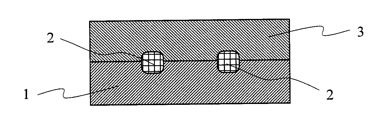
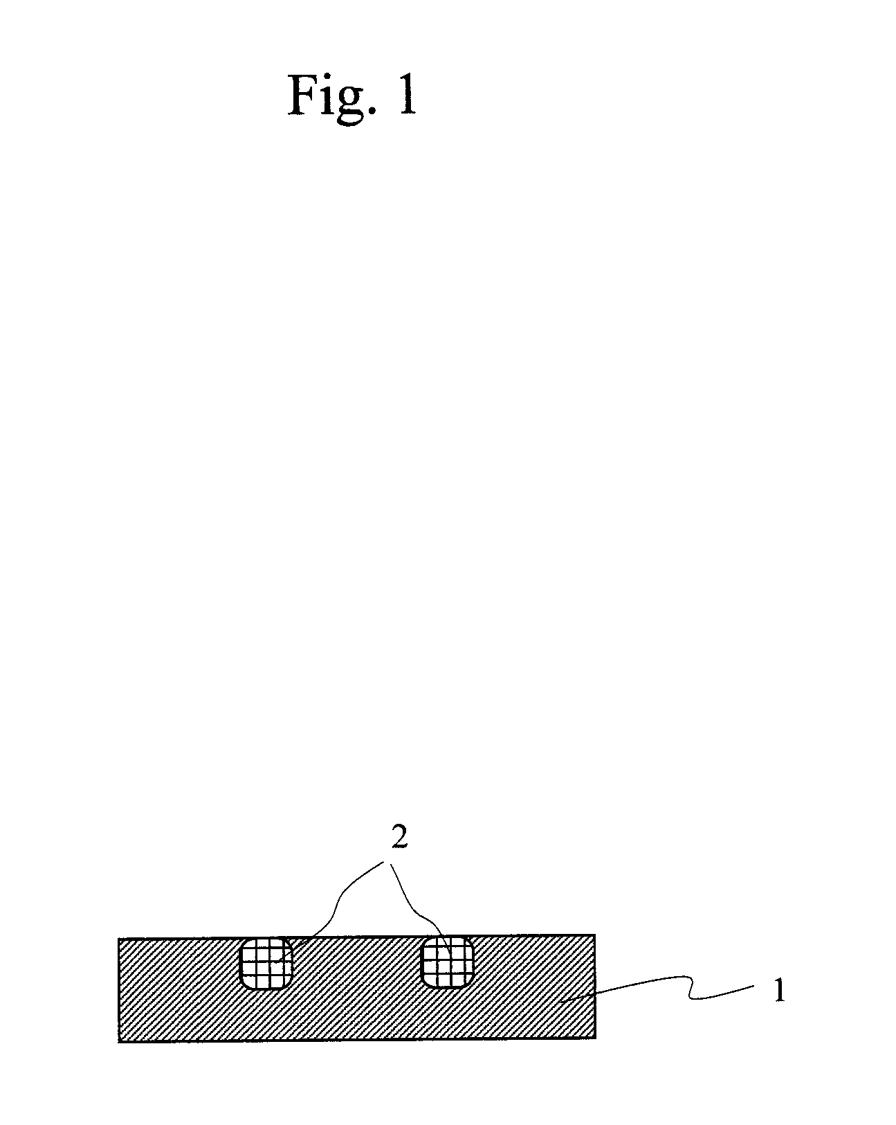
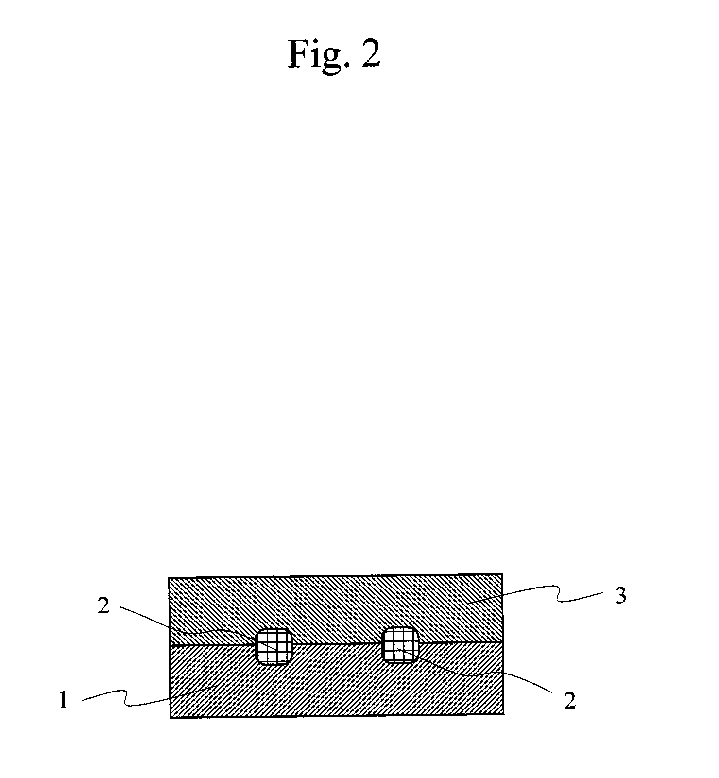
(Preparation of Substrate for Cell Culture)

(First-Step Reaction)

[0099]39.0 g of toluene and 2.25 g of TSL8350 (produced by GE Toshiba Silicone) were mixed. 450 μl of triethylamine was added to the solution while stirring. After several minutes of stirring of the solution at room temperature, the total volume of the solution was transferred to a glass plate. A 10-cm square glass substrate that had been washed with UV was immersed in the solution and then the substrate was allowed to stand at room temperature for 16 hours. Subsequently, the glass substrate was washed with ethanol and water and then dried by nitrogen blowing. The water contact angle of the substrate surface was approximately 53°.

(Second-Step Reaction)

[0100]25 μl of concentrated sulfuric acid was added dropwise to 50 g of tetraethylene glycol (TEG) while stirring. After several minutes of stirring of the solution, the total volume of the solution was transferred to a glass plate. The above substrate was immersed in the ...

example 2

(First-Step Reaction)

[0104]39.0 g of toluene and 1.20 g of TSL8350 (produced by GE Toshiba Silicone) were mixed. 450 μl of triethylamine was added to the solution while stirring. After several minutes of stirring of the solution at room temperature, the total volume of the solution was transferred to a glass plate. A 10-cm square glass substrate that had been washed with UV was immersed in the solution and then the substrate was allowed to stand at room temperature for 16 hours. Subsequently, the glass substrate was washed with ethanol and water and then dried by nitrogen blowing. The water contact angle of the substrate surface was approximately 51°.

(Second-Step Reaction)

[0105]25 μl of concentrated sulfuric acid was added dropwise to 50 g of tetraethylene glycol (TEG) while stirring. After several minutes of stirring of the solution, the total volume of the solution was transferred to a glass plate. The above substrate was immersed in the solution, followed by 20 minutes of reactio...

example 3

(First-Step Reaction)

[0109]39.0 g of toluene and 0.56 g of TSL8350 (produced by GE Toshiba Silicone) were mixed. 450 μl of triethylamine was added to the solution while stirring. After several minutes of stirring of the solution at room temperature, the total volume of the solution was transferred to a glass plate. A 10-cm square glass substrate that had been washed with UV was immersed in the solution and then the substrate was allowed to stand at room temperature for 16 hours. Subsequently, the glass substrate was washed with ethanol and water and then dried by nitrogen blowing. The water contact angle of the substrate surface was approximately 50°.

(Second-Step Reaction)

[0110]25 μl of concentrated sulfuric acid was added dropwise to 50 g of TEG while stirring. After several minutes of stirring of the solution, the total volume of the solution was transferred to a glass plate. The above substrate was immersed in the solution, followed by 2 hours of reaction at 80° C. After reaction...


PUM


Abstract

The present invention provides a method for performing a biological test under conditions in which an artificially prepared cell pattern with initial position coordinates that can be determined is three-dimensionally cultured within a gelled matrix. The present invention relates to a biological test method that comprises testing a biological indicator with reference to at least one selected from the group consisting of cell proliferation, cell movement, and cell differentiation in a cell pattern substantially embedded in gel. The present invention also relates to a kit for the biological test method.

Description

CROSS REFERENCE TO RELATED APPLICATION[0001]This application is a Divisional of application Ser. No. 11 / 936,836 filed Nov. 8, 2007, which is based on Japanese Patent Application No. 2006-305769 filed on Nov. 10, 2006, the disclosure of which is incorporated herein by reference in its entirety.BACKGROUND OF THE INVENTION[0002]1. Field of the Invention[0003]The present invention relates to a biological test method using cells, which is used in the fields involving pharmaceuticals, medical care, foods, medicine, pharmacy, biology, and the like. The present invention particularly relates to a biological test method using three-dimensional culture considered to enable obtainment of information that is more useful than that obtained via conventional two-dimensional culture. The biological test method is technology that is worthwhile as an alternative to animal testing.[0004]2. Background Art[0005]In recent years, lower investment efficiency in research and development has become an issue ...

Claims


Application Information

Patent Timeline
IPC IPC(8): C12N5/071
CPCG01N33/5008G01N33/5029G01N33/5032
Inventor HATTORI, HIDESHIOKOCHI, NORIHIKOKURODA, MASATOSHIHASE, MASAHIKO
Owner DAI NIPPON PRINTING CO LTD
Why Eureka
Try Eureka