
Direct and Indirect Surface Coil Correction for Cardiac Perfusion MRI

a surface coil and correction technology, applied in the field of cardiac imaging, can solve the problems of inhomogeneity correction, inability to acquire an equal amount of interest in the field of cardiac mr imaging, and drifting of signal intensities, so as to improve the signal-to-noise ratio

Inactive Publication Date: 2011-08-25
SIEMENS AG +1
View PDF7 Cites 10 Cited by
  • Summary
  • Abstract
  • Description
  • Claims
  • Application Information

AI Technical Summary

Benefits of technology

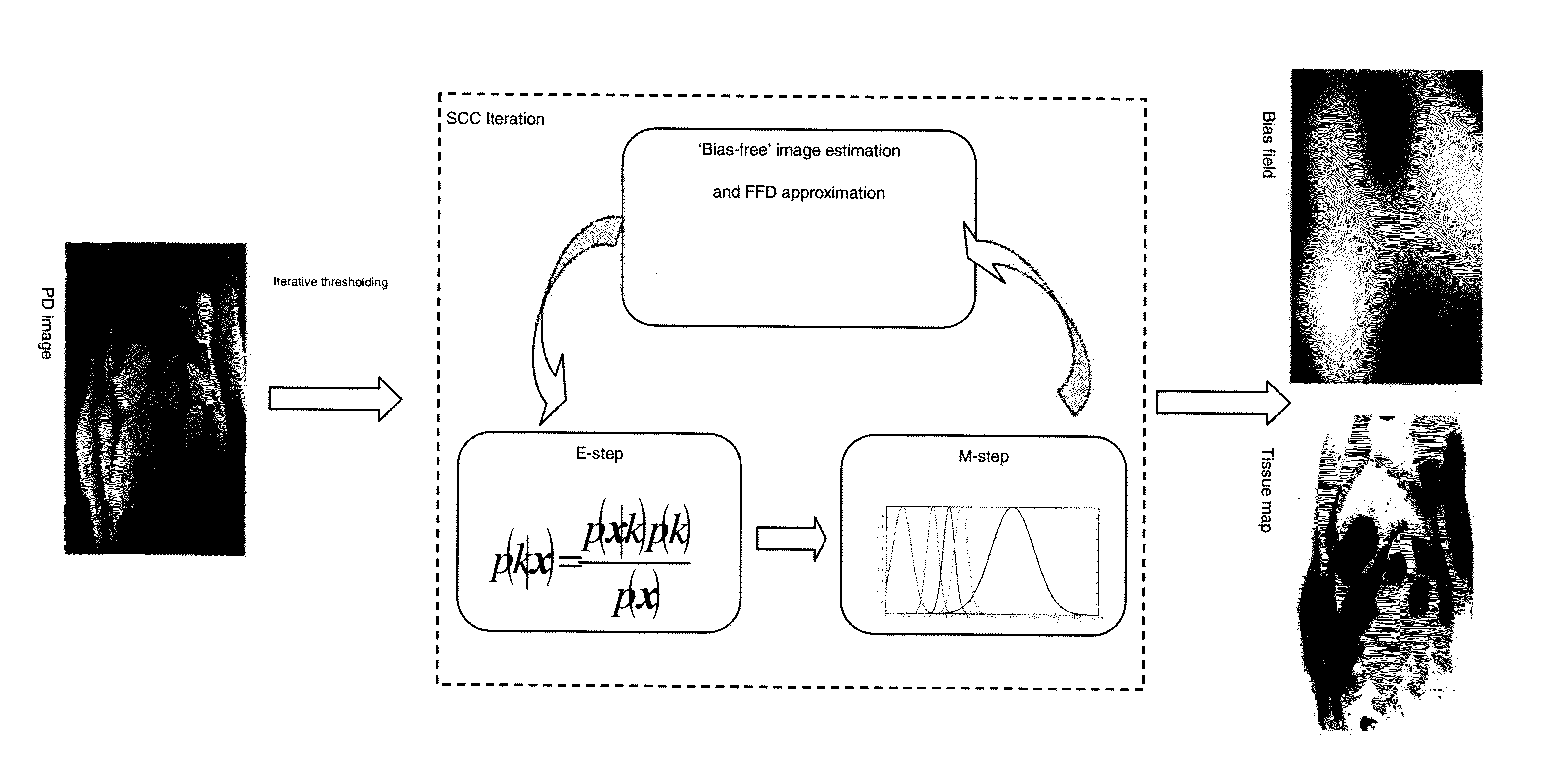
The present invention provides a method of cardiac perfusion magnetic resonance imaging (MRI) that can compensate for intensity variations in the image caused by the receiver coil and the motion of the patient. This is achieved by estimating the intensity variations in the image and using an approximation of the B-Spline free-form deformation (FFD) to compensate for the variations. The estimated bias field is then applied to the image series of the target cardiac region to improve the signal-to-noise ratio. The invention also provides a method of evaluating MRI images by estimating the intensity variations in the image and using the estimated bias field to compensate for the variations. The estimated bias field can be generated using an interleaving method or an iterative optimization method. The invention can help to improve the accuracy and reliability of cardiac perfusion MRI.

Problems solved by technology

However, the clinical routine to evaluate myocardial perfusion MR images still relies on qualitative visual reading by a health practitioner which is subjective and suffers from inter-observer variability.
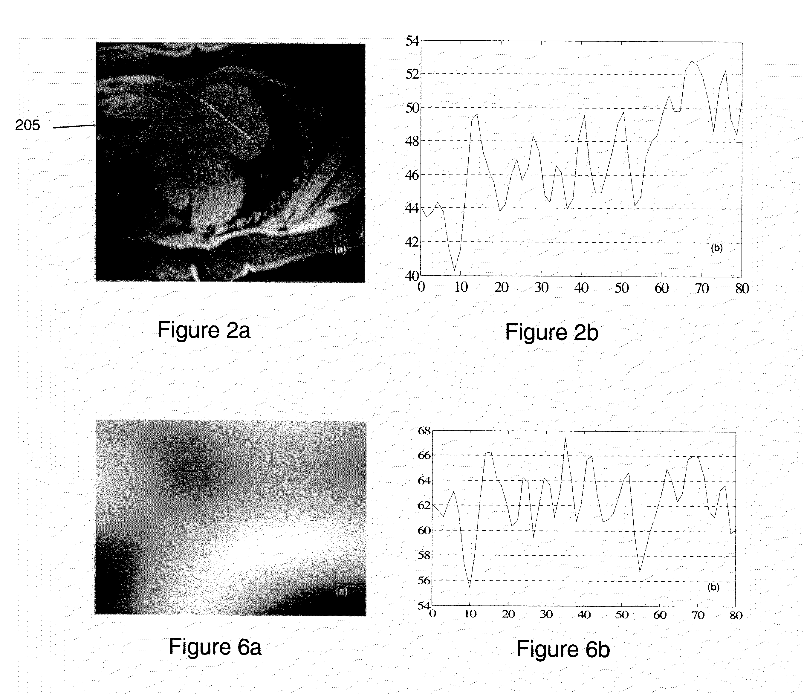
Unfortunately, the B1-field inhomogeneity caused by non-uniform characteristics of the receiver coils employed in conventional MR imaging systems causes signal intensity variation which will affect quantitative assessment (see, for example, FIG. 2b) and must be corrected before computing the perfusion quantitative maps (this is further described in the Kellman and Ara article cited above). FIG. 2a shows a proton density (PD) image of a heart acquired before normal perfusion acquisition.
238-251), it causes the drifting of signal intensities and leads to errors of quantitative perfusion analysis (as further described in the Kellman and Ara article cited above).
But, as indicated above, inhomogeneity correction has not acquired an equal amount of interest in the field of cardiac MR imaging.

Method used


Image

  • Direct and Indirect Surface Coil Correction for Cardiac Perfusion MRI
  • Direct and Indirect Surface Coil Correction for Cardiac Perfusion MRI
  • Direct and Indirect Surface Coil Correction for Cardiac Perfusion MRI

Examples


Embodiment Construction

[0026]FIG. 3 is a block diagram of a conventional MRI scanner 300 (simplified) that performs cardiac perfusion MR imaging in accordance with the present invention. A main magnet 312 generates a strong static magnetic field in an imaging region where the subject (i.e., patient) is introduced. The magnet 312 is used to polarize the target cardiac area, i.e., certain atoms in the target cardiac area that were previously randomly-ordered become aligned along the magnetic field. A gradient coil system 318, having a gradient coil subsystem 318a and a gradient coil control unit 319, generates a time-varying linear magnetic field gradient in respective spatial directions, x, y and z, and spatially encodes the positions of the polarized or excited atoms. An RF system 322, having an RF coil subsystem 324 and a pulse generation unit 326, transmits a series of RF pulses to the target cardiac region to excite the “ordered” atoms of the target cardiac area. The RF coil subsystem 324 may be adapte...


PUM


Abstract

Method of correcting cardiac perfusion MR imaging for inhomogeneities (430) caused by non-uniform receiver coil fields using proton density weighted images (410) and B-Spline Free-Form Deformation (425).

Description

CROSS-REFERENCE TO RELATED APPLICATIONS[0001]This application claims the benefit of Provisional U.S. Patent Application Ser. No. 61 / 306,593, entitled, “Direct and Indirect Surface Coil Correction for Cardiac Perfusion MRI”, filed in the name of Hui Xue, Jens Guehring, and Sven Zuehlsdorff, on Feb. 22, 2010, the disclosure of which is also hereby incorporated herein by reference.FIELD OF INVENTION[0002]The present invention relates to cardiac imaging. More particularly, the present invention relates to cardiovascular magnetic resonance imaging to assess or measure myocardial blood flow.BACKGROUND OF THE INVENTION[0003]Myocardial first pass perfusion magnetic resonance imaging (MRI) is a diagnostic imaging approach using cardiovascular magnetic resonance to assess or measure myocardial blood flow from the contrast enhancement observed during the first pass of a contrast agent bolus. It has proven its clinical significance in the diagnosis of known and suspected ischemic heart disease,...

Claims


Application Information

Patent Timeline
Patent Type & Authority Applications(United States)
IPC IPC(8): A61B5/055
CPCG01R33/246G01R33/5659G01R33/56366G01R33/5601
Inventor GUEHRING, JENSXUE, HUIZUEHLSDORFF, SVEN
Owner SIEMENS AG
Learn More