Methods for Detecting Tumor Origin Based on MUC1, MUC2, and CK-17 Expression Levels
a tumor origin and expression level technology, applied in the field of methods for detecting tumor origin based on muc1, muc2, and ck17 expression levels, can solve the problems of difficulty in accurately diagnosing pancreatic cancer, complex pancreatic cancer, etc., and achieve the effects of improving diagnosis, prognosis, and treatmen
- Summary
- Abstract
- Description
- Claims
- Application Information
AI Technical Summary
Benefits of technology
Problems solved by technology
Method used
Image
Examples
example 1
Immunohistochemical Procedures
[0106]Tissue samples were fixed in 10% neutral buffered formalin (NBF) and embedded in paraffin. Immunohistochemical procedures were performed using the automated BondMax system (Leica Microsystems Inc., Bannockburn, Ill.). Briefly, deparaffinization using BondMax was followed by an antigen retrieval step for 15 minutes using a high pH epitope retrieval buffer from Leica Microsystems Inc. (Bannockburn, Ill.). A polymer-based detection system used was the L-M Intense Kit with 3,3′-diaminobenzidine (DAB) as the chromogen (Leica Microsystems Inc., Bannockburn, Ill.). Slides were counterstained with hematoxylin. Appropriate positive and negative tissue control samples were used throughout the study. Mouse monoclonal antibodies, anti-MUC1 (1:250), anti-MUC2 (1:150), and anti-CK17 (1:200) were obtained from LeicaMicrosystems Novocastra Laboratories (Newcastle, UK). Mouse monoclonal antibody anti-CA19.9 (1:16000) was obtained from Covance (Princeton, N.J.).
[01...
example 2
Expression of MUC1, MUC2, and CK17 in Normal Tissues
[0108]Previous studies have reported that MUC1, MUC1, and CK17 are expressed in various tissue types (Goldstein N. S. et al. Am J Gin Patna 2001; 115:695-702; Higashi M. et al. Hepatology. 1999; 30:1347-1355; Hollingsworth M. A. et al. Nat Rev Cancer. 2004; 4:45-60). In the present Example, the expression patterns of these proteins were evaluated in multiple tissues by immunohistochemical analysis using the method described in Example 1.
[0109]MUC1 immunoreactivity was primarily cytoplasmic in most of the tissues examined including the following: the basal and para-basal cell layer of squamous epithelium, the stratified esophageal layer (which exhibited weak staining), the luminal borders of gastric glands in the stomach, the dendritic network of Peyer's Patches in the small intestine, luminal borders of hepatic bile ducts, distal renal tubules, alveolar lining cells and mesothelial cells in lung ducts and mammary lobules, the squam...
example 3
Expression of MUC1, MUC2, and CK17 in Adenocarcinomas
[0113]A primary reason pancreatic adenocarcinoma is among the deadliest malignancies is the lack of symptoms for early detection and diagnosis. In addition, early local invasion and metastatic spread of the tumor make both surgery and other conventional treatment strategies inadequate for most patients. Thus, the identification of a tumor's primary site of origin is critical for appropriate treatment of this disease.
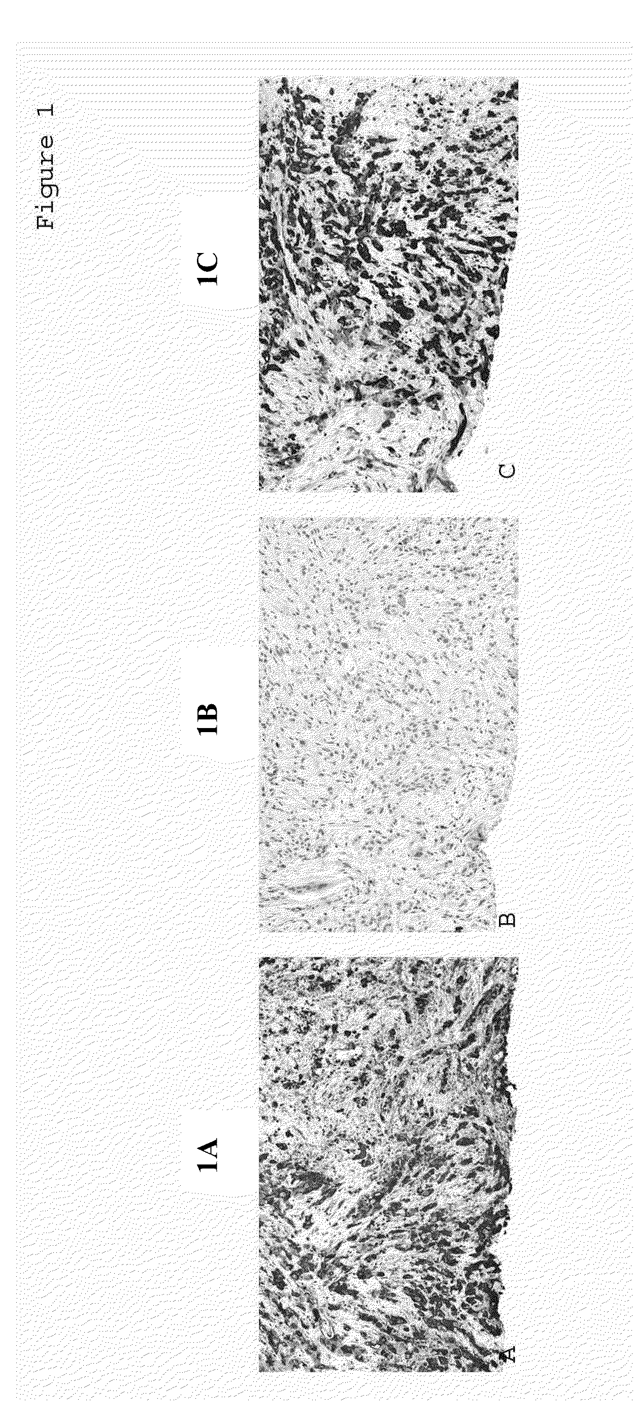
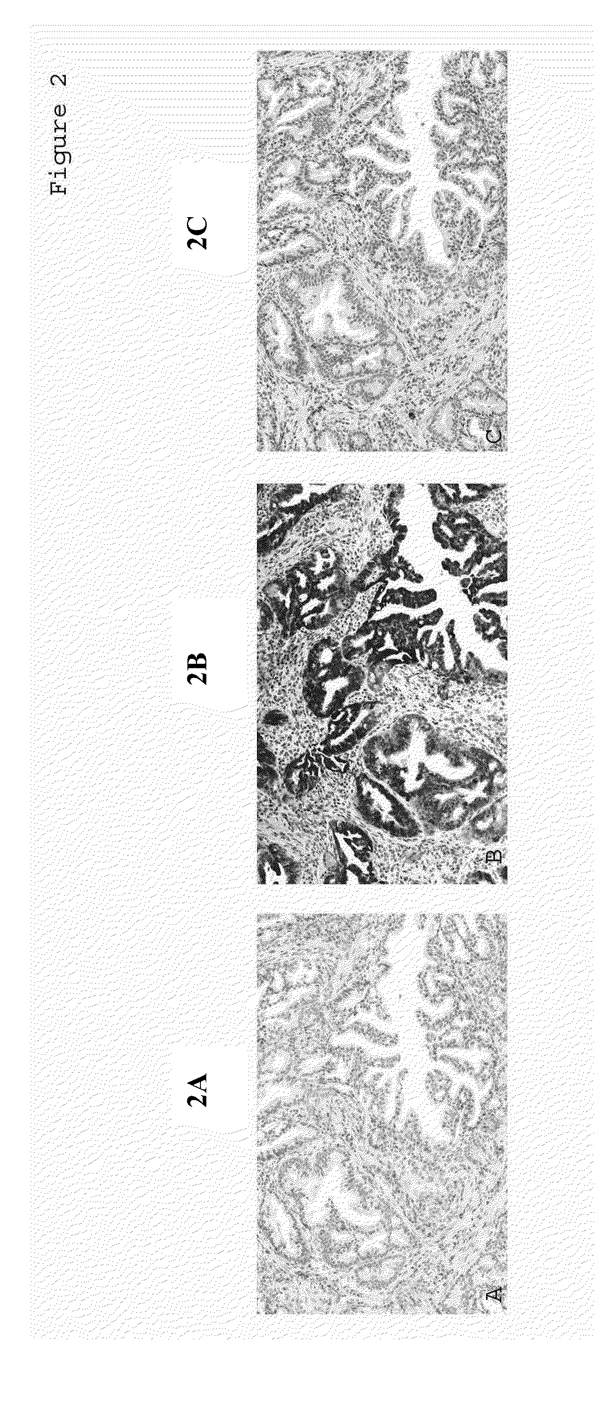
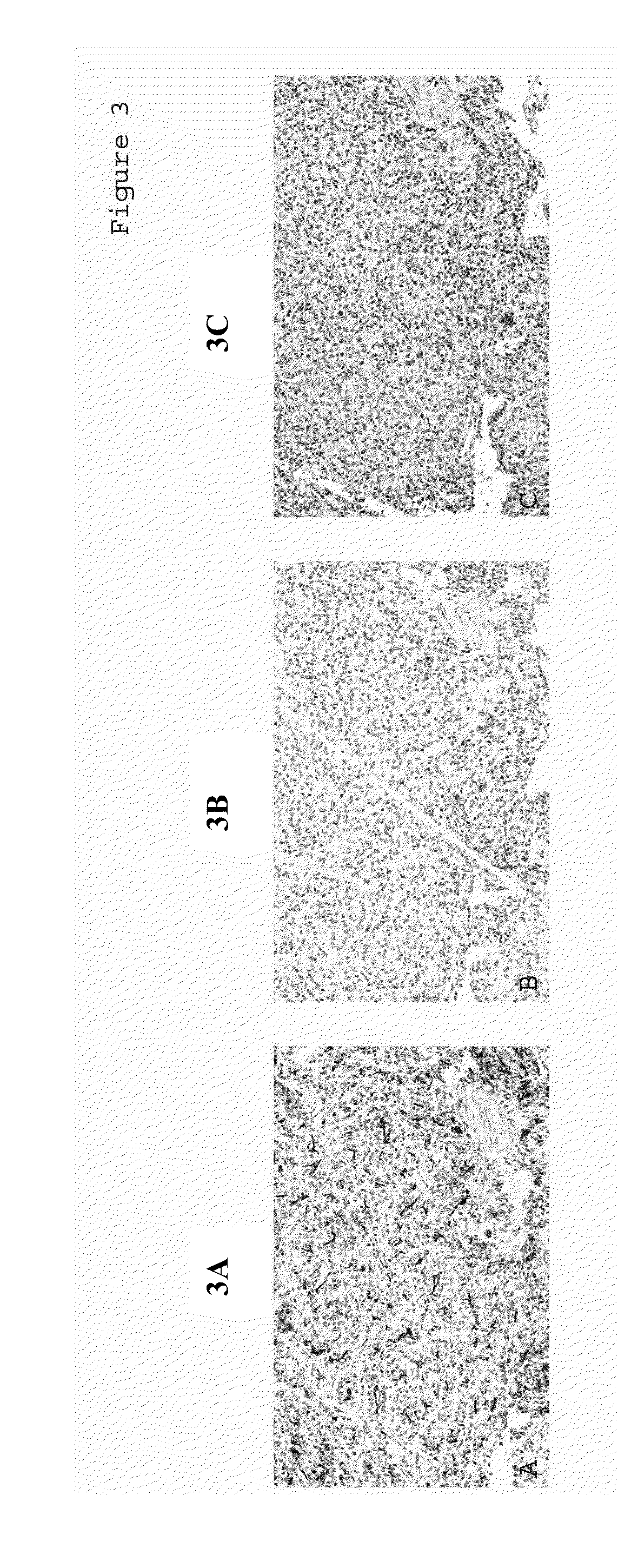
[0114]Adenocarcinomas from a variety of organs were collected and de-identified prior to their use. Tissue samples were fixed and stained as described above for Example 1. The immunoreactivity of MUC1, MUC2, or CK17 was evaluated based on the staining intensity of positive cells distributed in the carcinoma tissues. Intensity of staining was scored as 0 for no staining (negative), and as 1, 2, or 3 for weak (+), moderate (++), or strong staining (+++), respectively.
[0115]A MUC1, MUC2, and CK17 antigen expression panel ...
PUM
| Property | Measurement | Unit |
|---|---|---|
| Therapeutic | aaaaa | aaaaa |
| Level | aaaaa | aaaaa |
Abstract
Description
Claims
Application Information
 - R&D
- Intellectual Property
- Life Sciences
- Materials
- Tech Scout
- Unparalleled Data Quality
- Higher Quality Content
- 60% Fewer Hallucinations
Browse by: Latest US Patents, China's latest patents, Technical Efficacy Thesaurus, Application Domain, Technology Topic, Popular Technical Reports.
© 2025 PatSnap. All rights reserved.Legal|Privacy policy|Modern Slavery Act Transparency Statement|Sitemap|About US| Contact US: help@patsnap.com


