Eureka AIR delivers breakthrough ideas for toughest innovation challenges, trusted by R&D personnel around the world.

Surgical patch

Active Publication Date: 2011-10-20
SOFRADIM PROD SAS
View PDF7 Cites 63 Cited by
  • Summary
  • Abstract
  • Description
  • Claims
  • Application Information

AI Technical Summary

Benefits of technology

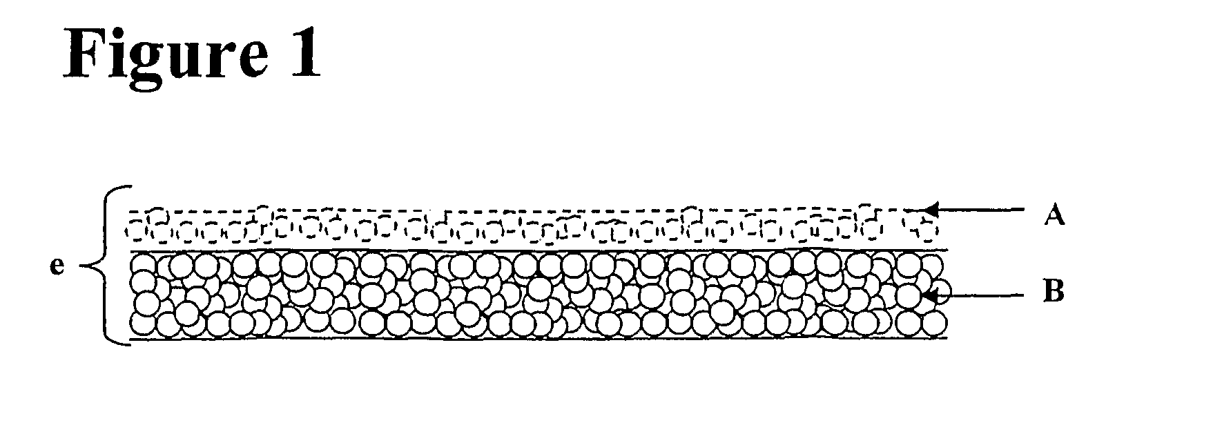
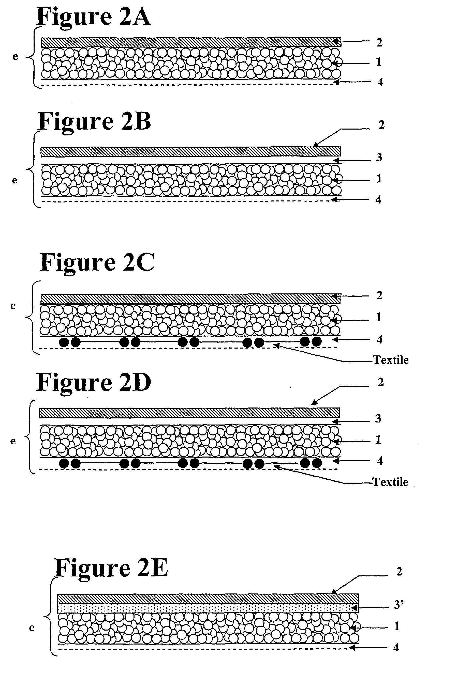
Surgical patches described herein are multi-layer structures that can, in embodiments, be self sticking and sealing and fully bioresorbable. In embodiments, the surgical patch is a bi-layered structure including a porous matrix layer (which optionally can be sub layered) that is loaded with a first hydrogel precursor and a layer made from a composition containing a second hydrogel precursor directly spread onto the porous matrix layer. In embodiments where the porous matrix layer is sub-layered, the porous matrix layer includes a first porous sublayer that is loaded with a first hydrogel precursor and a second porous sublayer that contains no hydrogel precursor onto which a layer of a second hydrogel precursor is directly spread. Generally, the first hydrogel precursor should be spatially separated from the second hydrogel precursor to prevent hydrogel precursors from reacting with each other until the implant is placed at the site of implantation and exposed to the physiological fluids of a patient.
Upon contact with tissue at the site of implantation, the implant will soak up physiological fluid. The second hydrogel precursor will be dissolved by the fluid. As the fluid wicks into and migrates across the implant, it will carry the dissolved second hydrogel precursor along through the implant. Eventually, the fluid will migrate through the implant sufficiently to reach the portion containing the first hydrogel precursor, thereby dissolving the first hydrogel precursor. The first and second hydrogel precursors will then react to form a biocompatible cross linked material, sticking the patch to the tissue at the site of implantation and sealing the defect. The biocompatible cross linked material produced by reaction of the first and second hydrogel precursors provides not only stickiness and sealant properties but also provides the implant with anti-adhesive properties between the defect and healthy surrounding tissues.
Methods for closing and healing visceral wall defects or incisions using the present surgical patch are also described. For example, the present surgical patch can improve the healing of gastro-intestinal anastomosis and be an effective approach to the management and prevention of fistula. Another example is the prevention by the present surgical patch of classic complications of polypectomy (e.g., bleeding and perforation). Another example is the use of embodiments of the present surgical patch that are reinforced with a mesh for the treatment of inguinal hernia and incisional hernia.

Problems solved by technology

Healing and sealing tissue wounds at the same time poses unique problems in visceral surgery.

Method used


Image

  • Surgical patch
  • Surgical patch
  • Surgical patch

Examples


example 1

Preparation of Coated Mesh Reinforcement Member

A knitted isoelastic, multifilament polyglycolic acid mesh reinforcement member is coated in a solution of porcine collagen at 0.8% w / v, by soaking it in the solution, spin-drying it and leaving it to dry under a laminar flow. This cycle of processes is repeated up to two times in order to obtain covering of the yarns.

The collagen used is porcine collagen type I, extracted from porcine dermis by solubilization at acidic pH or by digestion with pepsin, and purified by saline precipitations according to known techniques.

Dry collagen fibres obtained by precipitation of an acid solution of collagen by adding NaCl, and then washing and drying of the precipitate obtained with aqueous solutions of acetone having an increasing concentration of 80% to 100%, are preferably used.

At the end of the coating, the collagen deposited on the knit is crosslinked with glutaraldehyde at 0.5% w / v (aqueous solution of glutaraldehyde at 25%, w / v, sold by the c...

example 2

Preparation of the Porous Matrix

A suspension of collagen is prepared by mixing 60.5 g of glutaraldehyde-crosslinked collagen suspension at 1% w / w and 60.5 g oxidized collagen solution at 1% w / w. The pH of the collagen suspension thus obtained is then increased to 7 and tri-lysine is added to the blend as a first hydrogel precursor at a final concentration of 2.5 mg / ml. Then the suspension poured in a 17×12 cm box and is further lyophilized according to the following method: freezing is carried out as rapidly as possible, by decreasing the temperature of the product from 8° C. to −45° C., generally in less than 2 hours. Primary desiccation is initiated at −45° C., at a pressure of from 0.1 to 0.5 mbar. During this step, the temperature is gradually increased, with successive slopes and plateaux, to +30° C. The lyophilization ends with secondary desiccation, at +30° C., for 1 to 24 hours. Preferably, the vacuum at the end of secondary desiccation is between 0.005 and 0.2 mbar. The tot...

example 3

Preparation of the Porous Matrix

A suspension of collagen is prepared by mixing 60.5 g of glutaraldehyde-crosslinked collagen suspension at 1% w / w and 60.5 g oxidized collagen solution at 1% w / w. The pH of the collagen suspension thus obtained is then increased to 7. Then the suspension is poured in a 17×12 cm box and is further lyophilized according to the following method: freezing is carried out as rapidly as possible, by decreasing the temperature of the product from 8° C. to −45° C., generally in less than 2 hours. Primary desiccation is initiated at −45° C., at a pressure of from 0.1 to 0.5 mbar. During this step, the temperature is gradually increased, with successive slopes and plateaux, to +30° C. The lyophilization ends with secondary desiccation, at +30° C., for 1 to 24 hours. Preferably, the vacuum at the end of secondary desiccation is between 0.005 and 0.2 mbar. The total lyophilization time is from 18 to 72 hours.

Alternate Method for the Preparation of the Porous Matri...


PUM

PropertyMeasurementUnit
Structureaaaaaaaaaa
Bioabsorbableaaaaaaaaaa

Abstract

Multi-layer structures are useful as surgical patches.

Description

TECHNICAL FIELDThe present disclosure relates to a patch or implant for visceral surgery. More particularly, it concerns methods and compositions useful for sealing and / or healing damaged visceral tissues. In certain aspects, the present patches can also minimize the formation of post-surgical adhesions.BACKGROUNDHealing and sealing tissue wounds at the same time poses unique problems in visceral surgery. Devices (such as patches, meshes, plugs, etc.) that are used for the repair and / or regeneration of surrounding damaged visceral tissues need to be secured to tissue surrounding the wound. Sealing should be done without compromising the healing and by using the easiest techniques.Sealing tissue wounds has been achieved via suturing or stapling to surrounding tissues. Alternative to suturing and stapling have been developed for closing openings in tissue such as incisions, wounds, anastomosis and fistulae. They include the use of i) biological glues such as fibrin sealant, gelatine-r...

Claims


Application Information

Patent Timeline
IPC IPC(8): A61B17/03
CPCA61L27/48A61L27/52A61L27/56C08L89/06A61B2017/00004A61B17/0057A61B2017/00659
Inventor LADET, SEBASTIENBAYON, YVES
Owner SOFRADIM PROD SAS
Why Eureka
Eureka Blog
Learn More