Antibodies Against Hmgb1 and Fragments Thereof
a technology of hmgb1 and antibodies, applied in the field of antibodies against hmgb1 and, can solve the problems of elevated serum hmgb1 levels that are toxi
- Summary
- Abstract
- Description
- Claims
- Application Information
AI Technical Summary
Problems solved by technology
Method used
Image
Examples
example 1
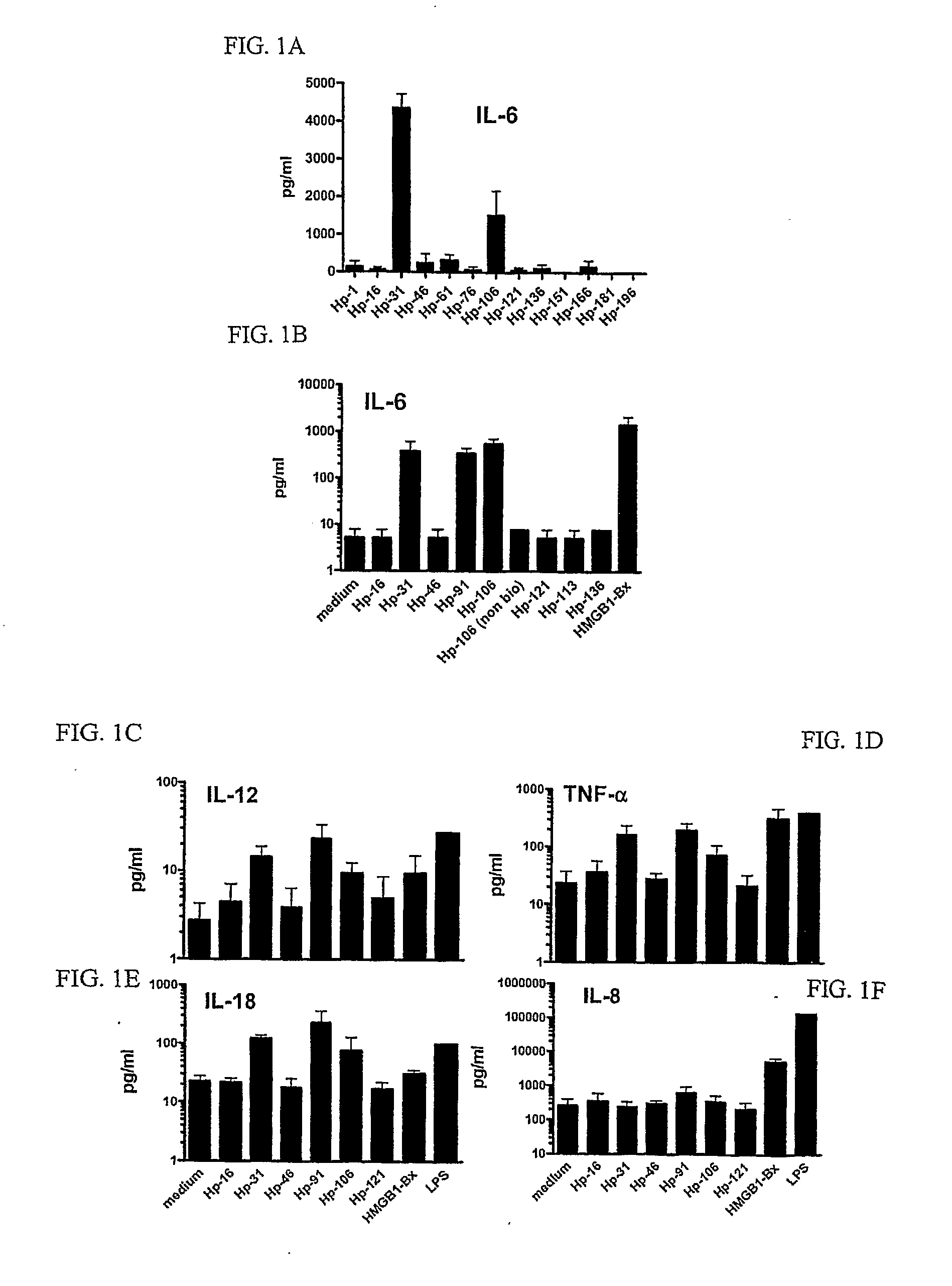
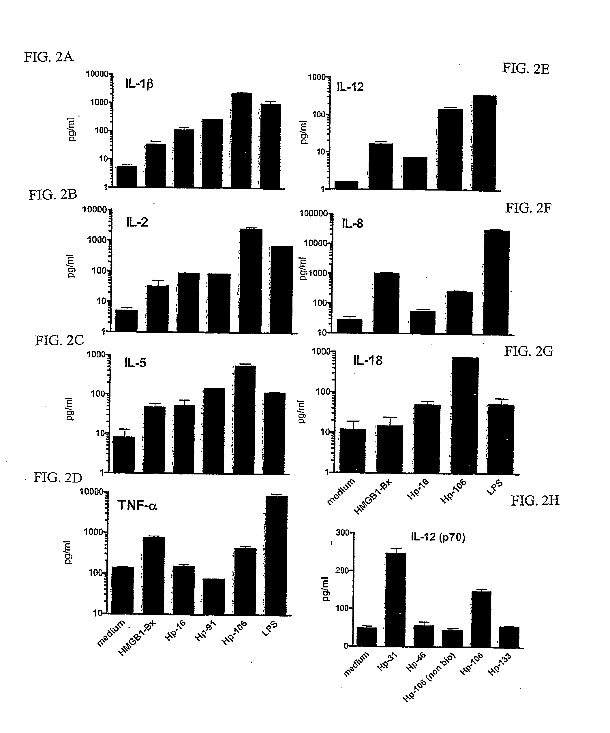
Four 18 Amino Acid HMGB1 Peptides Induce Cytokine Secretion in Human and Murine Dendritic Cells
Materials and Methods:
Generation of Human Dendritic Cells (DCs)
[0106]Peripheral blood mononuclear cells (PBMCs) were isolated from the blood of normal volunteers (Long Island Blood Services, Melville, N.Y.) over a Ficoll-Hypaque (Amersham Biosciences, Uppsala, Sweden) density gradient. CD14+ monocytes were isolated from PBMCs by positive selection using anti-CD14 beads (Miltenyi Biotech., Auburn, Calif.), following the manufacturer's instructions. To generate DCs, CD14+ cells were cultured in RPMI 1640 medium supplemented with 2 mM L-glutamine (GIBCO-BRL Life Technologies, Grand Island, N.Y.), 50 μM 2-mercaptoethanol (Sigma, St. Louis, Mo.), 10 mM HEPES (GIBCO-BRL), penicillin (100 U / ml), streptomycin (100 μg / ml) (GIBCO-BRL), and 5% human AB serum (Gemini Bio-Products, Woodland, Calif.). Cultures were maintained for 7 days in 6-well trays (3×10⁶ cells / well) supplemented with 1000 U GM-CSF ...
example 2
Select HMGB1 Peptides Induce Phenotypic Maturation of Murine BM-DCs
Materials and Methods
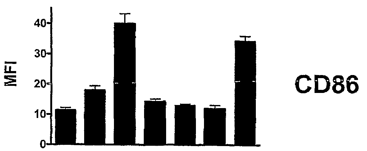
[0114]In order to determine whether HMGB1-Bx and / or particular HMGB1 peptides could induce phenotypic maturation of murine DCs, immature BM-DCs were exposed to either HMGB1-Bx, a particular HMGB1 peptide, or LPS (FIG. 3). Fluorescence activated cell sorting (FACS) analysis was performed on immature DCs that were cultured in the presence of either HMGB1-Bx (50 μg / ml), an HMGB1 peptide (200 μg / ml), or LPS (100 ng / ml). Untreated DCs (medium) were also tested as a control. DCs were gated on CD11c+ cells and analyzed for expression of specific maturation markers (e.g., CD86, MHC-II, CD40) by surface membrane immunofluorescence. In particular, 1×10⁴ DCs were reacted for at least 20 min at 4° C. in 100 ml of PBS / 5% FCS / 0.1% sodium azide (staining buffer) with fluorescein isothiocyanate (FITC)-conjugated IgG monoclonal antibodies (mAbs) that are specific for CD86, CD40 or MHC-II (eBioscience). Cells were...
example 3
HMGB1-Bx and Select HMGB1 Peptides Induce Functional Maturation of BM-DCs
Materials and Methods
[0116]Immature BM-DCs that were generated from C57 / BL6 mice (FIG. 4A) or Balb / c mice (FIG. 4B) were incubated for 48 h with either HMGB1-Bx (50 μg / ml), a particular HMGB1 peptide (200 μg / ml), LPS (100 ng / ml) or were left untreated (medium). T cells were isolated by negative selection using the mouse SpinSep antibody cocktail from StemCell Technologies (Vancouver, Calif.), according to the manufacturer's instructions. The cell purity of the isolated T cells was routinely ˜99% pure. In order to assess levels of T cell activation and proliferation, cells were plated at 105 cells per well in a round-bottomed 96-well tray at a DC:T cell ratio of 1:120 for 5 days in the medium described above. The microcultures were pulsed with (3H)-thymidine (1 mCi / well) for the final 8 h of culture. Cell cultures were harvested onto glass fiber filters with an automated multiple sample harvester and the amount ...
PUM
| Property | Measurement | Unit |
|---|---|---|
| concentration | aaaaa | aaaaa |
| stability | aaaaa | aaaaa |
| degree of homogeneity | aaaaa | aaaaa |
Abstract
Description
Claims
Application Information
 - R&D
- Intellectual Property
- Life Sciences
- Materials
- Tech Scout
- Unparalleled Data Quality
- Higher Quality Content
- 60% Fewer Hallucinations
Browse by: Latest US Patents, China's latest patents, Technical Efficacy Thesaurus, Application Domain, Technology Topic, Popular Technical Reports.
© 2025 PatSnap. All rights reserved.Legal|Privacy policy|Modern Slavery Act Transparency Statement|Sitemap|About US| Contact US: help@patsnap.com


