Gdf15 as molecular tool to monitor and enhance phenotypic stability of articular chondrocytes

a chondrocyte and phenotype technology, applied in the field of transforming growth factor (tgf), can solve the problems of limiting the function of joints, affecting the osteoarthritis of the involved joints, and rarely effective repair processes in healing defects, etc., and achieves the effect of enhancing the expression of matrix proteins

Inactive Publication Date: 2011-12-08
UNIV GENT
View PDF1 Cites 6 Cited by
  • Summary
  • Abstract
  • Description
  • Claims
  • Application Information

AI Technical Summary

Problems solved by technology

These lesions disrupt the congruence between the joint surfaces and therefore can lead to OA, which can be painful and severely limit the joint function.
Despite these attempts at repair, there is no appreciable increase in the bulk of cartilage matrix and the repair process is rarely effective in healing the defects.
Although initially sometimes painless, partial-thickness defects often degenerate into osteoarthritis of the involved joint.
This procedure has led to a proven at least symptomatic amelioration.
This conceptually promising approach has still wide margins for improvement, since it is known that in vitro expansion of chondrocytes results, after a limited number of cell divisions, in a loss of their phenotypic stability (as defined by the ability of chondrocytes to form hyaline cartilage in vivo) making the cell suspension to be injected unreliable.
To date, however, it is not known how far it is possible to expand chondrocytes without hampering their phenotypic stability and therefore their capacity to form stable hyaline cartilage in vivo, resistant to vascular invasion and endochondral bone formation.
However they do not measure the capacity of chondrocytes to form cartilage in vivo.

Method used


Image

  • Gdf15 as molecular tool to monitor and enhance phenotypic stability of articular chondrocytes
  • Gdf15 as molecular tool to monitor and enhance phenotypic stability of articular chondrocytes
  • Gdf15 as molecular tool to monitor and enhance phenotypic stability of articular chondrocytes

Examples


example 1

Results

Elevated GDF15 Concentrations in OA-Patients and OA-Chondrocyte Cultures

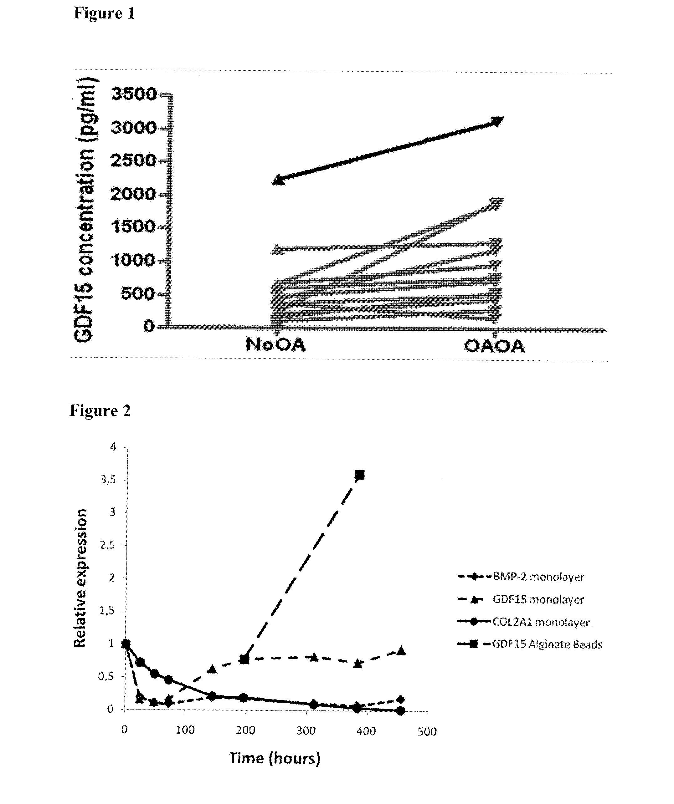
[0106]GDF15, secreted by human articular chondroctes isolated from visually intact (NoOA) and visually damaged (OAOA) zones of OA articular cartilage respectively, was analysed by measuring GDF15 concentrations in culture supernates of 13 different paired samples. FIG. 1A shows a consistent higher concentration in chondrocyte cell cultures from visually damaged zones compared to visually intact zones (p<0.01; Wilcoxon signed rank test).

example 2

Dedifferentiation Experiments

[0107]Articular Chondrocytes

[0108]Phenotypically stable articular chondrocytes isolated from 3 OA-patients were seeded in monolayer culture at low density (20.000 cells / cm2). When confluency was reached, cells were detached and reseeded at the initial density. At particular time points, cells were detached and encapsulated in alginate beads for 8 days, as described above. At indicated time points Trizol (Invitrogen) was added to the isolated cells, and RNA was extracted according to the manufacturer's instructions, followed by an additional purification step (RNeasy mini-kit (Qiagen)). This step included the digestion of DNA by deoxyribonuclease I (Invitrogen). cDNA was synthesized with oligo(dT) primers using the Superscript kit (Invitrogen).

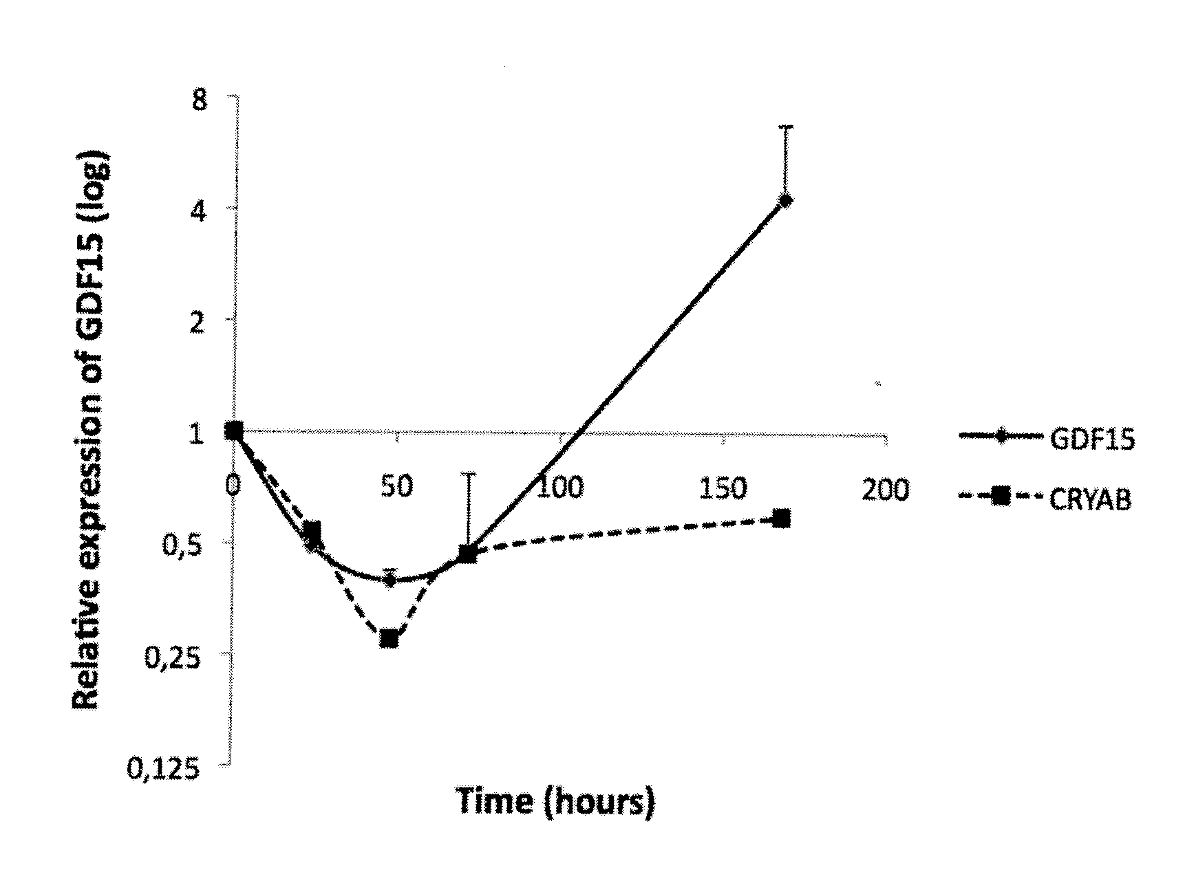
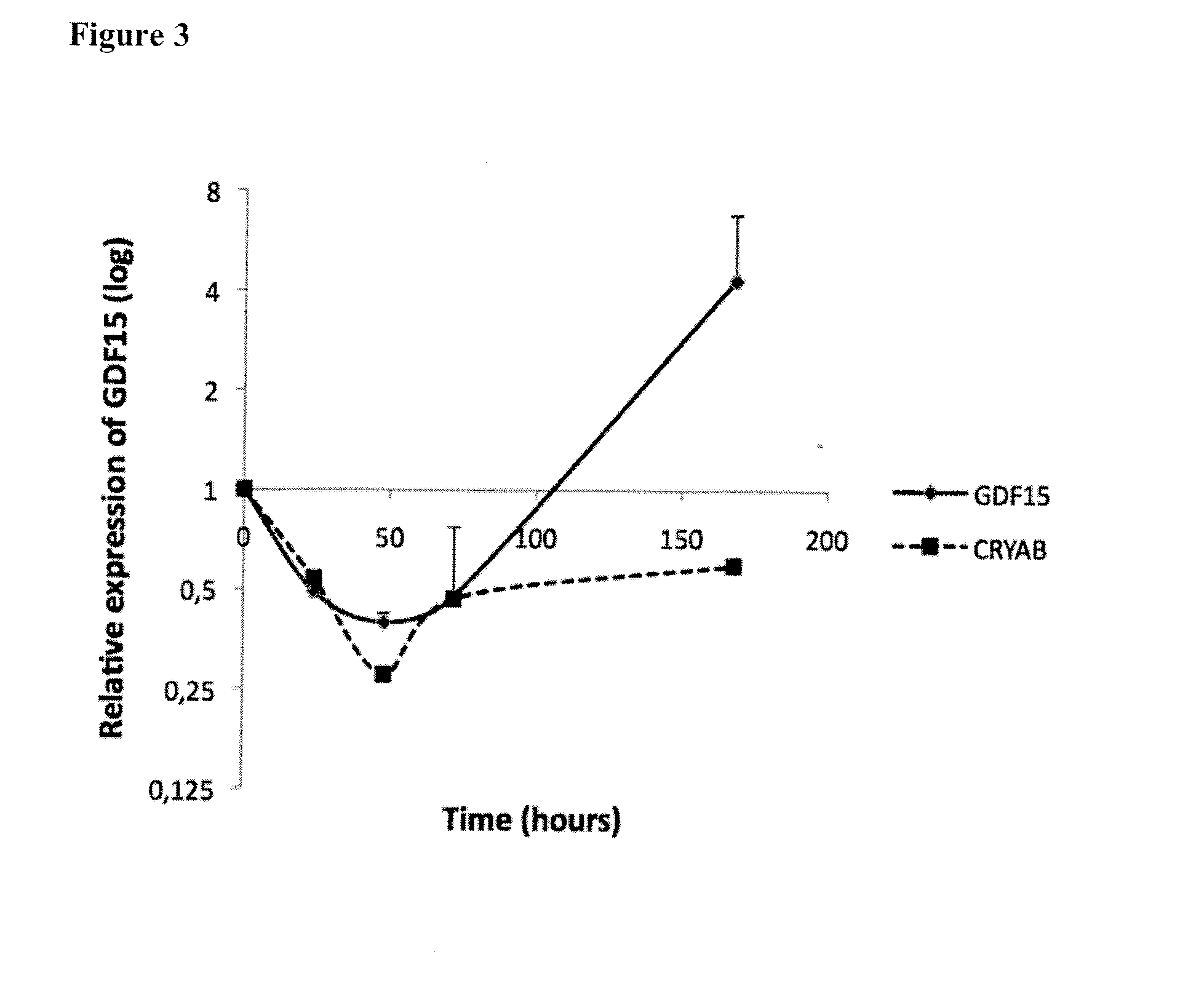
[0109]Meniscus Chondrocytes

[0110]Phenotypically stable meniscus chondrocytes, isolated from 2 OA-patients were seeded in monolayer culture at low density (20.000 cells / cm2) and allowed to expand for about 170 hours...

example 3

Western Blot Analysis

[0112]Cell lysates of articular chondrocytes were prepared by resuspending cell pellets in 40 mM Tris from the ReadyPrep Sequential Extraction Kit (Bio-Rad, Hercules, Calif., USA), supplemented with 0.1% SDS, containing protease inhibitors (Roche Diagnostics, Mannheim, Germany) and a phosphatase inhibitor-cocktail (Sigma-Aldrich, Steinheim, Germany). Cells were lysed by sonication and the proteins were isolated by centrifugation. Equal amounts (30 μg as determined by 2-D Quant kit, GE Healthcare, Fairfield, USA) were loaded on 10% SDS-PAGE gel. Equal loading was verified by Ponceau S staining (data not shown). MagicMark (Invitrogen, Paisley, UK) protein standards were run as molecular weight markers. Following 1-D gel electrophoresis, proteins were transferred to nitrocellulose membranes (Bio-Rad). The resulting membranes were immunostained with rabbit anti-GDF15 (Abcam, Cambridge, UK) followed by an anti-rabbit HRP-conjugated secondary antibody (Pierce, Rockfo...


PUM

PropertyMeasurementUnit
concentrationaaaaaaaaaa
timeaaaaaaaaaa
concentrationaaaaaaaaaa

Abstract

The present invention relates to GDF15 as a molecular marker in in vitro assays determining phenotypic stability of articular chondrocytes and predicting the outcome of chondrocyte transplantation.

Description

[0001]The invention relates generally to the field of tissue engineering. The present invention relates to GDF15 as a molecular marker in in vitro assays determining phenotypic stability of articular chondrocytes and predicting the outcome of chondrocyte transplantation.[0002]The invention further provides methods and compositions related to the generation of a population of cells suitable for the repair of cartilage, in particular in the repair of cartilage degeneration associated with osteoarthritis.BACKGROUND OF THE INVENTION [0003]The transforming growth factor-β (TGF-β) superfamily consists of an increasing number of molecules that regulate a variety of cellular processes such as growth, differentiation and oncogenesis. Members of the TGF-β superfamily have been classified into major family groupings which include TGF-β, morphogenic proteins (MP), bone morphogenic proteins (BMP), osteogenic proteins (OP), growth and differentiation factors (GDF), inhibins / activins, mullerian in...

Claims


Application Information

Patent Timeline
Patent Type & Authority Applications(United States)
IPC IPC(8): G01N33/566C12Q1/68C12N5/077C12Q1/02C12N5/071
CPCG01N33/6887
Inventor LAMBRECHT, STIJNDEFORCE, DIETERELEWAUT, DIRKVERBURGGEN, AUGUST
Owner UNIV GENT
Why Eureka
Try Eureka