

System and method for remote melanoma screening
- Summary
- Abstract
- Description
- Claims
- Application Information
AI Technical Summary
Benefits of technology
Problems solved by technology
Method used
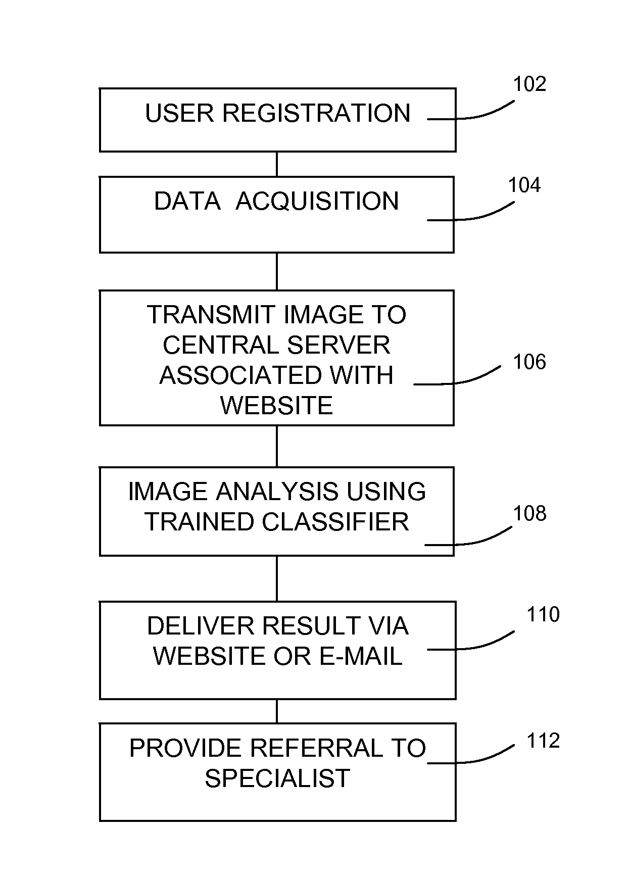
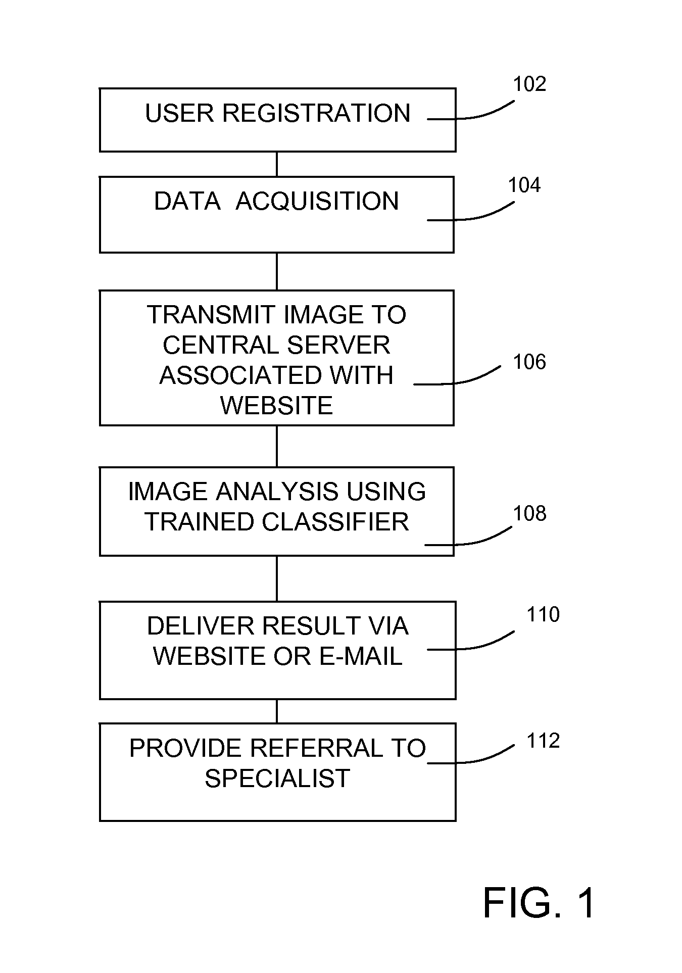
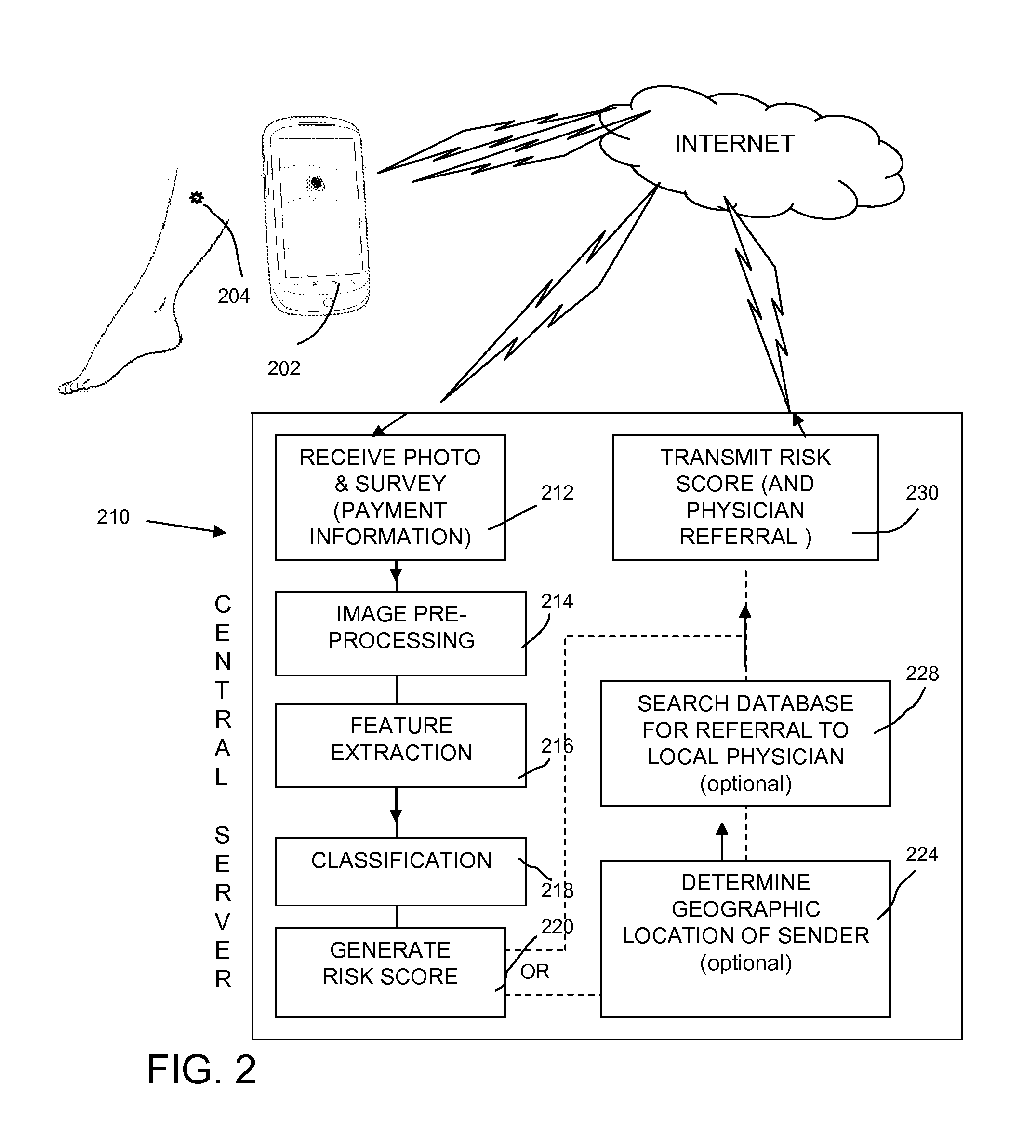
Image

Examples
example 1
Classifier Variation
[0219]Using a set of cropped images obtained from the DERMNET™ skin disease image atlas (available on the World Wide Web at dermnet.com), the recognizer that was used in the ABCD analysis described above with reference to FIG. 14, was re-trained and tested. Malignant samples (Malignant Melanoma and Lentigo Maligna) were separated from all other samples, including some very ambiguous ones that mimic melanoma.
[0220]Five-fold cross-validation experiments were performed using the same four classifiers that were used in the garbage image classification processing described above. The four classifiers were Naïve Bayes, Linear kernel, Polynomial—2nd degree (Poly 2) kernel, and RBF kernel, were used to process the previously-described data representation of ABC features. The best result was obtained using a linear kernel, with AUC=0.78. An ensemble classifier voting was tested on classifiers trained independently on A, B, and C features yielding AUC=0.75. An AUC of 0.80 ...
example 2
Modified Pre-Processing
[0245]The DERMATLAS image dataset from Johns Hopkins University (available on the World Wide Web at dermatlas.org) was used to extract 1000 images of skin diseases with various diagnoses, including herpes, acnea, drug reactions, insect bites, squamous cell carcinoma, basal cell carcinoma, congenital nevi, other benign moles, and melanoma. Associate with the images are annotations both in the form of MS access database and MS Excel spreadsheets. The annotations include age and gender of the patient, data source information, and diagnosis.
[0246]Software was written to crop the images into 1717 cropped pictures (some coming from the same image). The software also allows a framing box to be drawn around the lesion, such as described above with reference to FIG. 17a. The framing box facilitates segmentation of the mole from the background and is in a similar form to the data that would be collected using the smart phone application.
[0247]The original annotations do...
PUM
 Abstract
Description
Claims
Application Information