
Method and apparatus for minimizing thermal trauma to an organ during tissue ablation of a different organ

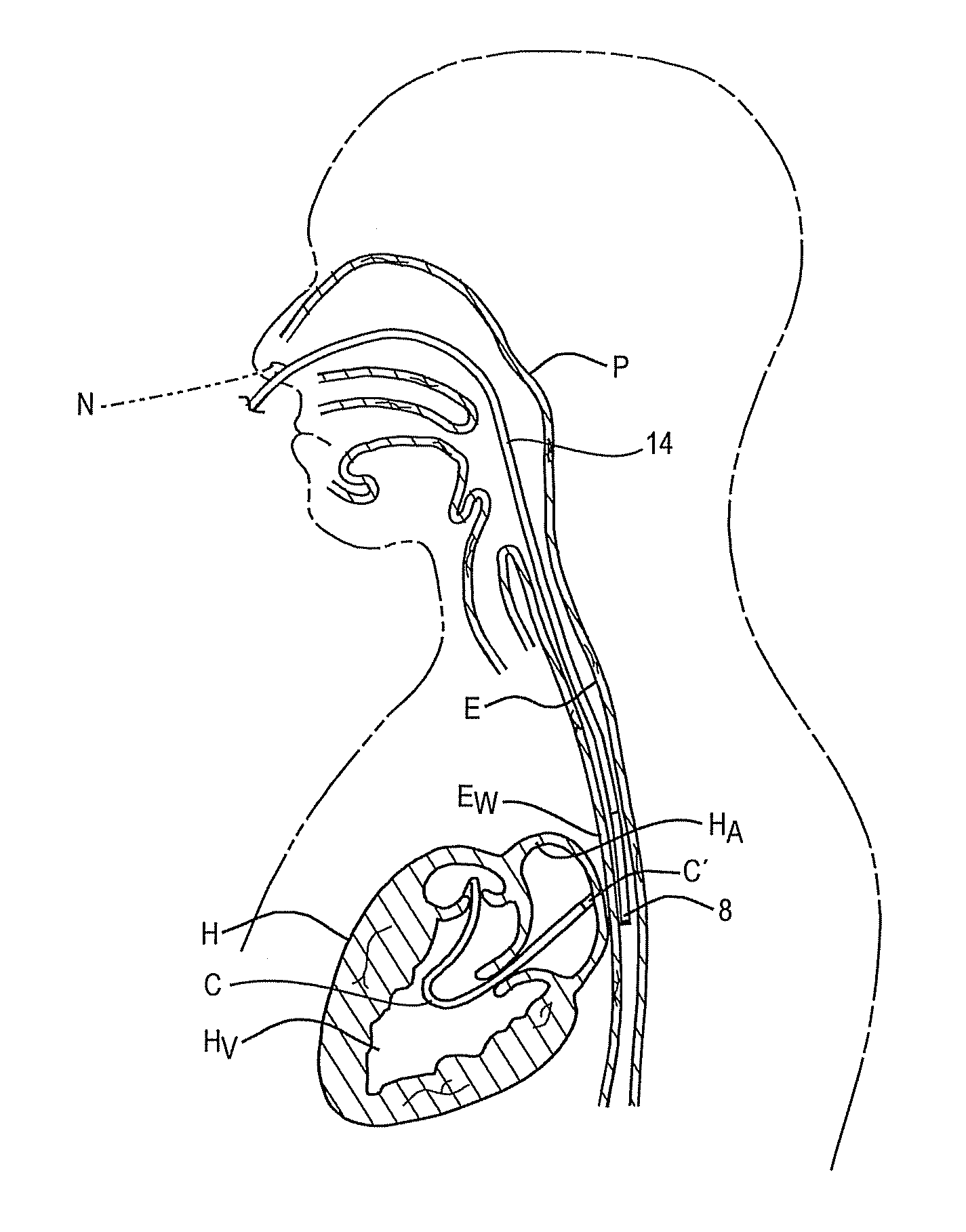
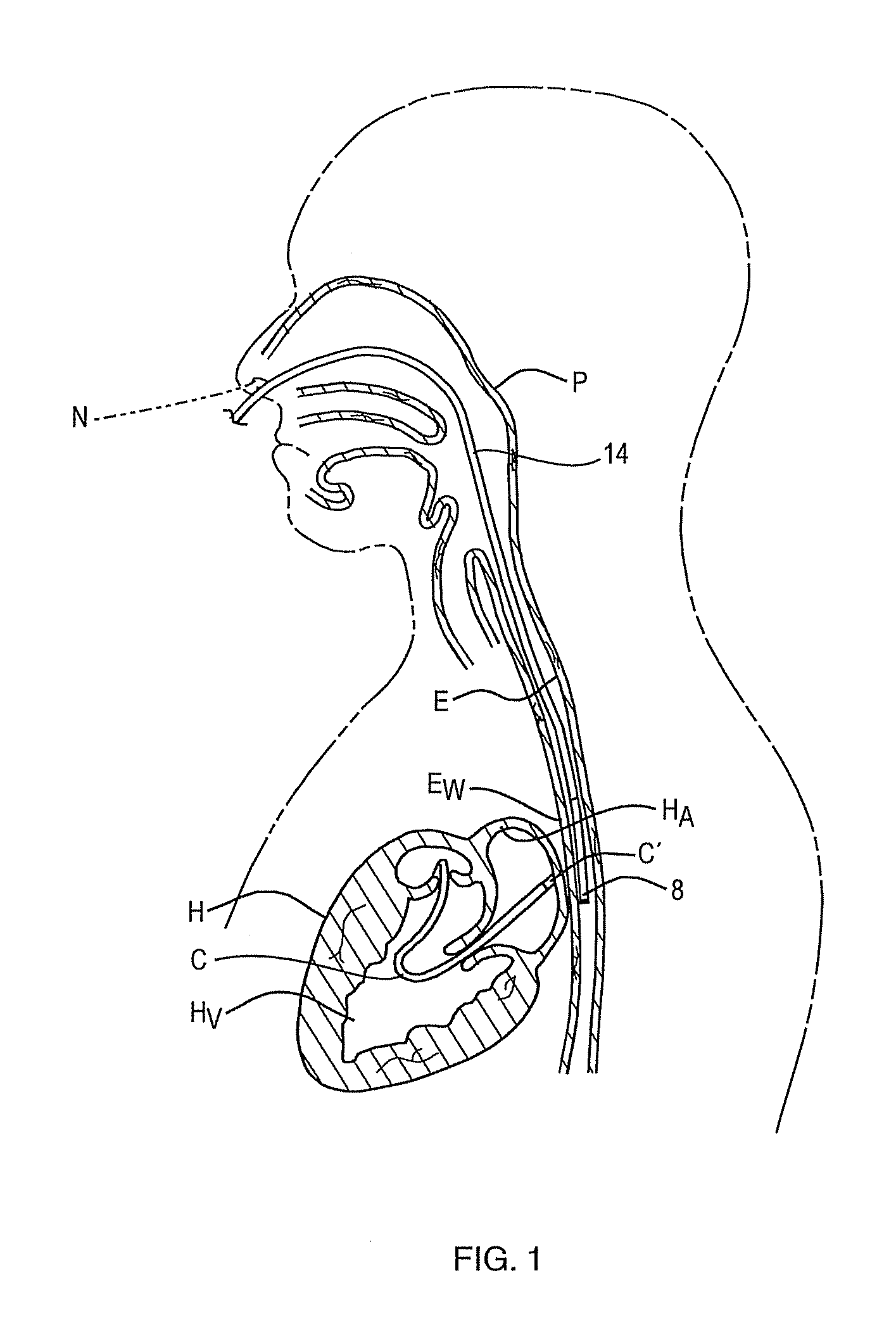
a tissue ablation and esophagus technology, applied in the field of methods and apparatus for minimizing thermal injury to the esophagus during a cardiac ablation procedure, can solve the problems of intentional, irreversible damage to the heart tissue, esophagus damage, severe consequences, etc., and achieve the effect of accurately measuring the temperature of the esophagus wall

Inactive Publication Date: 2012-02-09
ADVANCED CARDIAC THERAPEUTICS
View PDF10 Cites 69 Cited by
  • Summary
  • Abstract
  • Description
  • Claims
  • Application Information

AI Technical Summary

Benefits of technology

[0009]Accordingly, it is an object of this invention to provide a method for accurately measuring esophageal wall temperature at depth whether or not the esophagus is being cooled.
[0010]Another object of the invention is to provide a method for effectively cooling the inner surface of the esophagus during an ablation procedure in order to protect the esophagus from unintended thermal damage while accurately measuring the temperature at depth and at the outer surface of the esophageal wall.
[0011]A further object of the invention is to provide a method for accurately measuring the effectiveness of the overall cooling not only of the inner surface of the esophagus, but also deep in the esophagus wall and at the outer surface thereof.

Problems solved by technology

Thus, ablating certain regions of the left atrium to treat various arrhythmias in the heart can unintentionally cause thermal damage to the esophagus, often with severe consequences.
The point of highest resistance in this circuit, normally the interface between the catheter tip and the heart tissue, is the region which heats the most and thus may cause intentional, irreversible damage to the heart tissue to correct the arrhythmia.
A signal from the sensor is applied to a display in an external control unit, enabling the operating surgeon to adjust the power to the ablation catheter as needed to provide sufficient heating of the tissue to cause necrosis, but not enough to result in surface charring of the tissue that could cause a stroke and / or the formation of microbubbles (popping) that could rupture the heart vessel wall.
However, with this artificial cooling came much deeper lesions and, due to the relatively close position of the esophagus to a region of the left atrium which is often ablated during such procedures, there is a great risk that ablating parts of the left atrium which are intended to be heated and thus destroyed, could inadvertently overheat and injure the esophagus.
This can lead to serious complications, such as ulcers of the esophagus, bleeding, perforation of the esophagus wall and even the death of the patient.
In the case of the former type esophageal catheter which only cools the esophagus, even with constant irrigation of the inner surface of the esophagus, damage can still occur in the wall or on the outer surface of the esophagus, and in this type of instrument, there is no way to know if effective cooling of the wall of the esophagus is being achieved.
That is, as with many active cooling catheters, e.g. an RF ablation catheter, once a coolant is introduced, no conventional temperature sensors can be used to monitor tissue temperature because they only sense temperature at a point and not at depth.
Thus, even if such esophageal cooling catheters should allow for temperature measurement, they would not be able to measure accurately esophageal temperature once cooling is initiated.
Moreover, while surface cooling can be achieved with these catheters, there is no indication of the effectiveness of the cooling and there is no measurement of temperature rises at depth in the wall or at the outer surface of the esophagus.
The latter type esophageal catheter above, which has conventional temperature sensors on the outer surface thereof, is only capable of measuring the temperature of the inner surface of the esophagus and because it can only measure at a point and not at depth, it provides a very late indication of problems with overheating of the esophagus.

Method used


Image

  • Method and apparatus for minimizing thermal trauma to an organ during tissue ablation of a different organ
  • Method and apparatus for minimizing thermal trauma to an organ during tissue ablation of a different organ
  • Method and apparatus for minimizing thermal trauma to an organ during tissue ablation of a different organ

Examples


working example

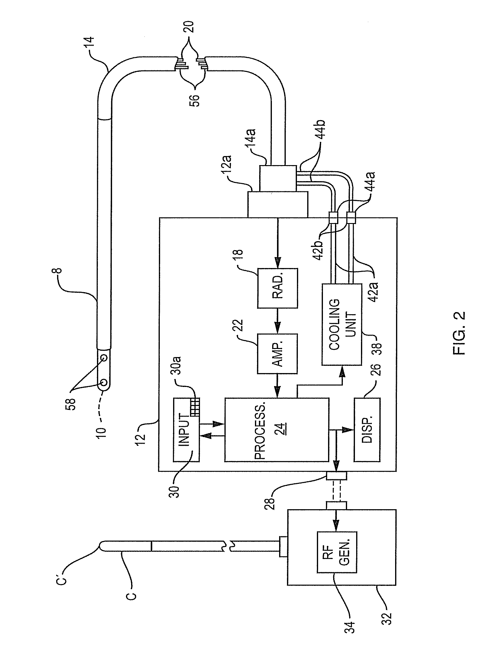
[0047]A test was performed using the temperature probe 8 depicted in FIG. 4 to verify that the temperature at depth in tissue can be recorded while part of the tissue is being cooled. Testing was done with the delivery of microwave power at 2.4 GHz via a catheter C to tissue which was actively cooled by body temperature saline solution running under the tissue to simulate blood flow and the probe 8 was positioned to record the temperature at depth in the tissue. The probe 8 in FIG. 4 is similar to probe 8 in FIG. 3 except that it has a body of low dielectric material above the antenna which causes the antenna to “look” down into the tissue as seen from the longitudinal sectional view of the antenna pattern in FIG. 4, i.e. the antenna is directional. The antenna in probe 8 operates at a frequency of 4 GHz. It should be noted that the antenna pattern in FIG. 4 was obtained with the antenna in the transmit or radiate mode rather than the receive mode because this is the usual custom si...


PUM


Abstract

A method of minimizing thermal trauma during tissue ablation includes the steps of placing an ablation catheter at an ablation site on a first organ in a patient's body, providing energy to the ablation catheter to heat first organ tissue at the ablation site, providing microwave radiometry apparatus including a probe containing a microwave antenna and a radiometer responsive to the antenna output for producing a temperature signal corresponding to the thermal radiation picked up by the antenna and positioning the probe in a body passage of a second organ in the patient's body having a wall portion adjacent to the ablation site so that the microwave antenna is located at a measurement site opposite the ablation site. Using the radiometry apparatus, the temperature at depth in the second organ tissue at the measurement site is measured to provide a corresponding temperature signal, and the ablation catheter is controlled in response to the temperature signal to maintain the temperature of the second organ tissue below a predetermined value that does not result in thermal trauma to the second organ tissue. Apparatus for carrying out the method is also disclosed.

Description

RELATED APPLICATION[0001]This application claims the benefit of U.S. Provisional Application Ser. No. 61 / 145,800, filed Jan. 20, 2009.BACKGROUND OF THE INVENTION[0002]This invention relates to method and apparatus for minimizing thermal injury to the esophagus during a cardiac ablation procedure. Anatomically, the esophagus is very close to, and often in contact with, part of the left atrium. Thus, ablating certain regions of the left atrium to treat various arrhythmias in the heart can unintentionally cause thermal damage to the esophagus, often with severe consequences. The present invention relates especially to a technique for measuring and monitoring the temperature of the esophagus wall at depth so as to avoid overheating that wall during cardiac ablation.[0003]During a typical cardiac ablation procedure, an electrode catheter is used to resistively heat heart tissue, usually at the left side of the heart, sufficiently to intentionally damage the target tissue in order to cure...

Claims


Application Information

Patent Timeline
IPC IPC(8): A61B18/18A61B18/04
CPCA61B5/01A61B90/04A61B18/00A61B18/10A61B18/1492A61B2017/00084A61B2017/00243A61B2017/00274A61B2018/00011A61B2018/00023A61B2018/00351A61B2018/00505A61B2018/00547A61B2018/00642A61B2018/00702A61B2018/00744A61B2018/00767A61B2018/00791A61B5/0507
Inventor LENIHAN, TIMOTHY J.
Owner ADVANCED CARDIAC THERAPEUTICS
Learn More