
System and method for estimating radiation dose and distribution using medium-dependent-correction based algorithms (MDC)

Inactive Publication Date: 2012-02-16
VANDERBILT UNIV
View PDF1 Cites 35 Cited by
  • Summary
  • Abstract
  • Description
  • Claims
  • Application Information

AI Technical Summary

Benefits of technology

[0008]In a second embodiment, a method for accurately calculating radiation doses in biological tissues exposed to a radiation source is provided. The method includes computing a dose deposition kernel using convolution / superposition dose calculations based on a configuration of the radiation source. The method further includes obtaining a raw geometry of the biological tissues expressed in computed tomography (CT) numbers. The method also includes determining the dose distribution (Dmedium-dependent-corrected) in the biological tissues by calculating:Dmedium-dependent-corrected(x,y,z)=Ddensity-corrected(x,y,z)fMDC(x,y,z)where Ddensity-corrected is the dose distribution calculated with a model-base calculation method, such as convolution / superposition method for example and fMDC is a Medium-Dependent-Correction factor, where fMDC is a function of effective bone depth dEB(x, y, z) matrix expressed by the following equation:fMDC(x,y,z)≡fc(dEB)where fc is a matrix of correction factor values for the system computed based on pre-defined correlation data between values for dEB(x, y, z) and values for fc.

Problems solved by technology

Frequent and repeated imaging procedures such as those performed in image-guided radiotherapy (IGRT) and diagnostic CT procedures may add significant dose to patients.
Imaging procedures are necessary due to their potential benefits, but the additional radiation also entails risk to patients.
Currently available model-based dose calculation methods are inaccurate and not suitable for low energy x-rays.
Although the Monte Carlo method is accurate; the long computational times have prevented it from practical use.
Furthermore, to the extent that the output distributions are an aggregate of the multitude of tested scenarios, current Monte Carlo simulation tools provided only a limited opportunity to interact with, and therefore develop a true understanding of, the simulation model.
An x-ray image procedure entails risk to patients because it exposes the radiation to radiosensitive organs.
However, heretofore, there has not been a dose calculation algorithm available that is fast and accurate for kV x-ray beams to meet the demand in clinical practice.
The Monte Carlo method is accurate but its calculation speed is too slow to meet the requirement.
The model based calculation algorithm is fast but its accuracy is unacceptable for kV x-ray beams.
Contrast between tissues in the body results from different rates of attenuation of x-rays within each tissue and is typically limited to differentiating between bone and soft-tissue, except in specialized applications such as mammography.
Though useful in many situations, the diagnostic information from conventional x-ray imaging is limited because the intensity of the transmitted x-rays at the detector is a result of the integrated attenuation of the x-rays along the path from the x-ray source to the detector.
While the benefit of diagnostic information provided by CT is often critical for patient care, the radiation exposure to patients also entails the risks of carcinogenesis from the increased x-ray procedures from CT imaging.
The radiation dose from imaging procedures is an increasingly important health issue as the public becomes aware of radiation exposure to patients, especially to pediatric patients, from repeated x-ray procedures in radiology.
Additional bone dose from repeated image guided procedures when added to the radiotherapy doses may further increase the risk of bone malformation and growth issues associated with pediatric radiotherapy treatment protocols.
The increased radiation exposure and the risk associated with it can no longer be ignored.
As discussed above, all currently available model-based dose calculation algorithms that may be adequate in the dose calculation for mega-voltage photon beams are incapable of calculating radiation dose from kilo-voltage x-rays leading to dose errors of up to 300% due to the increased photoelectric effect in doses to bone.
An earlier attempt of applying heterogeneity-corrected convolution dose calculations for kV x-rays was shown to be unsuccessful.
Although Monte Carlo techniques are capable of calculating patient dose resulting from x-rays, the long computation times (days) are prohibitive for practical clinical use.
The issues with Monte Carlo calculations include the very long computational times (many hours) required that limit “real time” calculations for clinical practice.
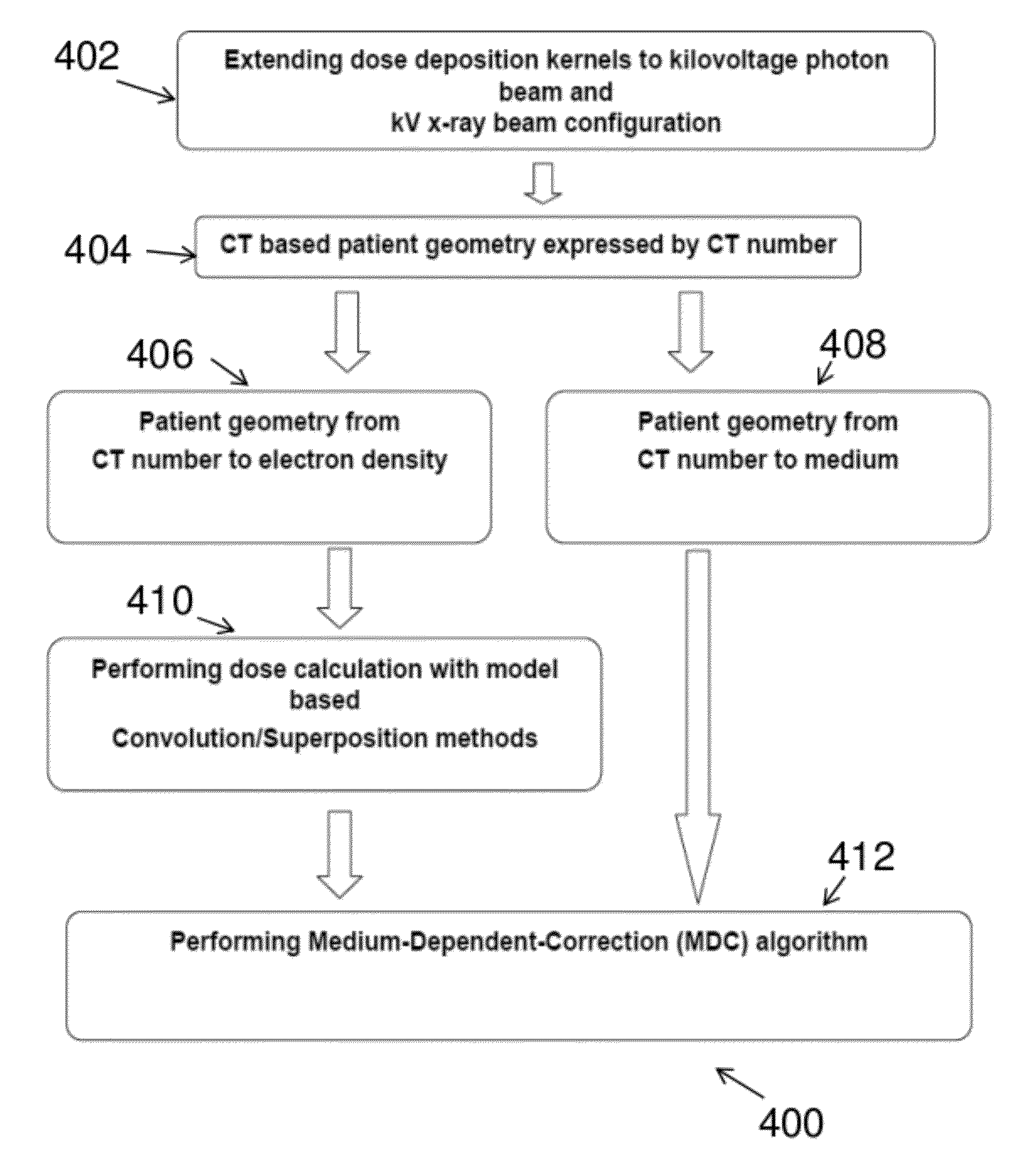
This process converts each voxel in the patient DICOM image from CT number to a specific material and density, which is not currently available in model-based commercial treatment planning systems.
We performed similar calculations with 4, 8, 32, 64, and 360 incident rays and found that calculations using less than 16 rays resulted in effective bone depth distributions that inadequately characterized the distribution of bone in the patient, and calculations using more than 16 incident rays did not show significant improvement to justify the additional calculation time.
The results of the model-based calculation (FIG. 9C) show that the currently available method is unable to calculate imaging dose from kV x-rays, most prevalently for bone anatomy.
This is in contrast to the model-based calculation, which resulted in significantly higher errors of up to −103% for bone, and 8% for soft-tissue.
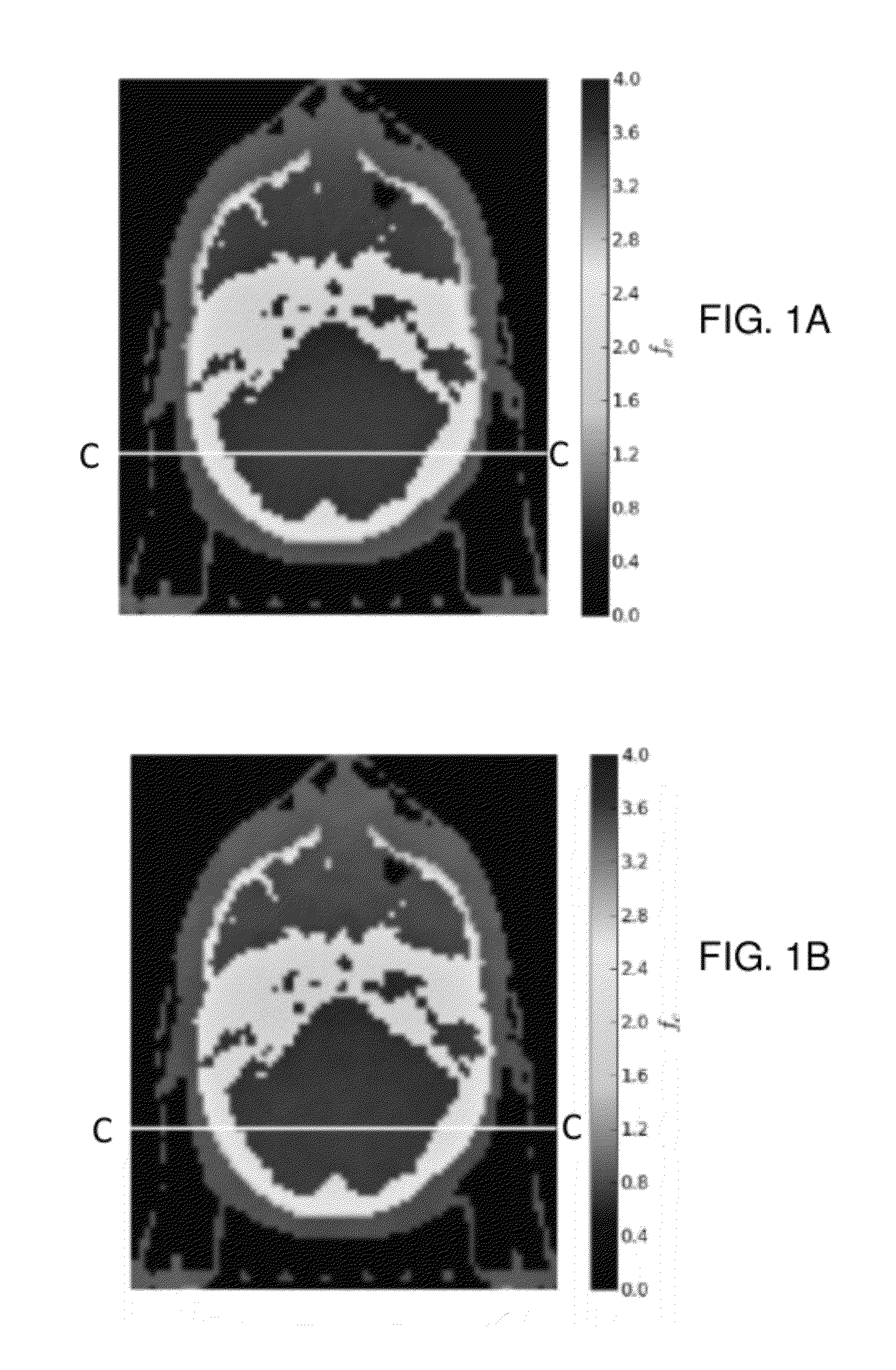
Similarly for the femoral head (FIG. 10C) the model-based correction is incapable of accurately calculating these doses.
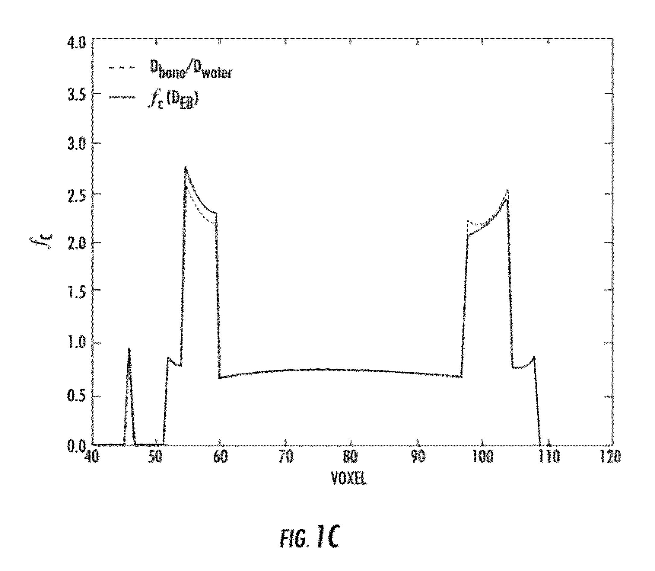
Simple application of a constant multiplicative correction factor would incorrectly predict the shape of the Monte Carlo distribution; however, the MDC-EA accurately models the dose-to-medium.
The density-corrected-only calculations result in mean bone dose errors up to −103%, with standard deviations of the distributions approaching nearly 40%.
The density-corrected-only calculation results in significant underestimation of bone dose and overestimates the soft-tissue structure dose.

Method used


Image

  • System and method for estimating radiation dose and distribution using medium-dependent-correction based algorithms (MDC)
  • System and method for estimating radiation dose and distribution using medium-dependent-correction based algorithms (MDC)
  • System and method for estimating radiation dose and distribution using medium-dependent-correction based algorithms (MDC)

Examples


Embodiment Construction

[0048]The present invention is described with reference to the attached figures, wherein like reference numerals are used throughout the figures to designate similar or equivalent elements. The figures are not drawn to scale and they are provided merely to illustrate the instant invention. Several aspects of the invention are described below with reference to example applications for illustration. It should be understood that numerous specific details, relationships, and methods are set forth to provide a full understanding of the invention. One having ordinary skill in the relevant art, however, will readily recognize that the invention can be practiced without one or more of the specific details or with other methods. The present invention is not limited by the illustrated ordering of acts or events, as some acts may occur in different orders and / or concurrently with other acts or events. Furthermore, not all illustrated acts or events are required to implement a methodology in ac...


PUM


Abstract

Systems and methods for accurately calculating radiation doses in biological tissues exposed to a radiation source are provided. In the systems and methods first, a radiation dose is computed to geometry of water equivalent medium of the biological tissues, expressed in computed tomography (CT) volumetric images. A Medium-Dependent-Correction factor that is a function of an effective bone depth matrix and the incident x-ray beam is obtained and tabulated. Using patient material and density data derived from CT images, the effective bone thickness can be calculated from a specific x-ray source. Finally, a Medium-Dependent-Correction factor that is a function of an effective bone depth matrix is used to accurately determine the radiation dose distributions to biological tissues.

Description

CROSS-REFERENCE TO RELATED APPLICATION[0001]This application claims the benefit of Provisional Application Ser. No. 61 / 363,695 entitled “SYSTEM AND METHOD FOR ESTIMATING RADIATION DOSE AND DISTRIBUTION USING MEDIUM-DEPENDENT-CORRECTION BASED ALGORITHMS (MDC)”, filed Jul. 13, 2010, which is herein incorporated by reference in its entirety.FIELD OF THE INVENTION[0002]Embodiments of the invention provide systems and methods for estimating radiation dose and distributions in biological tissues.BACKGROUND[0003]Frequent and repeated imaging procedures such as those performed in image-guided radiotherapy (IGRT) and diagnostic CT procedures may add significant dose to patients. It has been shown that kilovoltage photon beams results in doses to bone, due to the photo-electric effect, are up to a factor of 3-4 higher than those in surrounding soft tissue. Imaging procedures are necessary due to their potential benefits, but the additional radiation also entails risk to patients. There have b...

Claims


Application Information

Patent Timeline
IPC IPC(8): G06F19/00
CPCA61B6/032A61N5/1031A61B6/542A61B6/501
Inventor DING, GEORGEPAWLOWSKI, JASON
Owner VANDERBILT UNIV
Features
  • Generate Ideas
  • Intellectual Property
  • Life Sciences
  • Materials
  • Tech Scout
  • Unparalleled Data Quality
  • Higher Quality Content
  • 60% Fewer Hallucinations
Learn More