Compositions and methods for micro-rna expression profiling of cancer stem cells

a cancer stem cell and expression profiling technology, applied in the field of compositions and methods for microrna (mirna) expression profiling of cancer stem cells, can solve the problems of inability to find suitable approaches to eliminate cancer, cancer still remains a major cause of death worldwide, and the expansion of malignant tissu

Inactive Publication Date: 2012-03-01
UNIV REGENSBURG
View PDF0 Cites 75 Cited by
  • Summary
  • Abstract
  • Description
  • Claims
  • Application Information

AI Technical Summary

Problems solved by technology

Cancer still remains a major cause of death worldwide.
First, most tumors arise from a single cell, but not all the cells within a tumor are identical.
Stem cell division in normal tissues is tightly related to functional needs and thus strictly controlled but this control is lost in malignancy and consequently there is ongoing expansion of the malignant tissue.
Hence, malignant stem cells could represent a target for therapeutic intervention but suitable approaches of how to eliminate them have not been available as these cancer stem cells appear to be protected by mechanisms that render them less susceptible to therapeutic killing (Al-Hajj, M. et al.
Another unsolved issue with respect to the exploitation of the above-mentioned therapeutic concept concerns the reliable targeting of cancer stem cells.
However, little is known about the cellular functions of any of these markers, particularly whether the expression of which is directly correlated with malignant tumor growth.
Many diagnostic assays are also hampered by the fact that they are typically based on the analysis of only a single molecular marker, which might affect detection reliability and / or accuracy.
In addition, a single marker normally does not enable detailed predictions concerning latency stages, tumor progression, and the like.
However, the mechanism of how miRNAs repress translation of their target mRNAs is still a matter of controversy.
However, to date only few of these aberrantly expressed miRNAs have been directly linked with clinically relevant prognostic factors for tumor development and / or progression.
Currently, very limited information is also available with regard to mRNA target sequences recognized by such aberrantly expressed miRNAs.
Currently, only very few studies on miRNA expression analysis in stem cells are available.
However, no such miRNA signatures have been generated for any type of cancer stem cells.

Method used


Image

  • Compositions and methods for micro-rna expression profiling of cancer stem cells
  • Compositions and methods for micro-rna expression profiling of cancer stem cells
  • Compositions and methods for micro-rna expression profiling of cancer stem cells

Examples


example 1

Materials and Methods

1.1. Cell Culture

[0184]The generation of R11, R20, R28, R40, R44, and R52 cell lines from glioblastoma samples was performed as previously described (Beier, D. et al. (2007) Cancer Res. 67, 4010-4015).

1.2. Antibodies

[0185]The following antibodies were used: mouse anti-Tuj1 MMS-435p (from Covance), mouse anti-β-actin AC15 (from Abcam), rabbit anti-GFAP (from DAKO), anti-CD133-2 293C3-PE (from Miltenyi), anti-rabbit-HRP, anti-mouse-HRP (both from Sigma).

1.3. Oligonucleotides

[0186]2′O methyl oligonucleotides were chemically synthesized using RNA phosphoramidites (from Pierce) on a Äkta Oligopilot 10 DNA / RNA synthesizer (from GE healthcare), according to the manufacturer's protocol. The sequences were as follows:

hsa-miR-9* antisense:5′-ACUUUCGGUUAUCUAGCUUUAdThsa-miR-17-5p antisense:5′-ACUACCUGCACUGUAAGCACUUUGdThsa-miR-106b antisense:5′-AUCUGCACUGUCAGCACUUUAdThsa-miR-9 antisense:5′-UCAUACAGCUAGAUAACCAAAGAdThsa-miR-122 antisense:5′-ACAAACACCAUUGUCACACUCCAdTIle tRNA pr...

example 2

miRNA Expression Profiles of CD133+ and CD133− Glioblastoma Cells

2.1. Sorting / Separation of CD133+ and CD133− Glioblastoma Cell Fractions

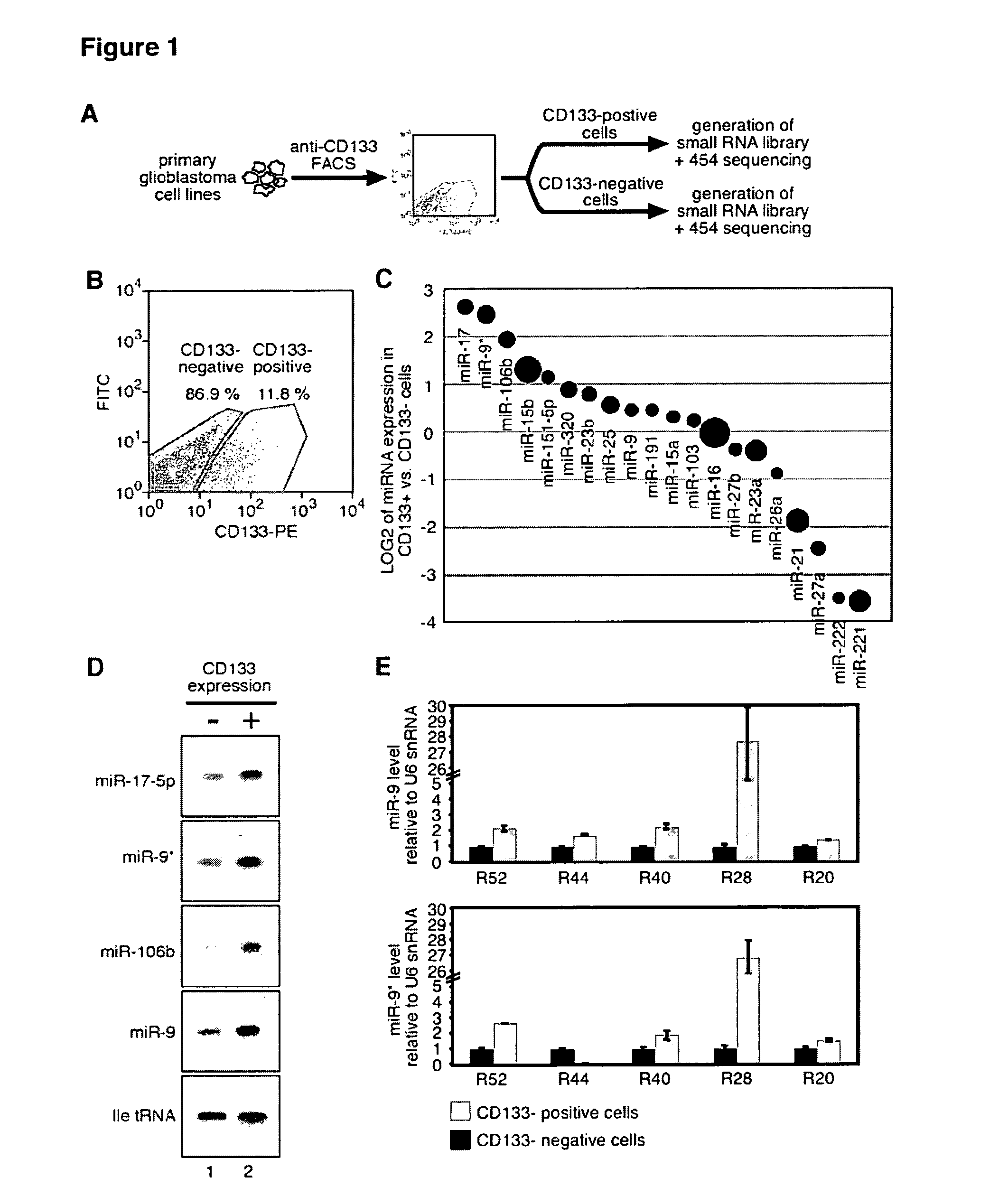
[0206]FIG. 1(A) schematically illustrates the experimental protocol employed for cell sorting. Primary human glioblastoma cell lines were incubated with ananti-CD133-phycoerythrin (PE) antibody and separated by fluorescence-assisted cell sorting (FACS) into CD133-positive and CD133-negative cell fractions. Subsequently, RNA was isolated and used to generate small RNA libraries, followed by 454 pyro-sequencing. A typical FACS profile of the primary glioblastoma cell line R11 is shown in FIG. 1(B). The dot-plot shows CD133-PE fluorescence on the x-axis and fluorescein isothiocyanate (FITC) on the y-axis as a control for auto-fluorescence. The regions indicate CD133-negative and CD133-positive cells, respectively. The CD133-positive (CD133+) and CD133-negative (CD133−) cell fractions were determined to be 11.8% and 86.9%, respectively.

[0207]Flow cytom...

example 3

Inhibition of hsa-miR-9 and hsa-miR-9* Expression Impairs Self-Renewal and Proliferation of Human Glioblastoma Cells

3.1. Effect on Cell Number / Cell Survival

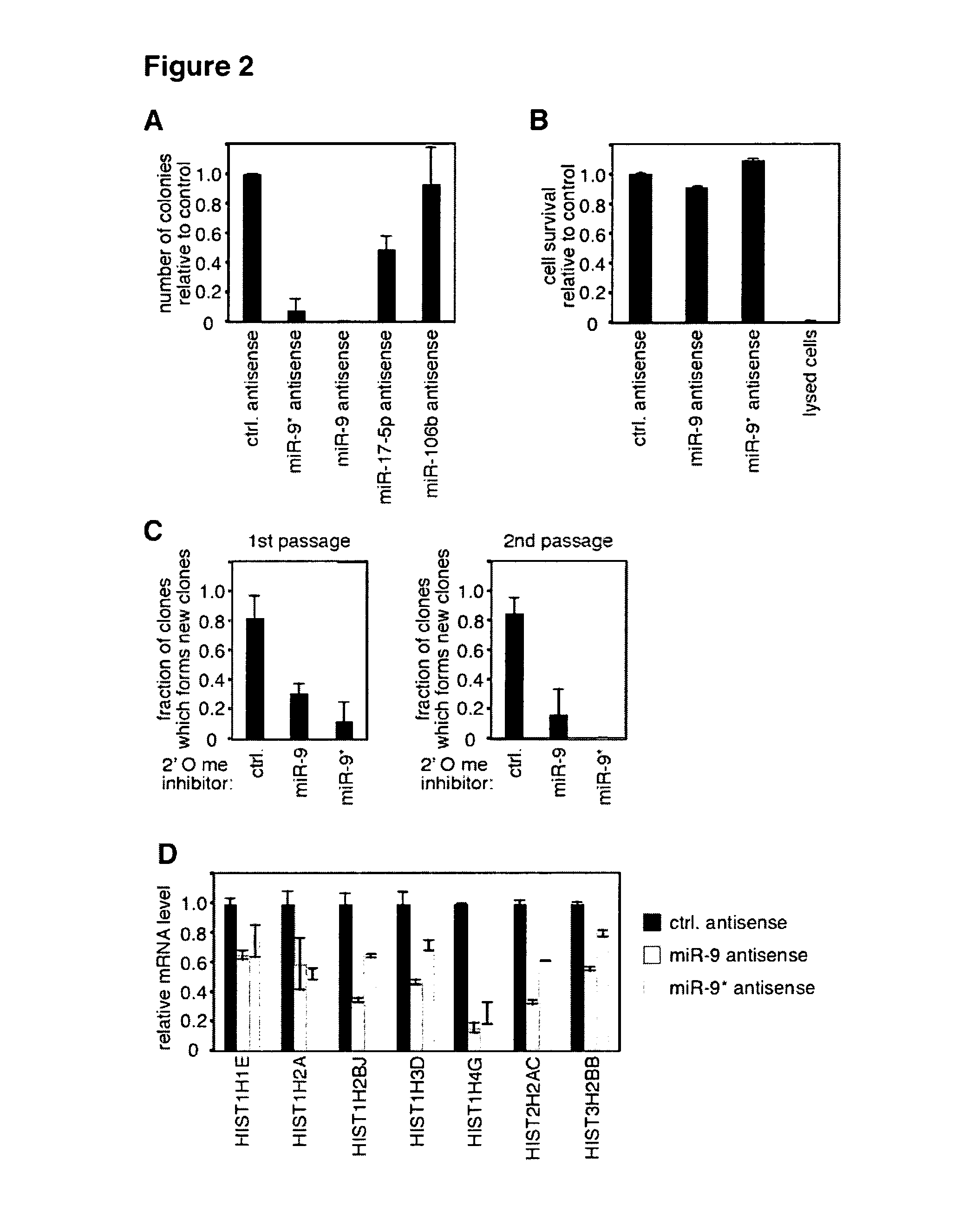
[0212]R11 cells were transfected twice with antagomirs (based on the antisense oligonucleotides described in section 1.3. above) and seeded to 48 well plates. Neurosphere-like colonies were counted 4 weeks after transfection. Transfection with hsa-miR-9 antisense completely blocked the formation of colonies, whereas transfection with hsa-miR-9* antisense resulted only in the formation of very few colonies. Transfection with hsa-miR-17-5p antisense and hsa-miR-106b resulted in less cell colonies than in the control, but the extent was much lower than with the other antagomirs (FIG. 2(A)).

[0213]In order to analyze the selectivity of the above results, cell survival rates were determined (FIG. 2(B)). R11 cells were transfected twice with antagomirs as described above (cf. section 1.15), and cell survival was assessed via measuring l...


PUM

PropertyMeasurementUnit
median survival timeaaaaaaaaaa
nucleic acid expressionaaaaaaaaaa

Abstract

The present invention relates compositions and methods for microRNA expression profiling of cancer stem cells. In particular, the invention relates to a method for identifying and / or diagnosing one or more cancer stem cells, the method comprising identifying from a plurality of nucleic acid molecules, each encoding a microRNA sequence, one or more nucleic acid molecules are differentially expressed in the cancer stem cells and in one or more control cells, wherein the one or more differentially expressed nucleic acid molecules together represent a nucleic acid expression signature that is indicative for the presence of cancer stem cells. The invention further relates to a corresponding diagnostic kit of molecular markers, namely the nucleic acid expression signature. Finally, the invention is directed to a method using such nucleic acid expression signatures for preventing the proliferation and / or self-renewal of such cancer stem cells as well as to a corresponding pharmaceutical composition.

Description

FIELD OF THE INVENTION[0001]The present invention relates to compositions and methods for microRNA (miRNA) expression profiling of cancer stem cells, particularly of stem cells derived from neuronal and / or glial tumors.BACKGROUND[0002]Cancer still remains a major cause of death worldwide. Although a better understanding of the regulatory mechanisms underlying tumor etiology and progression has enabled increasing therapeutic success for some types of malignancy, for others (e.g., stomach cancer, pancreas cancer, glioblastoma) there has been little or almost no improvement in rates of long-term patient survival.[0003]Cancerous cells develop from normal cells that gain the ability to proliferate aberrantly and to turn malignant. These malignant cells then grow clonally into tumors, eventually having the potential to metastasize. For decades, a central question in cancer biology has thus concerned the type of cells that can be transformed to form tumors.[0004]Two important observations ...

Claims


Application Information

Patent Timeline
Patent Type & Authority Applications(United States)
IPC IPC(8): A61K31/7088A61P35/00C12Q1/68
CPCA61K31/7105C12N15/113C12Q2600/178C12N2310/141C12Q1/6886C12N2310/113A61P35/00
Inventor MEISTER, GUNTERWEINMANN, LASSEBEIER, DAGMARBEIER, CHRISTOPH
Owner UNIV REGENSBURG
Why Eureka
Try Eureka